Original Article
miR-17-5p promotes proliferation and migration
of CAL-27 human tongue squamous cell carcinoma
cells involved in autophagy inhibition under hypoxia
Fawei Pang1,2, Chao Liu2,3, Yanjun Cui4, Kun Gong5, Guangping Liu1,6, Yuanyuan Bian1,2, Xiaoli Gao1,2, Dongsheng Zhang1,2
1Department of Oral and Maxillofacial Surgery, School of Stomatology, Shandong University, Jinan, Shandong,
China; 2Department of Oral and Maxillofacial Surgery, Shandong Provincial Hospital Affiliated to Shandong
University, Jinan, Shandong, China; 3Department of Oromaxillofacial Head and Neck Oncology, Shanghai Ninth
People’s Hospital, College of Stomatology, Shanghai Jiao Tong University School of Medicine, Shanghai, China;
4Linyi Traditional Chinese Medicine Hospital, Linyi, Shandong, China; 5Department of Oral and Maxillofacial
Surgery, Yantai Stomatological Hospital, Yantai, Shandong, China; 6Xintai People’s Hospital Affiliated of Taishan
Medical University, Xintai, Shandong, China
Received March 6, 2019; Accepted March 28, 2019; Epub June 1, 2019; Published June 15, 2019
Abstract: Autophagy contributes to head and neck squamous cell carcinoma (HNSCC) development and progres-sion. MiR-17-5p down-regulates Beclin-1 and thus plays an important role in autophagy, but little is known about the function and regulation of miR-17-5p in HNSCC autophagy. This study aimed to investigate the role of miR-17-5p on proliferation, migration, and autophagy under hypoxia in 27 human tongue squamous cell carcinoma cells. CAL-27 cells were transfected with 50 nmol miR-17-5p mimics to overexpress miR-17-5p. Cell proliferation and migration were determined by CCK-8 and wound healing assays, respectively, under hypoxia. Autophagy induced by hypoxia was detected by transmission electron microscope and Beclin-1 mRNA and protein expressions. The miR-17-5p mimics successfully increased the expression of miR-17-5p in CAL-27 cells by almost 700 fold compared with the miRNA mimic negative control. After 3 days, cells transfected with the miR-17-5p mimics showed higher prolifera-tion compared with controls (P < 0.05) under hypoxia. MiR-17-5p transfected CAL-27 cells had a higher migratory capacity compared with the control cells (P < 0.05) under hypoxia. Furthermore, transmission electron microscopy showed that miR-17-5p overexpression inhibited the formation of autophagosomes in hypoxic cells. Real-time quan-titative polymerase chain reaction (RT-qPCR) and western blot showed that miR-17-5p overexpression inhibited the mRNA and protein expression of Beclin-1 in CAL-27 cells submitted to hypoxia. MiR-17-5p overexpression promoted the proliferation and migration of the CAL-27 cells, but inhibited autophagy under hypoxia.
Keywords: miR-17-5p, autophagy, Beclin-1, head and neck squamous cell carcinoma, proliferation, migration
Introduction
Head and neck squamous cell carcinoma (HN- SCC) is the 6th cancer in term of worldwide inci-dence [1]. About 90% of all head and neck can-cers is HNSCC [2]. HNSCC may affect the lip, oral cavity, pharynx, and larynx. The risk factors for HNSCC include smoking, alcohol consump-tion, and human papilloma virus (HPV) infection [3]. Over the past decades, the diagnosis and treatment of HNSCC have made great progress, but mortality and prognosis have not been sig-nificantly improved [3]. A comprehensive multi-disciplinary approach is needed to treat HNSCC,
but the 5-year survival rate of patients with HNSCC is only about 50% [4]. HNSCC often shows factors of poor prognosis such as poor differentiation and lymph node invasion, but the molecular mechanisms are still poorly un- derstood. The study of HNSCC development at the molecular level could be of great signifi-cance for its prevention, control, and treat- ment.
ponse is critical [5]. Autophagy takes part in many physiologic and pathologic processes such as development, differentiation, tumor, and neurodegenerative diseases [6]. Data ab- out autophagy and its role in tumorigenesis are inconsistent among cancer types and organs [7]. In early cancer, autophagy can inhibit tumor formation; during cancer development, moder-ate autophagy will influence various death stim-uli, thereby promoting tumor survival, and ex- cessive autophagy can also induce autophagic cell death [8]. Cancer cells can enhance au- tophagy in order to survive to microenviron-ment stresses such as hypoxia and nutrient deprivation [9]. Indeed, hypoxia is commonly encountered in tumors since fast-growing tu- mors may lack the microcirculation necessary to provide oxygen to all cancer cells [10]. The hypoxia inducible factor (HIF) is central in the response to hypoxia and promotes autophagy, angiogenesis, metastasis, and resistance to chemotherapy and radiation therapy [10, 11]. In HNSCC, the autophagy induced by hypoxia is associated with an aggressive phenotype [12]. The complex network involved in the response to hypoxia and autophagy is still poorly under-stood. Nevertheless, Beclin-1 is recognized to play a critical role in autophagy and cell death by interacting with either BCL-2 or PI3K class III [13]. Zhao et al. [14] found that Beclin-1 and LC3B may contribute to the development of metastatic colorectal carcinoma. The study of autophagy in HNSCC and its regulation mecha-nism has wide clinical significance and good application prospects in the treatment of HN- SCC [15].
MicroRNAs (miRNAs) are short non-coding RNA molecules that play a pivotal role in the regula-tion of the expression of a number of genes. The abnormal expression of miRNAs plays a key role in the course of many diseases includ-ing cancer [16]. Previous studies have shown that miR-17-5p is involved in the development of colorectal, prostate, ovarian, pancreatic, ga- stric, esophageal, lung, and nasopharyngeal ca- ncers [17-20], but the role of miR-17-5p in the development of HNSCC remains elusive. The expression level of miR-17-5p is associated with cancer aggressiveness and therapy resis-tance [21]. In glioma cells, overexpression of miR-17-5p decreases Beclin-1-mediated auto- phagy and sensitizes the cells to radiation [22]. In non-small lung cancer, downregulation of miR-17-5p is associated with upregulation of
Beclin-1 and resistance to paclitaxel [23]. He- nce, miR-17-5p is involved in cancer develop-ment and progression by autophagy.
Despite vast research, the molecular mecha-nisms involved in hypoxia-induced autophagy of HNSCC cells are still poorly understood. Therefore, the aim of the present study was to transfect miR-17-5p mimics into CAL-27 human tongue squamous cell carcinoma cells to explore the role of miR-17-5p in CAL-27 cell pro-liferation, migration, and autophagy induced by hypoxia. The results could help unravel the mechanisms involved in hypoxia-induced au- tophagy and could provide hints for targeted treatments.
Materials and methods
Cells and treatment
The CAL-27 human tongue squamous cell carci-noma cell line was provided by the 9th People’s Hospital Affiliated to Shanghai Jiao Tong Uni- versity School of Medicine (Shanghai, China). Cells were cultured in Dulbecco’s modified ea- gle medium (DMEM) (Invitrogen, Carlsbad, CA, USA) supplemented with 10% fetal bovine se- rum (Thermo Fisher, Waltham, MA, USA) and 100 μM each of penicillin and streptomycin (Invitrogen, Carlsbad, CA, USA) in a humidified 5% CO2 environment at 37°C.
The cells were treated under hypoxic conditions that were achieved with a gas-controlled cham-ber (Thermo Electron Corp., Marietta, OH, USA) maintained at 1% O2, 94% N2, and 5% CO2 at 37°C [24].
MiRNA mimic transfection
We seeded CAL-27 cells on six-well plates at 1 × 10⁵ cells/well, then transfected miR-17-5p mimics (and mimic negative control) at 50 nM using the riboFECT CP reagent (Ribobio, Gu- angzhou, China) after the cells had reached 50-60% confluence, according to the manufac-turer’s instructions.
Real-time quantitative polymerase chain reac-tion (RT-qPCR)
agent (Takara, Otsu, Japan). The concentration and purity of RNA were determined spectropho-tometrically using the NanoDrop 2000 (Nano- Drop Technologies, Wilmington, DE, USA). The purity of total small RNA was analyzed by the ratio of A260:A280. The Mir-X miRNA First-Strand Synthesis Kit (Takara, Otsu, Japan) was used for converting miRNAs into cDNA to enable specific RNAs to be RT-qPCR. The total RNA was reverse-transcribed using the Prime- Script RT reagent Kit with gDNA Eraser (Perfect Real Time) (Takara, Otsu, Japan) according to the manufacturer’s instructions. RT-qPCR was performed using SYBR Premix Ex Taq II (Takara, Otsu, Japan) for miRNA and SYBR Premix Ex Taq (Takara, Otsu, Japan) for mRNA on a Light- Cycler (Roche Diagnostics, Basel, Switzerland). The relative quantification of miRNA or mRNA expression was calculated using the 2-ΔΔCt me- thod. The raw data are presented as the rela-tive quantity of target miRNA or mRNA, normal-ized with respect to U6 or GAPDH, and relative to a calibrator sample. The following primers were used: hsa-miR-17-5p forward 5’-CGG CGG CAA AGT GCT TAC AG-3’ and reverse 5’-TGG TGT CGT GGA GTC G-3’; U6 forward 5’-CTC GCT TCG GCA GCA CA-3’ and reverse 5’-AAC GCT TCA CGA ATT TGC GT-3’; BECN1 forward 5’-AAG GGT CTA AGA CGT CCA ACA A-3’ and reverse 5’-GCC TGG GCT GTG GTA AGT AAT G-3’; and GAPDH forward 5’-AAG GTG AAG GTC GGA GTC AAC-3’ and reverse 5’-CTT GAT TTT GGA GGG ATC TCG-3’.
Western blot
Forty-eight hours after transfection, Beclin-1 protein expression was detected by western blot. The cells were washed twice with cold PBS and lysed on ice in RIPA buffer (Beyotime, Shanghai, China) for 30 min. The cells were centrifuged at 12,000 × g for 15 min at 4°C. The supernatant was collected and the protein concentration was determined by the BCA Protein Assay Kit (Beyotime, Shanghai, China). Equal amounts of proteins (20 μg) were sepa-rated by SDS-PAGE using 10% polyacrylamide gel and transferred to polyvinylidenefluoride membrane (PVDF; Millipore corp., Billerica, MA, USA). After blocking with 5% nonfat milk in PBS, the membrane was immunoblotted overnight at 4°C with primary antibodies: anti-Beclin-1 monoclonal antibody (ab114071, Abcam, Cam- bridge, UK) and β-actin monoclonal anti-body (AA128, Beyotime, Shanghai, China). The
secondary antibody, HRP-labeled Goat Anti-Mouse IgG (H+L) (Beyotime, A0216, Shanghai, China), was incubated with the membrane for 1 h after three washes with TBST. The signals were detected using an Immobilon Western Chemiluminescence HRP substrate (WBKLS0- 500, Millipore corp., Billerica, MA, USA). The images were obtained and quantified using an Amersham Imager 600 system (GE Healthcare, Waukesha, WI, USA).
Cell counting kit-8 (CCK-8) assay
Cell proliferation was determined using the CCK-8 method. Briefly, cells were seeded in a 96-well microplate with 100 μL of growth medi-um per well and at a density of 2 × 10⁴/ml. After 24 h, the cells were transfected with miR-17-5p mimics and mimic negative control at 50 nM using the riboFECT CP reagent (Ribobio, Gu- angzhou, China). After transfection for 24, 48, and 72 h, the cells were treated using the cell proliferation CCK-8 Assay Kit (Beyotime, Sh- anghai, China). After adding the CCK-8 solution (10 μ/well) and incubating at 37°C for 1 h, the absorbance was measured using a microplate reader (Thermo Fisher Multiskan Go 1510-01981, Thermo Fisher Scientific, Waltham, MA, USA) at 450 nm.
Wound healing assay
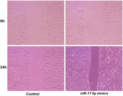
The wound healing assay was used to detect the ability of miR-17-5p transfected cells to repair scratches. We seeded CAL-27 cells on six-well plates at 1 × 10⁵ cells/well, then trans-fected miR-17-5p mimics or mimic negative control at 50 nM using riboFECT CP reagent (Ribobio, Guangzhou, China) after the cells re- ached 50-60% confluence. When the cells re- ached 90% confluence, scratches were made with a 200-μl pipette tip across the centre of the wells. After washing with culture medium to remove cell debris, the cells were allowed to migrate for 24 h. The wounds were photo-graphed at 0 and 24 h using an Olympus IX53 inverted phase contrast microscope (Olympus, Tokyo, Japan). Three random fields were marked and measured. The migration index was ex- pressed as the ratio of the migrating distance of the treated cells to that of the control cells. Transmission electron microscopy (TEM)
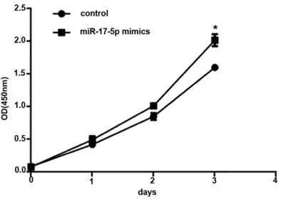
Figure 2. Overexpression of miR-17-5p promoted CAL-27 cell proliferation under hypoxia. CAL-27 cells were transfected with miR-17-5p mimics (50 nmol) or miRNA mimic negative control (50 nmol) for 1, 2, and 3 days under 1% O2, 94% N2, and 5% CO2 at 37°C. Cell proliferation was determined by the CCK-8 assay. Data are presented as mean ± SEM of three independent experiments. *P < 0.05 vs. the control group.
Chemical Industry Group, Co., Ltd., Beijing, China) in 0.1 M Sorensen buffer (pH 7.3; Beijing Chemical Industry Group, Co., Ltd., Beijing, China) for 1 h at 4°C, and post-fixed in 1% osmi-um tetroxide (Beijing Chemical Industry Group, Co., Ltd., Beijing, China) in 0.1 M cacodylate buffer (Beijing Chemical Industry Group, Co., Ltd., Beijing, China) for 1 h at room tempera-ture. The cells were dehydrated in solutions of ethanol (30-90%), then embedded in Epon resin (Beijing Chemical Industry Group, Co., Ltd., Beijing, China) and cut with an UC7 micro-tome (Leica, Wetzlar, Germany) to obtain 70-nm sections. The sections were placed on uncoat-ed copper grids. Sections were subsequently counterstained with 4% uranyl acetate (Beijing Chemical Industry Group, Co., Ltd., Beijing, Chi- na) and examined using transmission electron microscopy (TEM) (Model JEM-1200EX; JEOL, Tokyo, Japan) at 100 kV.
Statistical analysis
The Prism 5.0 software (GraphPad Software Inc., San Diego, CA, USA) was used for data analysis. All experiments were performed in duplicate with data averaged from at least three independent experiments. The data are presented as mean ± standard error of the mean (SEM). Statistical significance was evalu-ated by independent sample t-test. Two-sided P-values < 0.05 were considered significant.
Results
Effect of miR-17-5p mimics on the expression of miR-17-5p in CAL-27 cells
Figure 1 shows that the miR-17-5p mimics suc-cessfully increased the expression of miR-17-5p in CAL-27 cells by almost 700 fold compared with the miRNA mimic negative control.
MiR-17-5p promoted CAL-27 cell proliferation and migration
Figure 2 shows the effect of miR-17-5p overex-pression on CAL-27 cells proliferation under hy- poxia. After 3 days, cells transfected with the miR-17-5p mimics showed higher proliferation compared with controls (P < 0.05).
Figure 3 shows that miR-17-5p transfected CAL-27 cells had a higher migratory capacity com-pared with that of the control cells (P < 0.05) under hypoxia.
MiR-17-5p inhibits hypoxia-induced autophagy in CAL-27 cells
[image:4.612.89.289.71.218.2]CAL-27 cells were submitted to hypoxic condi-tions after transfection with miR-17-5p mimics or mimic negative control. TEM showed that miR-17-5p overexpression inhibited the forma-tion of autophagosomes in hypoxic cells (Figure 4). The role of Beclin-1 was explored in this observation. RT-qPCR and western blot showed
that miR-17-5p overexpression inhibited the mRNA and protein expressions of Beclin-1 in CAL-27 cells subjected to hypoxia (Figure 5). Discussion
Autophagy contributes to HNSCC development and progression [11]. MiR-17-5p downregulat-ed Beclin-1 and thus plays an important role in autophagy [18, 19, 21-23], but little is known about the function and regulation of miR-17-5p in HNSCC autophagy. Therefore, this study ai- med to investigate the role of miR-17-5p on the proliferation, migration, and autophagy under hypoxia of CAL-27 human tongue squamous cell carcinoma cells. The results suggest that miR-17-5p overexpression promoted the prolif-eration and migration of the CAL-27 cells, but inhibited autophagy under hypoxia.
Data about autophagy and its role in tumori-genesis are inconsistent among cancer types
and organs [7]. In addition, the degree of au- tophagy varies during cancer progression and may play differential roles [8]. Nevertheless, in HNSCC, the autophagy induced by hypoxia is associated with an aggressive phenotype [12]. In addition, autophagy in HNSCC has good ap- plication prospects in targeted treatment of HNSCC [15].
[image:5.612.91.524.70.407.2]Many miRNAs are associated with the occur-rence, invasion, and metastasis of oral cancer [25, 26]. Recent studies have reported that miRNAs can be used as biomarkers for predict-ing the prognosis and sensitivity of cancer to radiotherapy and chemotherapy [27]. MiR-17-5p imbalance has been reported to be associ-ated with the metastasis and invasion of hepa-tocellular carcinoma, ovarian cancer, breast cancer, and prostate cancer [17-20], but its role in HNSCC is still not clear. The expression levels of miR-17-5p is associated with cancer aggres-siveness and therapy resistance [21]. Furth-
ermore, hypoxia is an important factor associ-ated with HNSCC aggressiveness and resis-tance to treatments [11, 12]. MiR-17-5p has been shown to be involved in autophagy and cell death in response to hypoxia [28].
In the present study, miR-17-5p appears to have a dual role in HNSCC. Indeed, the overex-pression of miR-17-5p increased cancer cell proliferation and migration, but decreased au- tophagy under hypoxia. A previous study in
[image:6.612.90.523.70.305.2]gas-tric cancer cells showed that miR-17-5p binds to the TGFBR2 mRNA and that decreased ex- pression of TGFBR2 leads to uncontrolled cell growth and invasion [29], supporting the pres-ent study. Such a relationship between miR-17-5p has been shown in different cell types [30-32]. On the other hand, miR-17-5p has been shown to be a tumor suppressor in triple-nega-tive breast cancer [33], highlighting the differ-ential roles of miR-17-5p in different tumor types.
Figure 4. Overexpression of miR-17-5p inhibited the formation of autophagosomes induced by hypoxia in CAL-27 cells. CAL-27 cells were transfected with miRNA mimic negative control (50 nmol) or miR-17-5p mimics (50 nmol) under 1% O2, 94% N2, and 5% CO2 at 37°C for 48 h. Transmission electron microscope (TEM) was used to observe the formation of autophagosomes (magnification: × 6000).
[image:6.612.93.524.374.502.2]Regarding autophagy, miR-17-5p is important in regulating many genes involved in autopha-gy. Overexpression of miR-17-5p in glioma cells decreases Beclin-1-mediated autophagy and radioresistance [22], supporting the present study. In non-small lung cancer, downregulation of miR-17-5p is associated with upregulation of Beclin-1 and resistance to paclitaxel [23]. Nevertheless, this dual role of miR-17-5p could be the key to its eventual successful role in cancer treatment. Indeed, autophagy is a pro-cess by which cancer cells acquire resistance to adverse environmental conditions and can-cer treatments [5, 10, 12, 23]. In addition, tra-ditional chemotherapy and radiation therapy targets cells that are in active proliferation [34]. Hence, the increased cancer proliferation and invasion following miR-17-5p overexpression could be irrelevant in the context where the cells are more sensitive to therapy. In addition, the levels of miR-17-5p observed in the present study after mimic transfection are supraphysi-ological. Additional studies should be perfor- med in cells naturally overexpressing miR-17-5p. Nevertheless, these results strongly sug-gest that miR-17-5p could be used for HNSCC treatment.
In conclusion, miR-17-5p promoted the prolif-eration and migration of CAL-27 cells, but inhib-ited autophagy under hypoxia.
Acknowledgements
This study was funded by Key Research and Development project of Shandong Province (No. 2016GSF201174).
Disclosure of conflict of interest
None.
Address correspondence to: Dongsheng Zhang, Department of Oral and Maxillofacial Surgery, Sh- andong Provincial Hospital Affiliated to Shandong University, Jinan 250021, Shandong, China. Tel: +86-13791125330; Fax: +86-0531-68777980; E-mail: ds63zhang@sdu.edu.cn
References
[1] Jemal A, Bray F, Center MM, Ferlay J, Ward E and Forman D. Global cancer statistics. CA Cancer J Clin 2011; 61: 69-90.
[2] Brocic M, Kozomara R, Cerovic S, Jovic N, Vu- kelic-Markovic S and Stosic S. Clinical
signifi-cance of vascular endothelial growth factor expression in patients with carcinoma of the mouth floor and tongue. Vojnosanit Pregl 2009; 66: 440-448.
[3] Faraji F, Zaidi M, Fakhry C and Gaykalova DA. Molecular mechanisms of human papillomavi-rus-related carcinogenesis in head and neck cancer. Microbes Infect 2017; 19: 464-475. [4] Guidi A, Codeca C and Ferrari D. Chemotherapy
and immunotherapy for recurrent and meta-static head and neck cancer: a systematic re-view. Med Oncol 2018; 35: 37.
[5] Klionsky DJ. The molecular machinery of au-tophagy: unanswered questions. J Cell Sci 2005; 118: 7-18.
[6] Wu D, Wang X and Sun H. The role of mitochon-dria in cellular toxicity as a potential drug tar-get. Cell Biol Toxicol 2018; 34: 87-91.
[7] Lei Y, Zhang D, Yu J, Dong H, Zhang J and Yang S. Targeting autophagy in cancer stem cells as an anticancer therapy. Cancer Lett 2017; 393: 33-39.
[8] Tsuchihara K, Fujii S and Esumi H. Autophagy and cancer: dynamism of the metabolism of tumor cells and tissues. Cancer Lett 2009; 278: 130-138.
[9] White E. The role for autophagy in cancer. J Clin Invest 2015; 125: 42-46.
[10] Muz B, de la Puente P, Azab F and Azab AK. The role of hypoxia in cancer progression, an-giogenesis, metastasis, and resistance to ther-apy. Hypoxia (Auckl) 2015; 3: 83-92.
[11] Wu H, Huang S and Zhang D. Autophagic re-sponses to hypoxia and anticancer therapy in head and neck cancer. Pathol Res Pract 2015; 211: 101-108.
[12] Vigneswaran N, Wu J, Song A, Annapragada A and Zacharias W. Hypoxia-induced autophagic response is associated with aggressive pheno-type and elevated incidence of metastasis in orthotopic immunocompetent murine models of head and neck squamous cell carcinomas (HNSCC). Exp Mol Pathol 2011; 90: 215-225. [13] Kang R, Zeh HJ, Lotze MT and Tang D. The Be-
clin 1 network regulates autophagy and apop-tosis. Cell Death Differ 2011; 18: 571-580. [14] Zhao H, Yang M and Zhao B. Beclin 1 and LC3
as predictive biomarkers for metastatic color- ectal carcinoma. Oncotarget 2017; 8: 59058-59067.
[15] Zhang L, Zhang W, Wang YF, Liu B, Zhang WF, Zhao YF, Kulkarni AB and Sun ZJ. Dual induc-tion of apoptotic and autophagic cell death by targeting survivin in head neck squamous cell carcinoma. Cell Death Dis 2015; 6: e1771. [16] Lan J, Huang Z, Han J, Shao J and Huang C.
Redox regulation of microRNAs in cancer. Cancer Lett 2018; 418: 250-259.
D, Solmi R and Cheng JQ. Adenomatous pol-yposis coli (APC) regulates miR17-92 cluster through beta-catenin pathway in colorectal cancer. Oncogene 2016; 35: 4558-4568. [18] Gu J, Wang D, Zhang J, Zhu Y, Li Y, Chen H, Shi
M, Wang X, Shen B, Deng X, Zhan Q, Wei G and Peng C. GFRalpha2 prompts cell growth and chemoresistance through down-regulating tu-mor suppressor gene PTEN via Mir-17-5p in pancreatic cancer. Cancer Lett 2016; 380: 434-441.
[19] Chen P, Zhao H, Huang J, Yan X, Zhang Y and Gao Y. MicroRNA-17-5p promotes gastric can-cer proliferation, migration and invasion by di-rectly targeting early growth response 2. Am J Cancer Res 2016; 6: 2010-2020.
[20] Guo J, Mei Y, Li K, Huang X and Yang H. Down- regulation of miR-17-92a cluster promotes au-tophagy induction in response to celastrol tr- eatment in prostate cancer cells. Biochem Biophys Res Commun 2016; 478: 804-810. [21] Bobbili MR, Mader RM, Grillari J and Dellago H.
OncomiR-17-5p: alarm signal in cancer? On- cotarget 2017; 8: 71206-71222.
[22] Hou W, Song L, Zhao Y, Liu Q and Zhang S. Inhibition of Beclin-1-mediated autophagy by MicroRNA-17-5p enhanced the radiosensitivity of glioma cells. Oncol Res 2017; 25: 43-53. [23] Chatterjee A, Chattopadhyay D and Chakrabarti
G. miR-17-5p downregulation contributes to paclitaxel resistance of lung cancer cells thr- ough altering beclin1 expression. PLoS One 2014; 9: e95716.
[24] Dulloo I, Phang BH, Othman R, Tan SY, Vija- yaraghavan A, Goh LK, Martin-Lopez M, Mar- ques MM, Li CW, Wang de Y, Marin MC, Xian W, McKeon F and Sabapathy K. Hypoxia-inducible TAp73 supports tumorigenesis by regulating the angiogenic transcriptome. Nat Cell Biol 2015; 17: 511-523.
[25] Suh YE, Raulf N, Gaken J, Lawler K, Urbano TG, Bullenkamp J, Gobeil S, Huot J, Odell E and Tavassoli M. MicroRNA-196a promotes an on-cogenic effect in head and neck cancer cells by suppressing annexin A1 and enhancing ra-dioresistance. Int J Cancer 2015; 137: 1021-1034.
[26] Kinoshita T, Nohata N, Fuse M, Hanazawa T, Kikkawa N, Fujimura L, Watanabe-Takano H, Yamada Y, Yoshino H, Enokida H, Nakagawa M, Okamoto Y and Seki N. Tumor suppressive mi-croRNA-133a regulates novel targets: moesin contributes to cancer cell proliferation and in-vasion in head and neck squamous cell carci-noma. Biochem Biophys Res Commun 2012; 418: 378-383.
[27] Huang Y, Tan D, Xiao J, Li Q, Zhang X and Luo Z. miR-150 contributes to the radioresistance in nasopharyngeal carcinoma cells by targeting glycogen synthase kinase-3beta. J Cancer Res Ther 2018; 14: 111-118.
[28] Hao MX, Wang X and Jiao KL. MicroRNA-17-5p mediates hypoxia-induced autophagy and in-hibits apoptosis by targeting signal transducer and activator of transcription 3 in vascular smooth muscle cells. Exp Ther Med 2017; 13: 935-941.
[29] Qu Y, Zhang H, Duan J, Liu R, Deng T, Bai M, Huang D, Li H, Ning T, Zhang L, Wang X, Ge S, Zhou L, Zhong B, Ying G and Ba Y. MiR-17-5p regulates cell proliferation and migration by targeting transforming growth factor-beta re-ceptor 2 in gastric cancer. Oncotarget 2016; 7: 33286-33296.
[30] Shan SW, Fang L, Shatseva T, Rutnam ZJ, Yang X, Du W, Lu WY, Xuan JW, Deng Z and Yang BB. Mature miR-17-5p and passenger miR-17-3p induce hepatocellular carcinoma by targeting PTEN, GalNT7 and vimentin in different signal pathways. J Cell Sci 2013; 126: 1517-1530. [31] Wu Q, Luo G, Yang Z, Zhu F, An Y, Shi Y and Fan
D. miR-17-5p promotes proliferation by target-ing SOCS6 in gastric cancer cells. FEBS Lett 2014; 588: 2055-2062.
[32] Chen C, Lu Z, Yang J, Hao W, Qin Y, Wang H, Xie C and Xie R. MiR-17-5p promotes cancer cell proliferation and tumorigenesis in nasopharyn-geal carcinoma by targeting p21. Cancer Med 2016; 5: 3489-3499.
[33] Li J, Lai Y, Ma J, Liu Y, Bi J, Zhang L, Chen L, Yao C, Lv W, Chang G, Wang S, Ouyang M and Wang W. miR-17-5p suppresses cell proliferation and invasion by targeting ETV1 in triple-negative breast cancer. BMC Cancer 2017; 17: 745. [34] Liu H, Lv L and Yang K. Chemotherapy