Original Article
Prognostic value of human papillomavirus infection
and p53
,
p16
,
epidermal growth factor receptor
and p34
cdc2expression in patients with
salivary adenoid cystic carcinoma
Hong-Xia Liu1, Meng Wu2, Yu-Man Sun1, Feng-Yan Han1, Yi-Fei Liu1, Guang-Zeng Zhang1
1Tangshan Head and Neck Disease Pathology Research Base, Tangshan, P. R. China; 2Pathological Teaching and
Research Division, Department of Basic Medicine, Tangshan Vocational and Technical College, Tangshan, P. R. China
Received May 18, 2017; Accepted May 25, 2017; Epub July 1, 2017; Published July 15, 2017
Abstract: The aim of this study was to explore the correlation between HPV infection, p53, p16, epidermal growth factor receptor (EGFR), p34cdc2 protein expression and prognosis in patients with adenoid cystic carcinoma of
sali-vary gland. Totally 78 cases of adenoid cystic carcinoma of salisali-vary gland specimens were selected from January 1, 2004 to December 31, 2013 in Tangshan Union Hospital. PCR-reverse dot blot hybridization was used to de-tect infection of human papilloma virus (HPV), and SP immunohistochemical method was adopted to dede-tect the expression of p53, p16, EGFR and p34cdc2 protein in the carcinoma tissues. Clinical data were collected and the
patients were followed up. Results showed that the infection rate of HPV in adenoid cystic carcinoma tissues was 0% (0/78). The expression rate of p53, p16, EGFR and p34cdc2 protein in carcinoma tissues were 75.6% (59/78), 57.7%
(45/78), 60.1% (47/78) and 64.1% (50/78), respectively. Expression of p53, p16, EGFR and p34cdc2 proteins was
not significantly correlated with patients’ age, gender, disease location, TNM classification and histological type (P > 0.05). Kaplan-Meier analysis showed that positive patients had a lower median overall survival than EGFR-negative ones (58 months vs. 75 months, respectively. P = 0.001). The result of median progression-free survival was virtually the same for both EGFR-positive and EGFR-negative patients (43 months vs. 49 months, respectively. P = 0.002). p34cdc2-positive patients had a lower median overall survival than p34cdc2-negative ones (61 months vs.
71 months, respectively. P = 0.027). Median progression-free survival was also almost the same for both p34cdc2
-positive and p34cdc2-negative patients (44 months vs. 51 months, respectively. P = 0.011). Cox regression analysis
showed that expression of EGFR and p34cdc2 was independent risk factors for the prognosis of patients with adenoid
cystic carcinoma of salivary gland (relative risk = 13.199, 11.466, P < 0.001). In conclusion, HPV infection is not detected in salivary adenoid cystic carcinoma tissues. p53, p16, EGFR and p34cdc2 protein are positively expressed
in most salivary adenoid cystic carcinoma tissues. p16 is unsuitable as a surrogate for HPV infection status in pa-tients with adenoid cystic carcinoma of salivary gland. Expression of EGFR and p34cdc2 is independent risk factors
in the prognosis of patients with salivary gland adenoid cystic carcinoma. Patients with EGFR or p34cdc2 positive
expression should be followed up closely.
Keywords: Adenoid cystic carcinoma, HPV, EGFR, Cdc2, prognosis
Introduction
Adenoid cystic carcinoma (ACC) is one of the most common malignant tumors in salivary gland, which is a relatively rare tumor in the parotid gland with a 10% incidence, and its inci-dence rate in minor salivary glands is about 30% [1]. Histologically, the tumor shows the characteristics of local growth, perineural inva-sion, distant metastasis and high recurrence
ization. The paraffin-embedded salivary ade -noid cystic carcinoma specimens were cut to
extract HPV-DNA, PCR amplification, hybridiza
-tion, filter washing and color development
according to kit instructions. The positive qual-ity control was colored (blue spots) at the cor-responding and the IC membrane sites and other sites were not colored. In accordance with the order of the probe sequence and the color on the membrane the HPV genotypes were determined.
p53, p16, EGFR and p34cdc2 expression by
im-munohistochemistry
All the surgically removed tissue specimens
were fixed in 10% neutral formalin, embedded in paraffin, and one representative block from each patient was sectioned at 4 μm, stained
with hematoxylin and eosin (HE) and evaluated by immunohistochemistry according to the
pro-tocol described in the manufacturer’s guide
accompanying the kit. The mouse anti human p53 (1:100), EGFR (1:100) and p34cdc2 (1:300) monoclonal antibody were all purchased from Santa Cruz Biotechnology (USA). Mouse anti human p16 monoclonal antibody (1:100) and SP immunohistochemistry kit were all pur-chased from Beijing Zhongshan Golden Bridge Biotechnology Co., Ltd. Known positive sam-ples of p53, p16, EGFR and p34cdc2 were used as positive control. For the negative control, the primary antibodies were replaced with phos-phate-buffered saline (PBS).
Evaluation of immunohistochemical analysis
Positive signals of the immunohistochemical staining were brown or brownish yellow in color. Nuclear staining was considered positive on the immunostaining for p53 and p16, mem-brane and cytoplasm immunostaining was con-sidered positive for EGFR, while the cytoplas-mic and/or nuclear immunostaining was con- sidered positive for p34cdc2. 10 high magnifica -tion visions were selected randomly in each
stained section, 10 high power field represen -tatives were observed and the brown staining cells were counted. Positive staining in more than 10% of the cells was considered positive,
while less than 10% or colorless were defined
as negative.
Statistical analysis
The statistical analyses were performed with PASW Statistics 22.0 (SPSS Inc., Chicago, IL, questionable. The purpose of this study was to
evaluate the correlation of HPV infection and cyclin-dependent kinase inhibitor 2A (CDKN2A/ p16), tumor protein p53 (TP53), EGFR and cell cycle-dependent kinase p34cdc2 protein to determine the relationship between these fac-tors and the occurrence, development and prognosis of salivary gland adenoid cystic carcinoma.
Here, we analyzed the HPV DNA by PCR-DNA reverse dot blot and examined the expression of p53, p16, EGFR and p34cdc2 by immunohisto-chemistry in the adenoid cystic carcinoma of salivary gland, to investigate the relationship between HPV infection, the expression of the four proteins and prognosis of salivary gland adenoid cystic carcinoma patients.
Patients and methods
Clinical and pathological data
Our study involved 78 cases of salivary gland adenoid cystic carcinoma resection specimens selected at Tangshan Union Hospital from January 1, 2004 to December 31, 2013. All the cases underwent HPV-DNA, p53, p16, EGFR and p34cdc2 protein detection, including 41 males and 37 females, aged 41 to 70 years, with median age 51 years. Lesional location was in the parotid gland (26 cases), subman-dibular gland (24 cases), pars palatalis (21 cases) and sublingual gland (7 cases),
respec-tively. Histological classification included solid
type, sieve type and tubular type. According to the sixth edition of the American Joint Com- mittee on Cancer (AJCC) in 2002, they were all divided into four stages: including 6 cases in stage I, 16 cases in stage II, 20 cases in stage III and 36 cases in stage IV. All the 78 patients involved in the study have been informed con-tent and approved by the Ethics Committee of Tangshan Union Hospital. All the patients were followed up every 3 months by telephone to record the survival outcomes. Follow-up rate was 100% and date was up to January 6, 2016.
HPV DNA detection by PCR-DNA reverse dot blot hybridization
hybrid-itive rate of p53, p16, EGFR and p34cdc2 was all
irrelevant to the patient’s age, sex, disease
location, differentiation, TNM stage and (P > 0.05) (Figure 1, Table 1).
Prognosis of patients with salivary gland ad-enoid cystic carcinoma
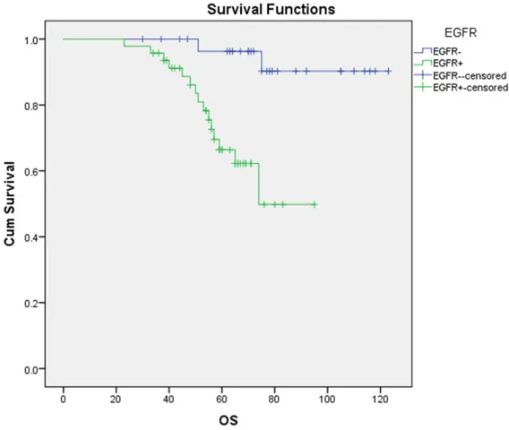
Follow-up to January 6, 2016, in total 78 sali-vary gland adenoid cystic carcinoma patients, 18 cases were dead, the total mortality rate was 24.36% (19/78). The median overall sur-vival (OS) was 64 months in all patients. The 5-year OS rate was 58.9% (46/78). Kaplan-Meier survival analysis showed that the me- dian OS of EGFR-negative and EGFR-positive patients was 58 months and 75 months res-
pectively, with statistically significant (P < 0.001, Figure 2). The median OS of p34cdc2 -neg-ative and p34cdc2-positive patients was 71 months and 61 months respectively, and these
difference was also statistically significant (P = USA). The correlations of HPV infection and
expression of p53, p16, EGFR and p34cdc2 with
the various clinicopathological findings were
evaluated using the Chi-square test, and the Kaplan-Meier method was used to analyze sur-vival rate. P values less than 0.05 were
consid-ered to be statistically significant.
Results
Infection rate of HPV and expression rate of p53, p16, EGFR and p34cdc2 in the salivary
gland adenoid cystic carcinoma tissues
PCR-DNA reverse dot blot hybridization show- ed that the infection rate of HPV was 0% (0/78) in the salivary gland adenoid cystic carcinoma tissues. Immunohistochemistry showed that expression rate of p53, p16, EGFR and p34cdc2 in the salivary gland adenoid cystic carcinoma tissues were 75.6% (59/78), 57.7% (45/78), 60.1% (47/78) and 64.1% (50/78), and the
pos-Figure 1. Expression of EGFR, p34cdc2, p53, p16 in salivary gland ACC tissues. SP method. A. EGFR staining diffusely
positive in ACC tissue (arrow). B. p34cdc2 staining is positive in ACC tissue (arrow). C. p53-positive staining in ACC
[image:3.612.92.523.71.396.2]Discussion
Adenoid cystic carcinoma is a malignant tumor originated from the gland ducts, its common histology is given priority to with cribriform, tubular, solid three types, with vary in amount 0.027, Figure 3). Median OS was irrelevant to
patients’ gender, age, lesion sites, TNM classifi -cation and histological type (P > 0.05). The overall median progression-free survival (PFS) of patients was 45 months, Kaplan-Meier sur-vival analysis showed that the PFS of
EGFR-Table 1. Correlation between expression of p53, p16, EGFR, p34cdc2 and clinical features in salivary gland adenoid cystic carcinoma tissues
Clinical Features Total No. p53+ P p16+ P EGFR+ P Cdc2+ P
Gender
Male 41 (52.6) 30 (73.1) 0.593 20 (48.8) 0.448 22 (53.7) 0.127 30 (73.2) 0.894
Female 37 (47.4) 29 (78.4) 25 (67.6) 25 (67.6) 20 (54.1)
Age
< 50 y 21 (26.9) 15 (71.4) 0.938 9 (42.9) 0.809 12 (57.1) 0.302 10 (47.6) 0.657
≥ 50 y 57 (73.1) 44 (77.2) 36 (63.2) 35 (61.4) 40 (70.2) Site
Parotid gland 26 (33.3) 19 (73.1) 0.922 12 (46.2) 1.000 17 (65.4) 0.807 16 (61.5) 0.234
Submandibular gland 24 (30.8) 18 (75.0) 14 (58.3) 14 (58.3) 15 (62.5)
Pars palatalis 21 (26.9) 17 (81.0) 14 (66.7) 12 (57.1) 11 (52.4)
Sublingual gland 7 (9.0) 5 (71.4) 5 (71.4) 4 (57.1) 5 (71.4)
TNM Staging
Stages I + II 22 (28.2) 17 (77.2) 0.818 15 (68.2) 0.384 9 (40.9) 0.050 10 (45.5) 0.894
Stages III + IV 56 (71.8) 42 (75.0) 30 (53.6) 38 (67.9) 40 (71.4)
Histological classification
Solid type 17 (21.8) 15 (88.2) 0.094 11 (64.7) 0.352 14 (82.3) 0.105 15 (88.2) 0.438
Sieve type 25 (32.0) 18 (72.0) 14 (56.0) 12 (48.0) 16 (64.0)
[image:4.612.92.522.96.348.2]Tubular type 36 (46.2) 26 (72.2) 20 (55.6) 21 (58.3) 19 (52.8)
Figure 2. Kaplan-Meier survival analysis showing the impact of EGFR
expres-sion on OS. χ2 = 11.232, P = 0.001.
negative and EGFR-positive patients was 49 months and 43 months respectively, with
statistically significant (P = 0.002, Figure 4). The median PFS of p34cdc2-negative and p34cdc2-positive patients was 51 months and 44 months respectively, and these dif- ference was also statistically
significant (P = 0.011, Figure 5). Median PFS was irrelevant
to patients’ gender, age, le-sion sites, TNM classification
[image:4.612.90.374.371.611.2]noid cystic carcinoma was 64%. Lassen et al
[8] found that the positive rate of p16 was 22% and was closely related with HPV infection in the pharyngeal and supraglottic carcinoma tis-sues in Denmark. Patients with p16 positive of mucus and pink colored basement
[image:5.612.91.375.73.312.2]mem-brane like substance. The tumor cells lined with ductal and myoepithelial epithelial, prone to nerve invasion and distant metastasis with poor prognosis. The related factors of the poor
Figure 3. Kaplan-Meier survival analysis showing the impact of p34cdc2
ex-pression on OS. χ2 = 4.881, P = 0.027.
Figure 4. Kaplan-Meier survival analysis showing the impact of EGFR
expres-sion on PFS. χ2 = 9.218, P = 0.002.
prognosis of the carcinoma are still under investigation. Data from International Ag- ency for Research on Cancer (IARC) in 2012 showed that the incidence of head and neck cancer induced by HPV increased with each passing year, especially in tumors of oromaxillo-facial region [3]. While, there were few studies about the HPV and its related factors in the process of in- ducing salivary gland adenoid cystic carcinoma and the con-clusion was different between researchers. Boland et al [4] found that no HPV positive cases detected in salivary gland adenoid cystic carcino-ma specimens by in situ hybridization. Bishop et al [5] found that HPV was negative in the ACC tissues of nasal cavity and paranasal sinuses. Hühns et al [2] in analyzing 17 samples of ACC displayed that the infection rate of HPV was 25%. Huo et al [6] report-ed HPV was negative in 27 cases of lung ACC by in situ hybridization detection. Our study showed that there was no HPV detected in 78 cases of salivary gland ACC, sug-gesting that the occurrence of ACC in salivary glands may not be associated with HPV infection.
[image:5.612.91.373.364.604.2]ade-ly in the ACC tissues, and the expression in crib-riform and tubular forms was higher than that in solid forms. Our study showed that the expression rate of EGFR in salivary gland ACC tissues was 60.5%, and the expression rate of EGFR in stage I and II tumor tissues was lower than that in stage III and IV carcinomas,
how-ever, the difference was not statistically signifi -cant and increase of the sample size was
need-ed to further confirm the finding. Multivariate
Cox regression analysis showed that the expression of EGFR was associated with short survival time, which was an independent prog-nostic factor in patients with salivary ACC. p34cdc2 is a member of the Ser/Thr protein kinase family coded by cell division cycle (cdc) gene 2 whose relative molecular weight is 34 KD. p34cdc2 is one of the most important regu-lated kinase of cell cycle and its main function is to monitor spindle microtubule assembly and
kinetochore’s proper connection of DNA in the
G2/M checkpoint. If an error occurs in adjust-had a higher tumor local control rate (58%:28%,
P = 0.0005), disease-specific survival rate
(72%:34%, P = 0.0006) and overall survival rate (62%:26%, P = 0.0003). Xing et al [9] reported that there was no correlation between high-risk HPV and p16 expression in vulvar and cervical ACC tissues. Boland et al [4] also
con-firmed that the detection of p16 instead of HPV
detection was not applicable in ACC specimens. In our study, the expression rate of p16 in 78 cases of salivary ACC tissues was 57.7% (45/78), however, HPV infection was not detect-ed. Therefore, the expression of p16 in salivary gland ACC cannot be used as an indicator of HPV infection.
[image:6.612.91.375.73.312.2]Tumor induced by HPV is correlation with wild-type p53 tumor suppressor gene. The wild wild-type p53 protein has a short half-life in vivo, which cannot be detected by immunohistochemistry. However, the mutant p53 protein has longer half-life that can be detected by immunohisto-chemistry. Therefore, the expression of p53
Figure 5. Kaplan-Meier survival analysis showing the impact of p34cdc2
ex-pression on PFS. χ2 = 6.418, P = 0.001.
Table 2. Multivariate analysis of prognosis for salivary gland ad-enoid cystic carcinoma patients by Cox model
B SE Wald P EXP (β) 95% CI
EGFR 3.260 0.866 14.167 < 0.01 26.042 3.256-55.286
p34cdc2 3.601 1.070 11.322 < 0.01 36.632 3.204-53.358
protein was negative in tum- ors induced by HPV. The con-clusion about the expression rate of p53 in adenoid cystic carcinoma is different. Jiang
et al [10] reported that the positive rate of p53 in the parotid gland ACC was 45.7%, but it was not related to the survival of the patients. Wang
et al [11] found that the expression rate of p53 in 36 cases of salivary adenoid cys-tic carcinoma was 69.44%. Our study found that the expression rate of p53 in sali-vary ACC tissues was 76.3%, p53 positive patients had shorter survival than p53 neg-ative ones, but there was no
significant difference in evalu -ation of the prognosis of patients with ACC and require larger sample size for further
research confirmed.
Epidermal growth factor rece- ptor plays an important role in the process of tumor growth and metastasis. Wang et al
[12] found that the expression
[image:6.612.88.375.388.430.2]-sion of molecular markers in adenoid cystic cancer of the salivary glands compared with lymph node metastasis: a retrospective study. World J Surg Oncol 2012; 10: 266.
[2] Hühns M, Simm G, Erbersdobler A, Zimpfer A. HPV Infection, but not EBV or HHV-8 infection, is associated with salivary gland tumours. Biomed Res Int 2015; 2015: 829349.
[3] Ferlay J, Soerjomataram I, Ervik M, Dikshit R, Eser S, Mathers C, Rebelo M, Parkin DM, For-man D, Bray F. Estimated cancer incidence, mortality and prevalence worldwide in 2012. International agency for research on cancer (IARC), globocan 2012 available online: http:// globocan.iarc.fr.
[4] Boland JM, McPhail ED, García JJ, Lewis JE, Schembri-Wismayer DJ. Detection of human papilloma virus and p16 expression in high-grade adenoid cystic carcinoma of the head and neck. Mod Pathol 2012; 25: 529-536. [5] Justin AB, Takenori O, Edward BS, Christopher
AM, Wayne MK, Sara IP, William HW. Human papillomavirus-related carcinoma with ade-noid cystic-like features: a peculiar variant of head and neck cancer restricted to the sinona-sal tract. Am J Surg Pathol 2013; 37: 836-844. [6] Huo Z, Meng Y, Wu H, Shen J, Bi Y, Luo Y, Cao J,
Liang Z. Adenoid cystic carcinoma of the tra-cheobronchial tree: clinicopathologic and im-munohistochemical studies of 21 case. Int J Clin Exp Pathol 2014; 7: 7527-7235.
[7] Isayeva T, Said-Al-Naief N, Ren ZY, Li R, Gnepp D, Brandwein-Gensler M. Salivary mucoepider-moid carcinoma: demonstration of transcrip-tionally active human papillomavirus 16/18. Head Neck Pathol 2013; 7: 135-148.
[8] Lassen P, Eriksen JG, Hamilton-Dutoit S, Tramm T, Alsner J, Overgaard J. Effect of HPV-associated p16INK4A expression on response to
radiotherapy and survival in squamous cell carcinoma of the head and neck. J Clin Oncol 2009; 27: 1992-1998.
[9] Xing D, Schoolmeester JK, Ren Z, Isacson C, Ronnett BM. Lower female genital tract tumors with adenoid cystic differentiation: P16 expres-sion and high-risk HPV detection. Am J Surg Pathol 2016; 40: 529-536.
[10] Jiang LC, Huang SY, Zhang DS, Zhang SH, Li WG, Zheng PH, Chen ZW, Zhang DS. Expres-sion of beclin 1 in primary salivary adenoid cystic carcinoma and its relation to Bcl-2 and p53 and prognosis. Braz J Med Biol Res 2014; 47: 252-258.
[11] Wang XF, Li SC, Fang DJ. Expression of survivin and p53 in salivary adenoid cystic carcinoma. Kouqiang Yixue Yanjiu Zazhi 2014; 30: 635-637.
[12] Wang WM, Zhao ZL, Zhang WF, Zhang L, Sun
SJ. Role of hypoxia-inducible factor-1α and
CD146 in epidermal growth factor
receptor-ment mechanism of p34cdc2, it will lead directly to cell differentiation disorders, disorder of cell cycle progression and induce abnormal cell proliferation or malignant transformation, thus promoting the occurrence and progression of tumor [13-15]. p34cdc2 overexpressed in many cancers, such as oral squamous cell carcinoma [16], tongue squamous cell carcinoma [17], supraglottic cancer [18], esophageal squa-mous cell carcinoma, esophageal adenocarci-noma [19], gastric cancer [20], liver cancer [21], colorectal cancer [22], breast cancer [23], ovarian cancer [24] and so on. Yang et al [25] found that the expression rate of p34cdc2 in
laryngeal carcinoma tissues was 70.6%, signifi -cantly higher than that in adjacent tissues and its negative margins. Patients with p34cdc2 posi-tive margins had higher recurrence rate than that of negative patients. However, its expres-sion in salivary gland tissues has not been reported. Our study shows that the expression rate of p34cdc2 in salivary gland ACC tissues was as high as 64.1% (50/78), the single factor analysis showed that the median OS of patients with p34cdc2 positive was 10 months shorter than that of negative ones, P = 0.001. Multi- variate analysis showed that the positive ex- pression of p34cdc2 was associated with short survival time of ACC, which was an independent factors affecting the prognosis of patients. In conclusion, HPV was not detected in the sali-vary gland ACC tissue. p53, p16, EGFR and p34cdc2 proteins were expressed in most sali-vary ACC tissues. p16 could not be used as a surrogate marker for HPV infection in patients with salivary ACC. Patients with positive expres-sion of EGFR and p34cdc2 had poor prognosis than the negative ones, and the patients with the expression of EGFR and p34cdc2 should be followed up closely.
Disclosure of conflict of interest None.
Address correspondence to: Dr. Guang-Zeng Zhang, Tangshan Head and Neck Disease Pathology Re- search Base, Tangshan, P. R. China. Tel: +86-1390-
3252461; Fax: +86-315-2320521; E-mail: zgz64-
45@163.com
References
expres-[20] Kim DH. Prognostic implications of cyclin B1, p34cdc2, p27 (Kip1) and p53 expression in
gas-tric cancer. Yonsei Med J 2007; 48: 694-700. [21] Ito Y, Takeda T, Sakon M, Monden M, Tsujimoto
M and Matsuura N. Expression and prognostic role of cyclin-dependent kinase 1 (cdc2) in he-patocellular carcinoma. Oncology 2000; 59: 68-74.
[22] Nozoe T, Honda M, Inutsuka S and Korenaga D. p34cdc2 expression is an independent
indica-tor for lymph node metastasis in colorectal car-cinoma. J Cancer Res Clin Oncol 2003; 129: 498-502.
[23] Kourea HP, Koutras AK, Scopa CD, Marangos MN, Tzoracoeleftherakis E, Koukouras D and Kalofonos HP. Expression of the cell cycle regu-latory proteins p34cdc2, p21waf1, and p53 in
node negative invasive ductal breast carcino-ma. Mol Pathol 2003; 56: 328-335.
[24] Barrette BA, Srivatsa PJ, Cliby WA, Keeney GL, Suman VJ, Podratz KC and Roche PC. Overex-pression of p34cdc2 protein kinase in epithelial
ovarian carcinoma. Mayo Clin Proc 1997; 72: 925-929.
[25] Yang JQ, Liu HX, Liang Z, Sun YM, Wu M. Over-expression of p53, p21 and Cdc2 in histologi-cally negative surgical margins is correlated with local recurrence of laryngeal squamous cell carcinoma. Int J Clin Exp Pathol 2014; 7: 4295-4302.
mediated angiogenesis in salivary gland ade-noid cystic carcinoma. Mol Med Rep 2015; 12: 3432-3438.
[13] Perez de Castro I, de Carcer G and Malumbres M. A census of mitotic cancer genes: new in-sights into tumor cell biology and cancer thera-py. Carcinogenesis 2007; 28: 899-912. [14] Barnabas S, Dana B. Premature Cdk1/Cdc5/
Mus81 pathway activation induces aberrant replication and deleterious crossover. EMBO J 2013; 32: 1155-1167.
[15] Liu P, Kao TP and Huang H. CDK1 promotes cell proliferation and survival via phosphoryla-tion and inhibiphosphoryla-tion of FOXO1 transcripphosphoryla-tion fac-tor. Oncogene 2008; 27: 4733-4744.
[16] Chang JT, Wang HM, Chang KW, Chen WH, Wen MC, Hsu YM, Yung BY, Chen IH, Liao CT,
Hsieh LL and Cheng AJ. Identification of
dif-ferentially expressed genes in oral squamous cell carcinoma (OSCC): overexpression of NPM, CDK1 and NDRG1 and underexpression of CHES1. Int J Cancer 2005; 114: 942-949. [17] Wada S, Yue L and Furuta I. Prognostic signifi
-cance of p34cdc2 expression in tongue
squa-mous cell carcinoma. Oral Oncol 2004; 40: 164-169.
[18] Yamamoto Y, Itoh T, Inoue I and Takahashi H. Expression of p34cdc2 protein kinase and p53
in supraglottic carcinomas. Auris Nasus Larynx 1996; 23: 105-110.