Original Article
miR-125b expression inovarian cancer cell lines
Tonghang Guo1, Guodong Shen1, Xianhong Tong1, Guixiang Zhou1, Yangyang Wan1, Daojing Li1, Xiuhuan Dai1, Xiaoke Sun1, Yongjie Tian2
1Department of Obstetrics and Gynecology, Anhui Provincial Hospital, Hefei 230001, Anhui, China; 2Department of Obstetrics and Gynecology, Provincial Hospital Affiliated to Shandong University, No.324 Jingwuweiqi Road, Jinan 250021, Shandong, China
Received January 15, 2016; Accepted March 26, 2016; Epub May 1, 2016; Published May 15, 2016
Abstract: Puerarin, a well-known isoflavone-C-glucosideisolated from Pueraria radix, has been demonstrated the anticancer activity in animal models as well as proliferation inhibition and apoptosis induction in a variety of can-cer cell lines in vitro. However, the effect and mechanisms of puerarin on ovarian cancan-cer (OC) have not been well understood. This study aimed to test whether puerarin could inhibit growth of ovarian cancer cells and reveal its underlying molecular mechanism. Cells viability was evaluated using cell counting Kit-8 in SKOV-3 and OVCAR-3 cells. Apoptosis of ovarian cancer cells was analyzed by flow cytometry. Bcl-2 levels were analyzed by Western blot and microRNAs levels were determined by real-time RT-PCR. Our study showed that puerarin inhibited SKOV-3 and OVCAR-3 cells growth in a concentration-dependent manner. Puerarin induced apoptosis by promoting expression of miR-125b, and then inhibiting expression of Bcl-2, a known target for miR-125b. Moreover, knockdown of miR-125b could reverse the reduction of cell viability induced by puerarin in SKOV-3 and OVCAR-3 cells. Taken together, our study unraveled the ability of puerarin to suppress ovarian cancer growth and elucidated the participation of miR-125b in the anti-cancer action of puerarin. Our findings suggest that puerarin can be considered to be a promising anticancer agent for ovarian cancer.
Keywords: Ovarian cancer, puerarin, miR-125b, proliferation, apoptosis
Introduction
Ovarian cancer is the fifth leading cause of can-cer-related death worldwide and is still the most lethal gynecologic malignancy among women [1]. Despite improved surgical technol-ogy and chemotherapy, the 5-year survival rate of patients with advanced ovarian cancer is only 25-35% [2]. One reason for this high mor-tality rate is lack of an effective early detection method forovarian cancer. To improve thera-peutics of ovarian cancer, it is important to explore the molecular mechanisms of ovarian cancer pathogenesis and to develop novel effective drugs for the treatment.
Currently, some natural bioactive phytochemi-cal are used to impede proliferation or metas-tasis of cancer cells [3]. Puerarin, a well-known isoflavone-C-glucoside, has been identified as a major constituent in Pueraria radix [4]. It has been shown to have beneficial effects on liver disease (cirrhosis) [5, 6], cardiovascular [7, 8],
neurological [9, 10], anti-platelet aggregation [11] and hyperglycemic disorders [12]. Recent studies have demonstrated that puerarin induc-es anticancer activity inanimal models as well as proliferation inhibition and apoptosis induc-tion in a variety of cancer cell lines in vitro [13]. Extensive studies have demonstrated that puerarin suppresses the growth of Human colon carcinoma cell line HT-29 [13]. However, limited studies have pay attention on the thera-peutic effects and underlying mechanisms of puerarin on ovarian cancer.
can-Puerarin inhibits proliferation of ovarian cancer cells via miR-125b
cer tissues in miRNA expression showed that there were significant changes of miRNA in ovarian cancer tissue [17, 18]. Lee et al. found that miR-125b inhibited ovarian cancer cells through post-transcriptional inactivation of EIF4EBP1 [19]. However, whether miR-125b participates in the anti-cancer effects of puera-rin is unknown.
In this study, we evaluated whether puerarin could exert anti-tumor effects on ovarian can-cer cells and further investigated the detailed mechanisms underlying this process. Con- sidering the important roles of miR-125b in carcinogenesis, we investigated whether miR-125b contributes to the anti-cancer effect of puerarin.
Materials and methods
Patient samples
All samples were handled and made anony-mous according to the ethical and legal stan-dards. Paired tissue specimens (tumor and adjacent normal tissues) from 20 patients with ovarian cancer were obtained and histologically confirmed by a pathologist at Department of Obstetrics and Gynecology, Anhui Provincial Hospital, China.
Reagents and antibodies
Puerarin was obtained from Nanjing Zelang Pharmaceutical Co., Ltd, China, and was dis-solved in DMSO to prepare a 200 μM stock solution which was stored at -20°C prior to use. CCK-8 and DMSO were purchased from Sigma Chemical (St. Louis, MO, USA). Antibodies against Bcl-2 and β-actin were purchased from Cell Signaling technologies (Danvers, MA). Rabbit antibodies conjugated with horseradish peroxidase (HRP) and sheep anti-mouse-HRP were purchased from Zhongsanjinqiao (Beijing, China). All others chemical reagents were pur-chased from Sinopharm Chemical Reagent Co., Ltd (Shanghai, China).
Cell culture and treatments
Ovarian cancer cell line SKOV-3, OVCAR-3, HO8901 and Caov-3 were obtained from the Cell Bank of Chinese Academy of Sciences (Shanghai, China), and cultured in DMEM/F12 (Invitrogen, Carlsbad, CA) containing 10% fetal
bovine serum (FBS) (Biowest, Nuaillé, France), 100 U/ml penicillin, and 100 mg/ml streptomy-cin (Hyclone). All cell lines were maintained in a humidified atmosphere of 5% CO2/air at 37°C. The cells treated with the indicated concentra-tions of puerarin were collected at 24 h, 36 h and 48 h for further measurements. Immor- talized normal ovarian surface epithelial cell line IOSE80 was obtained as a generous gift from the laboratory of Dr Nelly Auersperg (The University of British Columbia, Vancouver, Canada). The cells were grown in a 1:1 combi-nation of two media, Medium 199 (Invitrogen) and MCDB 105 (Cell Applications Inc., San Diego, CA) with 10% FBS in a humidified atmo-sphere containing 5% CO2 at 37°C.
Transfection
The DNA fragment encoding miR-125b mimics and miR-125b inhibitor were purchased from GenePharma (Shanghai, China). Cells were transfected with miR-125b inhibitor, miR-125b mimic or the nonspecific control using Lipo- fectamine 2000 (Invitrogen) according to the manufacturer’s instruction.
Cell proliferation assay
Cells were seeded in 96-well plates at 0.8-1×10³ per well. Cell proliferation was evaluated using Cell Counting Kit-8 (Beyotime, China) according to the manufacturer’s instructions. Briefly, 10 μl of CCK-8 solution was added to culture medium, and incubated for 2 h. The absorbance at 450 nm wavelength was deter-mined with a reference wavelength of 570 nm.
Quantitative real-time RT-PCR analysis
mer: 5’-gtcgtatccagtgcagggtccgaggtattcgcact- ggatacgactcacaa-3’. miR-125b forward, 5’-gc- cctccctgagacctcaa-3’. miR-125b reverse, 5’-gt- gcagggtccgaggt-3’.
Cell apoptosis detection by flow cytometry
Cell apoptosis was analyzed using an Annexin V-FITC/PI apoptosis detection kit (Beyotime Institute of Biotechnology China). The SKOV-3 and OVCAR-3 cells were seeded in 6-well plates overnight and then treated with puerarin at indicated concentration for 48 h. The following steps were conducted as the manufacturer’s instruction. The cells were subjected to Annexin V-FITC/PI staining and analyzed using flow cytometer [20].
Western blot analysis
Protein extracts from SKOV-3 and OVCAR-3 cells were subjected to 10% SDS-PAGE and subsequently transferred to a PVDF membrane. This was followed by probing with mouse pri-mary antibodies against Bcl-2. Anti-β-actin anti-body was used as an internal control. The immunoreactivity was detected using Odyssey Infrared Imaging System and analyzed using Odysseysoftware (Infrared Imaging System LI-COR Biosciences) [21].
Statistical analysis
[image:3.612.88.518.81.459.2]Statistical analyses were performed with SPSS 13.0 software. The results were evaluated by χ2
Puerarin inhibits proliferation of ovarian cancer cells via miR-125b
test and the other data were evaluated by Student’s t-test and expressed as the mean ± SD from three independent experiments. A
P-value of less than 0.05 was considered sta-tistically significant.
Results
Puerarin inhibited cell proliferation and in-duced cell apoptosisin OC cells
To investigate the effect of puerarin on the growth of OC cells, we conducted CCK-8 assay to measure cell survival of SKOV-3 and OVCAR-3 cells after puerarin treatment for 72 h. Our results showed puerarin treatment caused cell proliferation inhibition in a dose-dependent manner in both SKOV-3 and OVCAR-3 cells (Figure 1A). The IC50 value of puerarin after 72 h of incubation was 40.2 μM and 56.7 μM for
puerarin treatment caused a significant eleva-tion of miR-125b expression in both SKOV-3 and OVCAR-3 cells (P<0.05), whereas the ex- pression of miR-124, let-7i, miR-200, miR-9 and miR-100 have no significant change (Figure 2A-F, P>0.05). In the following studies, we chose miR-125b for further investigation.
Enforced expression of miR-125b inhibited cell proliferation in OC cells
[image:4.612.91.365.73.385.2]To assess the role of miR-125b in ovarian can-cer progression, we first examined the miR-125b expression levels in ovarian cancer tis-sues and ovarian cancer cell using quantitative real-time PCR. We verified that miR-125b expression levels were significantly downregu-lated in ovarian cancer tissues and ovarian cancer cell lines (Figure 3A). Subsequently, we also detected the expression of miR-125b in 4
Figure 2.Puerarin treatment increased 125b in OC cells. A-F. miR-214, let-7i, miR-200, miR-9, miR-215b, miR-100 mRNA levels were detected by qRT-PCR in puerarin treated SKOV-3 and OVCAR-3 cells re-spectively. GAPDH gene served as an internal control. The relative gene expression was calculated using 2-ΔΔCt method. Data represent the mean
± SD of three independent experiments, **P<0.01, compared to the DMSO group.
SKOV-3 and OVCAR-3 cells, respectively. In the following studies, we chose puerarin with the concentration of 50 μM for further investigation.
To determine whether apoptosis contributes to cell growth inhibi-tion, flow cytometry assay was applied to detectapoptotic rate of SKOV-3 and OVCAR-3 cells treated with puerarin. As shown in Figure 1B, the percentage of apoptosis cells was increased by puerarin treatment in both SKOV-3 and OVCAR-SKOV-3 cells. It suggests that the puerarin may induce cell apoptosis of ovarian cancer cells.
Puerarin promotes expression of miR-125b
human ovarian cancer cell lines including SKOV-3, OVCAR-3, HO8901 and Caov-3 using qRT-PCR. Immortalized normal ovarian surface epithelial cell line IOSE80 used as a negative control. Low expression of miR-125b in ovarian cancer cell lines were observed, especially in SKOV3 and OVCAR-3 cells (Figure 3B).
To examine the effect of miR-125b on cell pro-liferation, we transfected miR-125b mimics into SKOV3 and OVCAR-3 cells (Figure 3C). Indeed, we observed that miR-125b mimics decreased the cell viability and increased percentage of apoptotic cells in SKOV3 and OVCAR-3 cell cells (Figure 3D, 3E). And, forced expression of
[image:5.612.90.521.73.495.2]Puerarin inhibits proliferation of ovarian cancer cells via miR-125b
125b by transfection markedly reduced the level of Bcl-2 protein, a known target for miR-125b (Figure 3F). These data suggest that upregulated expression of miR-125b is related to the progression of ovarian cancer.
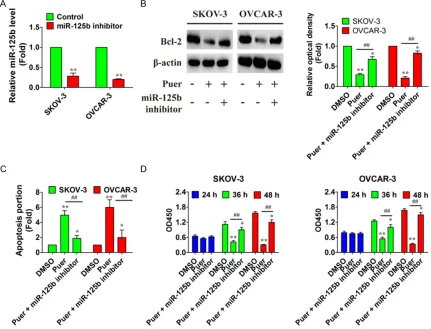
Knockdown of miR-125b alleviates inhibition of cell proliferation induced by puerarin
To examine whether the up-regulation of miR-125b contributes to the puerarin-induced cell inhibition in SKOV-3 and OVCAR-3 cells, we knocked down the expression of miR-125b by transfection of its specific inhibitor, miR-125b inhibitor, and then observed the alteration of puerarin-induced inhibition in OC cells (Figure 4A). The combination of miR-125b inhibitor transfection and puerarin treatment reversed
the dysregulation of Bcl-2 caused by puerarin alone in SKOV-3 and OVCAR-3 cells and allevi-ates the proapoptotic action of puerarin (Figure 4B and 4C). Moreover, the combination of miR-125b inhibitor transfection and puerarin treat-ment restored the reduction of cell viability induced by puerarin alone (Figure 4D). These results suggested that puerarin induced cell proliferation partly due to up-regulation of miR-125b in OC cells.
Discussion
In the present study, we demonstrated that puerarin could inhibit the growth of ovarian cancer cell via up-regulating the expression of miR-125b. And, we investigated the altered expression pattern of miR-125b in ovarian
[image:6.612.92.523.74.403.2]cer cells and its indication of cancer cell apop-tosis through suppression of Bcl-2 expression. Moreover, we found knock down of miR-125b could alleviate puerarin-induced proliferation. These findings should provide a new insight into the use of puerarin in the treatment of ovarian cancer.
Several reports revealed that puerarin exerted an intriguing role in inducing cell apoptosis and suppressing cell proliferation especially in tumor cells. More evidences have found that puerarin inhibited cell growth through inducing cell cycle arrest in breast cancer and glioblas-toma cells [22, 23]. And, recent studies have found that puerarin can inhibit the adhesion, invasion and migration of HO-8910 cells, plays an antagonist effect against the stimulation of estrogen on the malignant behavior of tumor cells [24]. But there is little literature material about its antitumor property onovarian cancer cells. Our data reinforce this anti-tumor action of puerarin by showing that the ability of puera-rin to inhibit ovarian cancer cells growth, sug-gesting that puerarin could be used for the treatment of ovarian cancer.
Dysregulated expression of miRNAs has been observed in numerous human cancers includ-ing ovarian cancer. Fu X et al. found that miR-613 overexpression suppressed ovarian can-cer cell proliferation, colony formation, and invasion by directly targeting KRAS [25]. A study from Sun KX et al. demonstrates that miR-186 may sensitize ovarian cancer cell to paclitaxel and cisplatin by targeting ABCB1 and modulat-ing the expression of GST-π [26]. Modulation of miRNA levels by a number of agents has been demonstrated to function as cancer chemopre-ventive agents, such as curcumin, isoflavone genistein, revesterol and so on [27-29]. How- ever, it is unknown whether miRNAs mediated the anti-cancer effect of puerarin.
MiR-125b is a tumour-suppressing miRNA downregulated in several cancers [30, 31]. For example, miR-125b was found to suppress the oncoproteins MUC1, ERBB2, and ERBB3, inhib-iting the growth of breast cancer cells [31]. Recently, deregulation of miR-125b has been observed in invasive breast cancer, ovarian car-cinoma, hepatocellular carcar-cinoma, and thyroid carcinoma [32-35], and it is also associated with clinical outcome in liver cancer patients [34]. To date, Bak1, CYP24, and Bcl2 have been
identified as targets of miR-125b [36]. For fur-ther study, we chose to focus on Bcl-2, an important anti-apoptotic protein that is well characterized in many tumors. In this study, we found that reduced miR-125b expression is a frequent event in OVARIAN CANCER, which is consistent with other published studies [34, 37]. Moreover, our data showed that puerarin promoted the expression of miR-125b, which suppressed cancer growth by promoting cell apoptosis through the down-regulation of Bcl-2 expression. This is consistent with the current view that a single miRNA can regulate gene expression by targeting multiple mRNAs, which is collectively named a “targetome” [38]. In conclusion, our findings demonstrated that miR-125b was involved in the anti-cancer action of puerarin. However, emerging reports showed that other miRNAs participated in the progression of ovarian cancer. Maybe this is the reason why the dysregulation of Bcl2 was not fully recovered when we knocked down of miR-125b before treating with puerarin in OVARIAN CANCER cells. In addition, the evi-dences of puerarin negatively regulating the growth of ovarian cancer cells in xenograft models in the future will provide more convinc-ing basis to support that puerarin may be a can-didate to treat ovarian cancer.
Disclosure of conflict of interest
None.
Address correspondence to: Yongjie Tian, Depart- ment of Obstetrics and Gynecology, Provincial Hospital Affiliated to Shandong University, 324 Jingwuweiqi Road, Shandong, China. Tel: +86 531-68776384; E-mail: tianyongjie@sdu.edu.cn
References
[1] Lengyel E. Ovarian cancer development and metastasis. Am J Pathol 2010; 177: 1053-1064.
[2] Scholz HS, Tasdemir H, Hunlich T, Turnwald W, Both A and Egger H. Multivisceral cytoreduc-tive surgery in FIGO stages IIIC and IV epithelial ovarian cancer: results and 5-year follow-up. Gynecol Oncol 2007; 106: 591-595.
[3] Li Y, Kong D, Wang Z and Sarkar FH. Regulation of microRNAs by natural agents: an emerging field in chemoprevention and chemotherapy research. Pharm Res 2010; 27: 1027-1041. [4] Tian F, Xu LH, Zhao W, Tian LJ and Ji XL. The
Puerarin inhibits proliferation of ovarian cancer cells via miR-125b
puerarin treatment of spinal cord ischemia-re-perfusion injury in rats. J Ethnopharmacol 2011; 134: 892-896.
[5] Liu XJ, Zhao J and Gu XY. The effects of genis-tein and puerarin on the activation of nuclear factor-kappaB and the production of tumor necrosis factor-alpha in asthma patients. Pharmazie 2010; 65: 127-131.
[6] Xiao C, Li J, Dong X, He X, Niu X, Liu C, Zhong G, Bauer R, Yang D and Lu A. Anti-oxidative and TNF-alpha suppressive activities of puerarin derivative (4AC) in RAW264.7 cells and colla-gen-induced arthritic rats. Eur J Pharmacol 2011; 666: 242-250.
[7] Wu L, Qiao H, Li Y and Li L. Cardioprotective effects of the combined use of puerarin and Danshensu on acute ischemic myocardial in-jury in rats. Phytother Res 2007; 21: 751-756. [8] Zhang SY, Chen G, Wei PF, Huang XS, Dai Y,
Shen YJ, Chen SL, Sun-Chi CA and Xu HX. The effect of puerarin on serum nitric oxide con-centration and myocardial eNOS expression in rats with myocardial infarction. J Asian Nat Prod Res 2008; 10: 373-381.
[9] Gu L, Yang Y, Sun Y and Zheng X. Puerarin in-hibits acid-sensing ion channels and protects against neuron death induced by acidosis. Planta Med 2010; 76: 583-588.
[10] Zhu J, Wang X, Shang Y, Xie X, Zhang F, Chen J and Fu G. Puerarin reduces endothelial pro-genitor cells senescence through augmenta-tion of telomerase activity. Vascul Pharmacol 2008; 49: 106-110.
[11] Hu W, Zhang Q, Yang X, Wang Y and Sun L. Puerarin inhibits adhesion molecule expres-sion in tnf-alpha-stimulated human endotheli-al cells via modulation of the nuclear factor kappaB pathway. Pharmacology 2010; 85: 27-35.
[12] Meng XH, Ni C, Zhu L, Shen YL, Wang LL and Chen YY. Puerarin protects against high glu-cose-induced acute vascular dysfunction: role of heme oxygenase-1 in rat thoracic aorta. Vascul Pharmacol 2009; 50: 110-115. [13] Yu Z and Li W. Induction of apoptosis by
puera-rin in colon cancer HT-29 cells. Cancer Lett 2006; 238: 53-60.
[14] Bezerra JE and Latronico AC. MicroRNA era: the importance for diagnosis and prognosis of adrenocortical tumors. Biomed Res Int 2014; 2014: 381917.
[15] Krol J, Loedige I and Filipowicz W. The wide-spread regulation of microRNA biogenesis, function and decay. Nat Rev Genet 2010; 11: 597-610.
[16] Ambros V. microRNAs: tiny regulators with great potential. Cell 2001; 107: 823-826. [17] Hu X, Macdonald DM, Huettner PC, Feng Z, El
Naqa IM, Schwarz JK, Mutch DG, Grigsby PW,
Powell SN and Wang X. A miR-200 microRNA cluster as prognostic marker in advanced ovar-ian cancer. Gynecol Oncol 2009; 114: 457-464.
[18] Shih KK, Qin LX, Tanner EJ, Zhou Q, Bisogna M, Dao F, Olvera N, Viale A, Barakat RR and Levine DA. A microRNA survival signature (MiSS) for advanced ovarian cancer. Gynecol Oncol 2011; 121: 444-450.
[19] Lee M, Kim EJ and Jeon MJ. MicroRNAs 125a and 125b inhibit ovarian cancer cells through post-transcriptional inactivation of EIF4EBP1. Oncotarget 2016; 7: 8726-42.
[20] Yang Q, Zhang F, Ding Y, Huang J, Chen S, Wu Q, Wang Z, Wang Z and Chen C. Antitumour ac-tivity of the recombination polypeptide GST-NT21MP is mediated by inhibition of CXCR4 pathway in breast cancer. Br J Cancer 2014; 110: 1288-1297.
[21] Liang H, Zhang C, Ban T, Liu Y, Mei L, Piao X, Zhao D, Lu Y, Chu W and Yang B. A novel re-ciprocal loop between microRNA-21 and TGFbetaRIII is involved in cardiac fibrosis. Int J Biochem Cell Biol 2012; 44: 2152-2160. [22] Lin YJ, Hou YC, Lin CH, Hsu YA, Sheu JJ, Lai CH,
Chen BH, Lee Chao PD, Wan L and Tsai FJ. Puerariae radix isoflavones and their metabo-lites inhibit growth and induce apoptosis in breast cancer cells. Biochem Biophys Res Commun 2009; 378: 683-688.
[23] Yang JA, Li JQ, Shao LM, Yang Q, Liu BH, Wu TF, Wu P, Yi W and Chen QX. Puerarin inhibits pro-liferation and induces apoptosis in human glio-blastoma cell lines. Int J Clin Exp Med 2015; 8: 10132-10142.
[24] Han J, Yu CQ and Shen W. [Inhibitory effects of puerarin on invasion and metastasis of oopho-roma cells HO-8910]. Zhongguo Zhong Xi Yi Jie He Za Zhi 2009; 29: 632-635.
[25] Fu X, Cui Y, Yang S, Xu Y and Zhang Z. MicroRNA-613 inhibited ovarian cancer cell proliferation and invasion by regulating KRAS. Tumour Biol 2015; [Epub ahead of print]. [26] Sun KX, Jiao JW, Chen S, Liu BL and Zhao Y.
MicroRNA-186 induces sensitivity of ovarian cancer cells to paclitaxel and cisplatin by tar-geting ABCB1. J Ovarian Res 2015; 8: 80. [27] Haldar S and Basu A. Modulation of MicroRNAs
by Chemical Carcinogens and Anticancer Drugs in Human Cancer: Potential Inkling to Therapeutic Advantage. Mol Cell Pharmacol 2011; 3: 135-141.
[28] Tsang WP and Kwok TT. Epigallocatechin gal-late up-regulation of miR-16 and induction of apoptosis in human cancer cells. J Nutr Biochem 2010; 21: 140-146.
human pancreatic cancer cells. Mol Cancer Ther 2008; 7: 464-473.
[30] Chen H and Xu Z. Hypermethylation-Associated Silencing of miR-125a and miR-125b: A Potential Marker in Colorectal Cancer. Dis Markers 2015; 2015: 345080.
[31] Scott GK, Goga A, Bhaumik D, Berger CE, Sullivan CS and Benz CC. Coordinate suppres-sion of ERBB2 and ERBB3 by enforced expres-sion of micro-RNA miR-125a or miR-125b. J Biol Chem 2007; 282: 1479-1486.
[32] Iorio MV, Ferracin M, Liu CG, Veronese A, Spizzo R, Sabbioni S, Magri E, Pedriali M, Fabbri M, Campiglio M, Menard S, Palazzo JP, Rosenberg A, Musiani P, Volinia S, Nenci I, Calin GA, Querzoli P, Negrini M and Croce CM. MicroRNA gene expression deregulation in hu-man breast cancer. Cancer Res 2005; 65: 7065-7070.
[33] Visone R, Pallante P, Vecchione A, Cirombella R, Ferracin M, Ferraro A, Volinia S, Coluzzi S, Leone V, Borbone E, Liu CG, Petrocca F, Troncone G, Calin GA, Scarpa A, Colato C, Tallini G, Santoro M, Croce CM and Fusco A. Specific microRNAs are downregulated in hu-man thyroid anaplastic carcinomas. Oncogene 2007; 26: 7590-7595.
[34] Li W, Xie L, He X, Li J, Tu K, Wei L, Wu J, Guo Y, Ma X, Zhang P, Pan Z, Hu X, Zhao Y, Xie H, Jiang G, Chen T, Wang J, Zheng S, Cheng J, Wan D, Yang S, Li Y and Gu J. Diagnostic and prognos-tic implications of microRNAs in human hepa-tocellular carcinoma. Int J Cancer 2008; 123: 1616-1622.
[35] Nam EJ, Yoon H, Kim SW, Kim H, Kim YT, Kim JH, Kim JW and Kim S. MicroRNA expression profiles in serous ovarian carcinoma. Clin Cancer Res 2008; 14: 2690-2695.
[36] Zhao A, Zeng Q, Xie X, Zhou J, Yue W, Li Y and Pei X. MicroRNA-125b induces cancer cell apoptosis through suppression of Bcl-2 ex-pression. J Genet Genomics 2012; 39: 29-35. [37] Su H, Yang JR, Xu T, Huang J, Xu L, Yuan Y and
Zhuang SM. MicroRNA-101, down-regulated in hepatocellular carcinoma, promotes apoptosis and suppresses tumorigenicity. Cancer Res 2009; 69: 1135-1142.