Original Article
Overexpression of IL-12 reverses the phenotype and
function of M2 macrophages to M1 macrophages
Xiao-Lin Yu, Bi-Tao Wu, Ting-Ting Ma, Yan Lin, Feng Cheng, Hai-Yu Xiong, Chang-Li Xie, Cui-Ying Liu, Qin Wang, Zi-Wei Li, Zhi-Guang Tu
College of Laboratory Medicine, Key Laboratory of Laboratory Medical Diagnostics of Education Ministry, Chongqing Medical University, Chongqing, China
Received July 15, 2016; Accepted July 22, 2016; Epub September 1, 2016; Published September 15, 2016
Abstract: The characteristic of M2-polarized macrophage is IL-10highIL-12low. Reports have shown that the combined application of CpG and anti-interleukin-10 receptor antibody could switch M2 to M1, while there are few literatures about the impact of over-expression of IL-12 on M2. After infected with the recombinant adenovirus Ad-IL-12-GFP for 60 h, the generated M2 macrophages were harvested for analysis of PCR/FCM/WB, and the supernatants were col-lected as conditioned media. HepG2 cells were examined by MTT assay and FCM following incubated with different conditioned media. After overexpression of IL-12, The PCR analysis showed that the M2 macrophages exerted a high level of M1 macrophage specific markers (such as IL-23, IL-1β, et al), a low level of M2 macrophage specific mark -ers (IL-10, TGF-β), the FCM analysis showed that CD86 was up-regulated while CD206 was down-regulated, and the WB showed a similar change trend. The conditioned medium of IL-12 overexpressed macrophages suppressed the proliferation, induced apoptosis, and caused G0/G1 cell cycle arrest in HepG2 cells compared to the control groups (P<0.05). To conclude, these results have indicated that the phenotype and function of M2 macrophages could be reversed by the overexprression of IL-12. It may be beneficial to explore the anti-cancer immunotherapy strategy switching the M2 macrophages to M1 macrophages.
Keywords: Recombinant adenovirus Ad-IL-12, macrophage, M2, M1, polarization
Introduction
Macrophages, the major inflammatory compo -nent infiltrating the tumor microenvironment, play an indispensible role in the process of tumor growth and progression. Heterogeneity and plasticity are the remarkable hallmarks of macrophages. The heterogeneity means that macrophages include a variety of activation status or phenotypes, and the plasticity means that the phenotype of macrophages could be reverted responding to its local microenviron-ment [1-3].
Of the numerous phenotypes, M1- and M2- polarized macrophages are the two extreme groups. Classically activated macrophages (M1), stimulated by IFN-γ alone or with LPS, are characterized by IL-12highIL-23highIL-10low. In
con-trast, alternatively activated macrophages (M2), promoted by IL-4 and IL-13, and are
char-acterized by IL-10highIL-12lowIL-23low. Additionally,
Interleukin-12 (IL-12), linking the innate and adaptive immune responses, belongs to the IL-12 family of cytokines. Consisting of the subunit p35 (a light chain) and p40 (a heavy chain) [9], IL-12 is mainly produced by dendr- itic cells (DCs), monocytes and macrophages, and exerts various aspects of roles [10-12]. Although a majority of researches have demon-strated that IL-12 is one of the most potential anti-tumor cytokine, but until now IL-12 has not successfully translated into the clinics owning to its non-negligible side-effects when admi- nistrated systemically [13, 14].
Based on the pro-tumoral effects of M2, a mul-titude of researches have focused on the potential anti-tumor strategies targeting on M2 macrophages [15-17]. Guiducci C and his col-leagues found that combined application of CpG and anti-interleukin-10 receptor antibody could promptly switch infiltrating macrophages from M2 to M1 within 16 h [18]. Our recent study has proven that IL-12 overexpressed monocytes could directionally polarize to M1- like macrophages [19], and treatment with IL-12 would induce the differentiation of mono-cytic tumor cells [20]. Considering the charac-teristic of M2 macrophages (IL-10highIL-12low), it
will be of great interest to hypothesize that M2 macrophages could be also reverted after over-expression of IL-12. Therefore, the objective of the current study was to explore whether the phenotype and function of M2 macrophages could be altered by overexprression of IL-12.
Materials and methods
Main materials and reagents
Recombinant adenovirus Ad-IL-12-GFP and Ad-CMV-GFP were constructed by our laborato-ry [18]. Phorbol-12myristate13-acetate (PMA) and LPS were purchased from Sigma-Aldrich
Cell culture
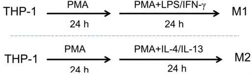
THP-1 (human acute monocytic leukemia cell line) and HepG2 (human hepatocellular carci-noma cell line) were purchased from Chinese Type Culture Collection (Shanghai, China). Cells were cultured in RPMI-1640 and DMEM (Inv-itrogen, USA), respectively, both supplemented with 10% FBS (Gibco, USA) and 100 mg/ml penicillin-streptomycin, and incubated at 37°C in a humidified atmosphere with 5% CO2. For M1 and M2 polarization, the following pro-cedures were performed according to the method described [21]: THP-1 cells were tre- ated with 50 ng/ml PMA for 48 h, and then stimulated with 20 ng/ml IL-4 (added at 24 h later) to generate M2-polarized macrophages (M2), with 20/ng ml IFN-γ+100 ng/ml LPS (added at 24 h later) to generate M1-polarized macrophages (M1) positive control (Figure 1). The phenotypes were identified with the exp-ression of surface maker and phenotype-spe-cific cytokines.
Defining experimental groups and collecting conditioned medium
[image:2.612.89.346.74.151.2]The established M2-polarized macrophages were washed thoroughly 3 times with PBS and then propagated in different media, briefly, in complete medium only (group Blank), compl- ete medium with GFP (group Ad-CMV-GFP), or complete medium with Ad-IL-12-GFP (group Ad-IL-12-GFP). After above treatment for 24 h, macrophages of three groups were grown in complete medium for another 48 h, then the supernatants from the three groups were collected, filtered, aliquoted and stored at -20°C until use [22-24]. The cells were harvest-ed for different use (PCR/FCM/WB) describharvest-ed below.
Figure 1. The schematic method to differentiate THP-1 to M1/M1 macrophages. Treated with PMA for 24 h and then cultured with PMA plus LPS/IFN-γ for another 24 h, THP-1 was induced to M1-polarized macrophages (“M1”). Treated with PMA for 24 h and then cultured with PMA plus IL-4/IL-13 for another 24 h, THP-1 was differentiated to M2-polarized macrophages (“M2”).
For cell cycle and apoptosis of HepG2
The protocol was described previously [20, 25], briefly, HepG2 were treated with different con -ditioned media. Following examinations were performed (1) Cells were washed 3 times with PBS (pH 7.4), and fixed by 75% ethyl alcohol for 1 h, and then immersed in propidium iodide (PI) staining solution for cell cycle analysis by the flow cytometer (BD company, NJ, USA). (2) Cells were washed 3 times with PBS (pH 7.4), and stained by Annexin V/PI staining for cell apoptosis analysis by the flow cytometer (BD company, NJ, USA).
Proliferation assay
The HepG2 cells were seeded into 96-well plates at a density of 1,600 cells per well and cultured with different conditioned media des- cribed above for indicated time. The cells were then incubated with complete media plus 10% Cell Counting Kit-8 (CCK-8) solution for addi-tional 1 h. The optical density (OD) was read at 450 nm as recommended by the manufacturer (Dojindo, Kumamoto, Japan). Experiments were performed in triplicate from three independent tests.
Western blotting
After transfected with recombinant adenovirus for 60 h, macrophages were treated with lysis buffer containing protease and phosphatase inhibitors. The protein concentrations were detected by BCA protein assay kit (Beyotime, Jiangsu, China). Protein samples were subject-ed to SDS-PAGE, and the separatsubject-ed proteins To evaluate the effect of M2 macrophages
infected by Ad-IL-12-GFP on cancer cells, HepG2 cells were treated with different condi-tioned medium described above for indicated time and grouped as HepG2, HepG2/CMV and HepG2/Ad-IL-12, respectively.
RNA isolation, reverse transcription-PCR (RT-PCR) and quantitative real-time PCR (q(RT-PCR)
Total RNA was extracted from macrophages using Trizol reagent (TaKaRa, Japan) according to the manufacturer’s protocol. Reverse tran-scription was performed with 1,000 ng total RNA from each sample using PrimeScript RT Reagent Kit (TaKaRa, Japan) in accordance with the manufacturer’s protocol. qPCR was carried out following the protocol supplied with TaKaRa kit (TaKaRa, Japan), and the relative expression of target genes was determined using the 2-ΔΔct method by normalizing target
gene Ct values to those for reference gene β-actin. The appropriate primers used were shown in Table 1 and samples were analyzed in triplicates.
Flow cytometry analyses
[image:3.612.92.522.84.243.2]For cell surface markers of macrophages: After the M2-polarized macrophages were transfe-cted with recombinant adenovirus (Ad-IL-12-GFP, Ad-CMV-GFP) or without for 60 h, cells were collected and washed 3 times with cold PBS, followed by incubation with human CD86-PE, CD206-CD86-PE, or isotype IgG2A-PE for 30 min at 4°C. Samples were then analyzed by a flow cytometer (BD Company, NJ, USA).
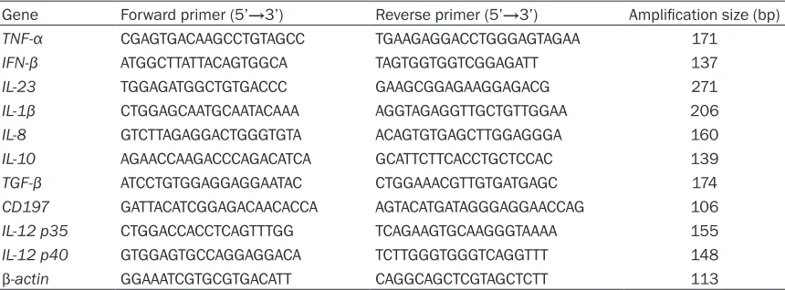
Table 1. Primer sequences for qPCR
Gene Forward primer (5’→3’) Reverse primer (5’→3’) Amplification size (bp)
TNF-α CGAGTGACAAGCCTGTAGCC TGAAGAGGACCTGGGAGTAGAA 171
IFN-β ATGGCTTATTACAGTGGCA TAGTGGTGGTCGGAGATT 137
IL-23 TGGAGATGGCTGTGACCC GAAGCGGAGAAGGAGACG 271
IL-1β CTGGAGCAATGCAATACAAA AGGTAGAGGTTGCTGTTGGAA 206
IL-8 GTCTTAGAGGACTGGGTGTA ACAGTGTGAGCTTGGAGGGA 160
IL-10 AGAACCAAGACCCAGACATCA GCATTCTTCACCTGCTCCAC 139
TGF-β ATCCTGTGGAGGAGGAATAC CTGGAAACGTTGTGATGAGC 174
CD197 GATTACATCGGAGACAACACCA AGTACATGATAGGGAGGAACCAG 106
IL-12 p35 CTGGACCACCTCAGTTTGG TCAGAAGTGCAAGGGTAAAA 155
IL-12 p40 GTGGAGTGCCAGGAGGACA TCTTGGGTGGGTCAGGTTT 148
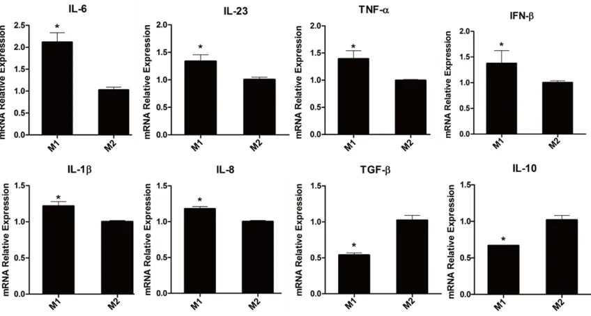
TNF-α, IFN-β, IL-23, IL-1β, CD197 and IL-8 was significantly decreased, while cytokines IL-10, TGF-β were markedly increased in group M2 (Figure 2).
Overexprression of IL-12 in M2 macrophages after transfected with Ad-IL-12-GFP
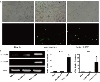
Recombinant adenovirus transduction was evi-denced by GFP expression (Figure 3A). The expression of subunits of IL-12 p35 and p40 were examined by RT-PCR and qPCR. As shown in Figure 3B and 3C), M2 macrophages trans-fected with recombinant adenovirus (Ad-IL-12-GFP) for 36 h had a significantly higher expres -sion of p35 and p40 than those of control groups (Ad-CMV-GFP or M2 blank). These results have indicated that IL-12 over-expressed M2 macrophages were established by Ad-IL- 12-GFP transfection.
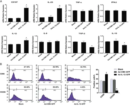
Phenotypical changes of M2 macrophages af-ter transfected with Ad-IL-12-GFP
To explore the effects of overexpression of IL-12 on the phenotype of M2 macrophages, some M1 and M2 macrophage specific markers were assessed by qPCR (Figure 4A). Following transfection of recombinant adenovirus for 60 h, M1 macrophage specific markers IL-23, CD197, IL-1β elevated significantly, while M2 were transferred onto the PVDF membranes.
The membranes were blocked with 5% skim milk, incubated with different primary antibod-ies (anti-p-stat3, anti-p-stat1, anti-wnt2, β-catenin, c-myc, NF-к BP50 and anti-NF-к BP65 antibodies) overnight, followed by incubation with HRP-conjugated secondary antibody, and visualized with the ECL reagents (Millpore, USA). To ascertain equal loading of proteins, house-keeping protein actin was uti-lized as a loading control.
Statistical analysis
Experimental results were analyzed by Stu- dent’s t test or One-Way ANOVA with SPSS ver-sion 19.0 software, P<0.05 was considered to be statistically significant. All experiments were repeated at least three times. Values were shown as mean ± SD.
Results
Established M1 and M2 macrophages
[image:4.612.94.518.72.299.2]In accordance with the method described by Tjiu et al, THP-1 was polarized to M1 and M2 macrophages with minor modifications, as shown in Figure 1. Some pro- and anti-inflam -matory cytokines were analyzed by qPCR, com-pared with group M1, the mRNA expression of
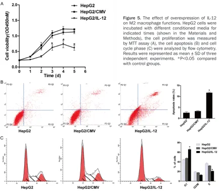
tioned media, the proliferation of HepG2 was assessed by MTT. AS shown in Figure 5A, the proliferation of HepG2 was dramatically inhib-ited from the day 3 in group HepG2/IL-12 com-pared with group HepG2 and group HepG2/ CMV.
In addition, the apoptosis and cell cycles of HepG2 were examined by FCM (Figure 5B and
5C). After incubation with conditioned media for 72 h, the early apoptosis rate of group HepG2/IL-12 was 11.33%, while the early apoptosis rates in group HepG2 and group HepG2/CMV were 2.91% and 3.01%, respec-tively. The cell cycle distribution of HepG2 was also altered following cultured with different conditioned media. The proportion of G1 phase population was far higher in group HepG2/ IL-12 than those in control groups. While com-pared the proportion of cells in S phase, group macrophage specific markers IL-10, TGF-β
exhibited markedly reduction in group Ad-IL-12-GFP compared to group Ad-CMV-Ad-IL-12-GFP and group Blank. Furthermore, some cell surface markers were detected by FCM (Figure 4B), compared to control groups, group Ad-IL-12-GFP had a high-er expression of M1 surface markhigh-er CD86 and lower expression of M2 surface marker CD206. These results have suggested that overexprres-sion of IL-12 could reverse the phenotype of M2 macrophages to M1-like macrophages.
Functional changes of M2 macrophages after infected with Ad-IL-12-GFP
To investigate the effects of Ad-IL-12-GFP on the function of M2 macrophages, some biologi-cal behaviors of HepG2 were analyzed includ-ing the growth, apoptosis and cell cycle distri-bution. After treatments with different
[image:5.612.93.519.69.418.2]those in group Blank and group Ad-CMV-GFP (Figure 6).
Discussion
In the current study we investigated the effects of Ad-IL-12-GFP on the phenotype and function of M2 macrophages. It was found that after over-expression of IL-12, the phenotype of M2 macrophages was re-directed to that of M1-like macrophages, accompanied with alteration of the phenotype characteristic markers and related signaling molecules. And the function of M2 macrophages was also changed from pro-tumor to anti-pro-tumor.
IL-12 is one of the most extensively studied cytokines in the field of anti-tumor immunother -apy [13]. Watkins and his colleagues have found that the functional features of tumor-HepG2/IL-12 was much lower than the control
groups. Similarly G2/M phase was also lower in group HepG2/IL-12 although did not reach the significant level. These findings have revealed that overexprression of IL-12 could change the function of M2 macrophages, which is from tumor promotion to tumor suppression.
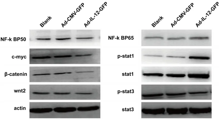
Alteration of some signaling molecules related to macrophage polarization
[image:6.612.93.519.74.422.2]Majorities of signaling molecules were involved in the process of macrophage polarization. Parts of these molecules were analyzed by Western blotting following transduction of recombinant adenovirus. The signaling mole-cules p-stat3, wnt2, β-catenin, c-myc and NF-к BP50 were down-regulated in group Ad-IL-12-GFP, while signaling molecules p-stat1 and NF-к BP65 were up-regulated, compared to
some inflammation factors and cell surface antigens were applied as markers to distin-guish M1 and M2 macrophages in our research. The results showed that “M2” exhibited a high level of IL-10, TGF-β, whereas a low level of CD197 TNF-α, IFN-β, IL-23, IL-1β, and IL-8, sug -gesting that the M2 macrophage model was successfully established, which was also in line with the reports [21].
Overexpression of target gene is usually accom-plished with viral-vector to transfer target genes to interesting cells. Frequently, the viral-vector carries target genes as well as green fluores -cent protein (GFP) gene. The expression of GFP was used as a visible indicator of the viral-vec-tor successful transfection. In our study, the transfection of Ad-IL-12-GFP vector was identi-fied by fluorescence microscopy, and the expression of target gene IL-12 was proved by the high expression of IL-12 subunits P35 and associated and tumor-infiltrating macrophages
could be rapidly altered by IL-12 carried through microcapsules [26]. Our recent researches have also revealed that IL-12 overexpressed monocytes would directionally differentiate to M1-like macrophages through downregulation of p-stat-3 and result in the inhibition of HCC growth in vitro and vivo [20]. Furthermore, IL-12 treatment could induce monocytic tumor cells differentiation and its possible mechanism is related with the up-regulation of c-fms expres-sion and the phosphorylation of CSF-1R Tyr-809 site [21].
[image:7.612.91.524.71.448.2]THP-1 was the widely utilized cell lines to explore the features of monocytes and macro-phages in vitro [25]. In accordance with a vari-ety of documents [21, 27, 28], especially the report by Tjiu et al [21], we induced THP-1 to M1 and M2 macrophages, respectively. Based on the characteristics of polarized macrophages,
P40 through RT-PCR and qPCR. These results have indicated that M2 macrophage model of over-expression of IL-12 is well constructed. In the aspect of M2 macrophage phenotype influenced by over-expression of IL-12, some related markers including cytokines, chemo-kines and cell surface antigens were measured. It was found that the expression of genes relat-ed to M1 specific markers IL-23, CD197 and IL-1β were significantly elevated, while M2 mac -rophage specific markers IL-10 and TGF-β exhibited markedly reduction (Figure 4). In con-sistent with above results, the cell surface anti-gens CD86, a marker of M1 macrophages [29], was up-expressed, whereas M2 macrophages marker CD206 [30] showed a down-expres -sion. Furthermore, in the aspect of M2 macro-phage function, some biological behaviors of HepG2, such as HepG2 cell proliferation, cell apoptosis and cell cycle were examined. It was found that the HepG2 cells exhibited prolifera-tion suppression, apoptosis inducprolifera-tion and G0/ G1 cell cycle arrest following treatment with conditioned medium of over-expression IL-12 macrophages culture compared with those of two control groups. The reason that we used conditioned medium rather than the Transwell co-culture model was due to the M2 macro-phage redirected in phenotype until infected with recombinant adenovirus for as long as 60
[image:8.612.94.519.73.305.2]findings have suggested that over-expression of IL-12 inducing the phenotype and function of M2 macrophages into M1-like macrophages involve above signaling molecules. Neverthe- less, further analysis with the effect of each individual signal molecule and their interac-tions in the regulation mechanism of macro-phage re-direction by overexpression IL-12 is needed.
In conclusion, present study reveals that over-expression of IL-12 could reverse the pheno-type and function of M2 macrophages into M1-like macrophages; it means from tumor pro-motion action conversion to tumor suppression effect. This may contribute to the understand-ing of anti-tumor immune therapy strategies targeting the polarization of M2 macrophages and TAM to M1 macrophages with IL-12 over-expression.
Acknowledgements
This work was supported by Grant 81172016 from the National Natural Science Foundation of China.
Disclosure of conflict of interest
None.
Address correspondence to: Zhi-Guang Tu, College of Laboratory Medicine, Chongqing Medical Univ-ersity, Chongqing 400016, China. Tel: 86-23-68485759; Fax: 86-23-68485239; E-mail: tuzhi-guang@aliyun.com
References
[1] Sica A and Mantovani A. Macrophage plasticity and polarization: in vivo veritas. J Clin Invest 2012; 122: 787-795.
[2] Mosser DM and Edwards JP. Exploring the full spectrum of macrophage activation. Nat Rev Immunol 2008; 8: 958-969.
[3] Mantovani A, Sozzani S, Locati M, Allavena P and Sica A. Macrophage polarization: tumor-associated macrophages as a paradigm for polarized M2 mononuclear phagocytes. Trends Immunol 2002; 23: 549-555.
[4] Adams DO and Hamilton TA. The cell biology of macrophage activation. Annu Rev Immunol 1984; 2: 283-318.
[5] Stein M, Keshav S, Harris N and Gordon S. In-terleukin 4 potently enhances murine macro-phage mannose receptor activity: a marker of alternative immunologic macrophage activa-tion. J Exp Med 1992; 176: 287-292.
[6] Martinez FO, Gordon S, Locati M and Man-tovani A. Transcriptional profiling of the human monocyte-to-macrophage differentiation and polarization: new molecules and patterns of gene expression. J Immunol 2006; 177: 7303-7311.
[7] Martinez FO, Sica A, Mantovani A and Locati M. Macrophage activation and polarization. Front Biosci 2008; 13: 453-461.
[8] Sica A, Schioppa T, Mantovani A and Allavena P. Tumour-associated macrophages are a dis-tinct M2 polarised population promoting tu-mour progression: Potential targets of anti-cancer therapy. Eur J Cancer 2006; 42: 717-727.
[9] Jalah R, Rosati M, Ganneru B, Pilkington GR, Valentin A, Kulkarni V, Bergamaschi C, Chowd-hury B, Zhang GM, Beach RK, Alicea C, Broder-ick KE, Sardesai NY, Pavlakis GN and Felber BK. The p40 Subunit of Interleukin (IL)-12 Pro-motes Stabilization and Export of the p35 Sub-unit. J Biol Chem 2013; 288: 6763-76. [10] Gee K, Guzzo C, Che Mat NF, Ma W and Kumar
A. The IL-12 Family of Cytokines in Infection, Inflammation and Autoimmune Disorders. In-flamm Allergy Drug Targets 2009; 8: 40-52. [11] Colombo MP and TrinchieriG. Interleukin-12 in
anti-tumor immunity and immunotherapy. Cy-tokine Growth Factor Rev 2002; 13: 155-168. [12] Xu M, Mizoguchi I, Morishima N, Chiba Y, Mizu-guchi J and Yoshimoto T. Regulation of Antitu-mor Immune Responses by the IL-12 Family Cytokines, IL-12, IL-23, and IL-27. Clin Dev Im -munol 2010; 1-9.
[13] Tugues S, Burkhard SH, Ohs I, Vrohlings M, Nussbaum K, Vom Berg J, Kulig P and Becher B. New insights into IL-12-mediated tumor sup-pression. Cell Death Differ 2015; 22:237-246. [14] Marshall E. Cancer trial of interleukin-12
halt-ed. Science 1995; 268: 1555.
[15] Wahl L and Kleinman HK. Tumor-associated macrophages as targets for cancer therapy. J Natl Cancer Inst 1998; 90: 1583-1584. [16] Mantovani A and Locati M. Tumor-Associated
Macrophages as a Paradigm of Macrophage Plasticity, Diversity, and Polarization. Arterio -scler Thromb Vasc Biol 2013; 33: 1478-1483. [17] Guiducci C, Vicari AP, Sangaletti S, Trinchieri G
and Colombo MP. Redirecting in vivo elicited tumor infiltrating macrophages and dendritic cells towards tumor rejection. Cancer Res 2005; 65: 3437-3446.
[19] Wang Q, Cheng F, Ma TT, Xiong HY, Li ZW, Xie CL, Liu CY, Tu ZG. Interleukin-12 inhibits the hepatocellular carcinoma growth by inducing macrophage polarization to the M1-like pheno-type through downregulation of Stat-3. Mol Cell Biochem 2016; 415: 157-168.
[20] Ma TT, Wu BT, Lin Y, Xiong HY, Wang Q, Li ZW, Cheng F and Tu ZG. IL-12 could induce mono-cytic tumor cells directional differentiation. Mol Cell Biochem 2015; 402: 157-69.
[21] Tjiu JW, Chen JS, Shun CT, Lin SJ, Liao YH, Chu CY, Tsai TF, Chiu HC, Dai YS, Inoue H, Yang PC, Kuo ML and Jee SH. Tumor-Associated Macro-phage-Induced Invasion and Angiogenesis of Human Basal Cell Carcinoma Cells by Cycloox-ygenase-2 Induction. J Invest Dermatol 2009; 129: 1016-1025.
[22] Engström A, Erlandsson A, Delbro D, Wijkander J. Conditioned media from macrophages of M1, but not M2 phenotype, inhibit the prolifer-ation of the colon cancer cell lines HT-29 and CACO-2. Int J Oncol 2014; 44: 385-392. [23] Hedbrant A, Erlandsson A, Delbro D and Wi
-jkander J. Conditioned media from human macrophages of M1 phenotype attenuate the cytotoxic effect of 5-fluorouracil on the HT-29 colon cancer cell line. Int J Oncol 2015; 46: 37-46.
[24] Guihard P, Danger Y, Brounais B, David E, Brion R, Delecrin J, Richards CD, Chevalier S, Rédini F, Heymann D, Gascan H and Blanchard F. In -duction of Osteogenesis in Mesenchymal Stem Cells by Activated Monocytes/Macrophages Depends on Oncostatin M Signaling. Stem Cells 2012; 30: 762-772.
[25] Qin Z. The use of THP-1 cells as a model for mimicking the function and regulation of monocytes and macrophages in the vascula-ture. Atherosclerosis 2012; 221: 2-11. [26] Watkins SK, Egilmez NK, Suttles J and Stout
RD. IL-12 rapidly alters the functional profile of tumor-associated and tumor-infiltrating macro -phages in vitro and in vivo. J Immunol 2007; 178: 1357-1362.
[27] Laskar A, Eilertsen J, Li W and Yuan XM. SPION primes THP1 derived M2 macrophages to-wards M1-like Macrophages. Bicchem Biophy Res Commun 2013; 441: 737-742.
[28] Jeong SK, Yang K, Park YS, Choi YJ, Oh SJ, Lee CW, Lee KY, Jeong MH and Jo WS. Interferon gamma induced by resveratrol analog, HS-1793, reverses the properties of tumor associ-ated macrophages. Int Immunopharmacol 2014; 22: 303-310.
[29] Mantovani A, Sozzani S, Locati M, Allavena P and Sica A. Macrophage polarization: tumor-associated macrophages as a paradigm for polarized M2 mononuclear phagocytes. Trends Immunol 2002; 23: 549-555.
[30] Hamilton TA, Zhao C, Pavicic PG Jr and Datta S. Myeloid colony-stimulating factors as regula-tors of macrophage polarization. Front Immu-nol 2014; 5: 554.
[31] Takaishi K, Komohara Y, Tashiro H, Ohtake H, Nakagawa T, Katabuchi H and Takeya M. In-volvement of M2-polarized macrophages in the ascites from advanced epithelial ovarian carcinoma in tumor progression via Stat3 acti-vation. Cancer Sci 2010; 101: 2128-2136. [32] Yu H, Kortylewski M and Pardoll D. Crosstalk
between cancer and immune cells: role of STAT3 in the tumour microenvironment. Nat Rev Immunol 2007; 7: 41-51.
[33] Lang R. Tuning of macrophage responses by Stat3-inducing cytokines: molecular mecha-nisms and consequences in infection. Immu-nobiology 2005; 210: 63-76.
[34] Siapati EK, Papadaki M, Kozaou Z, Rouka E, Michali E, Savvidou I, Gogos D, Kyriakou D, An -agnostopoulos NI and Vassilopoulos G. Prolif-eration and bone marrow engraftment of AML blasts is dependent on β-catenin signaling. Br J Haematol 2011; 152: 164-174.
[35] Pello OM, De Pizzol M, Mirolo M, Soucek L, Zammataro L, Amabile A, Doni A, Nebuloni M, Swigart LB, Evan GI, Mantovani A and Locati M. Role of c-MYC in alternative activation of hu-man macrophages and tumor-associated mac-rophage biology. Blood 2012; 119: 411-421. [36] Porta C, Rimoldi M, Raes G, Brys L, Ghezzi P, Di
Liberto D, Dieli F, Ghisletti S, Natoli G, De Baet -selier P, Mantovani A and Sica A. Tolerance and M2 (alternative) macrophage polarization are related processes orchestrated by p50 nuclear factor B. Proc Natl Acad Sci U S A 2009; 106: 14978-14983.
[37] Cheng F, Wang HW, Cuenca A, Huang M, Ghansah T, Brayer J, Kerr WG, Takeda K, Akira S, Schoenberger SP, Yu H, Jove R and Sotomay-or EM. A critical role fSotomay-or Stat3 Signaling in im-mune tolerance. Immunity 2003; 19: 425-436.
[38] Williams L, Bradley L, Smith A and Foxwell B. Signal transducer and activator of transcrip-tion 3 is dominant mediator of the anti-inflam -matory effects of IL-10 in human macrophages. J Immunol 2004; 172: 567-576.