Original Article
Overexpression of ADAMTS-13 and neuronal nitric
oxide synthase relates with neuropathology in
streptozotocin-induced type 1 diabetic rats
Gungor Cagdas Dincel1, Serkan Yildirim2
1Laboratory and Veterinary Health Program, Eskil Vocational School, University of Aksaray, Aksaray, Turkey; 2
De-partment of Pathology, Faculty of Veterinary Medicine, University of YuzuncuYil, Van, Turkey
Received January 9, 2016; Accepted March 25, 2016; Epub April 1, 2016; Published April 15, 2016
Abstract: Hyperglycemia plays a critical role in the development and progression of diabetic encephalopathy. A few studies have focused on a disintegrin and metalloprotease with thrombospondin type I repeats-13 (ADAMTS-13) expression in the central nervous system (CNS), and its function continues to remain unclear. The purpose of this
study was to compare the expression of ADAMTS-13, neuron specific enolase (NSE), neurofilament (NF), neuronal nitric oxide synthase (nNOS) and glial fibrillary acidic protein (GFAP) in brain tissues of experimental streptozotocin
(STZ)-induced diabetic rats. Expression of ADAMTS-13 (P < 0.001), nNOS (P < 0.001), NSE (P < 0.001) and NF (P < 0.001) expressions in the brain tissue markedly increased while GFAP decreased (P < 0.001) in diabetic animals
versus controls. The most prominent finding of our study was that ADAMTS-13 expression increased significantly,
suggesting that it may play an important function/s in the regulation and protection of the blood-brain barrier integ-rity and central nervous system microenvironment in diabetes. The results also suggested that nitric oxide produc-tion may increase due to increased nNOS expression and this also might contribute to neuropathology related with
diabetes. Furthermore, increased expression of ADAMTS-13, NSE, NF and decreased expression of GFAP may give an idea of the disease progress and thus may have a critical diagnostic significance. To the best of the authors’ knowledge, this is the first report on ADAMTS-13 expression in the CNS of STZ-induced diabetic animals.
Keywords: ADAMTS-13, neuropathology, nitric oxide, diabetes mellitus, neurofilament, neuron specific enolase
Introduction
Diabetes mellitus (DM) is a metabolic disease
that is characterized by dysfunctions in the glu -cose metabolism [1, 2]. Hyperglycemia causes
brain edema, increases size of brain infarct [1,
3] through triggering of neuronal cell death and also induces some vascular complications [4, 5]. The most severe complication of diabetes is diabetic encephalopathy [6-8]. Although the research about the diabetic encephalopathy has been intensive and comprehensive, the cause of pathophysiology of degeneration is still unknown.
ADAMTS13 is a large zinc-containing metallo
-protease enzyme that is involved in blood clot
-ting [9]. It is mainly synthesized in hepatic stel -late cells of the liver [10], but is also expressed in endothelial cells [11, 12] and the cells of
many other organs including brain [11, 13-15]. ADAMTS-13 plays a role in the regulation of
inflammation and prevention of microvascular
thrombus formation by decreasing thrombotic activity through destruction of the Ultra Large
von Willebrand Factor (UL-vWF) multimers into less active forms [16-18]. UL-vWF is essential in
the induction of platelet adhesion after vascu-lar injury [19, 20]. Moreover, it was also dis-cussed that severe ADAMTS-13 expression in the brain and cerebellum decreases infarct risk by preventing thrombus formation [15]. These studies show that ADAMTS-13 has essential roles in the homeostasis of brain and in
throm-bosis as well as in the regulation of inflamma -tory processes.
hyperglycemia triggers disruption of the blood-brain barrier (BBB) and increases in its perme-ability [27-31]. Impairment of tight junctions, which plays a role in the transmembrane pro-tein interactions between the BBB and the endothelial cells, also increases the permeabil-ity of the BBB in diseases with myelin disorders
such as multiple sclerosis and idiopathic inflam -matory demyelination [32]. In a previous study of Dincel and Kul, in Border Disease that is
characterized by demyelination, it was men -tioned that there might be a positive correlation between ADAMTS-13 and demyelination [11]. Depending on the level of expression, nitric oxide (NO) has dual effects in the CNS. High level of NO that is produced by nNOS and other types of NOS was shown to be responsible for
the irregularities in cerebral blood flow [33-36].
It is also known that high-level NO that is pro-duced by neuronal and glial cells (astrocytes, microglia and oligodendrocytes) causes degen-eration and apoptosis in CNS [35, 37, 38]. This situation is explained by cytochrome c release that is caused by the loss of the mitochondrial membrane potential [39-42]. It is also known that apoptosis plays an important role in the pathogenesis of DM and some common neuro-degenerative diseases [1, 2, 43, 44].
The principle objective of this research is to investigate whether there is a correlation between degeneration of central nervous
sys-tem (CNS) of streptozotocin (STZ)-diabetic rats
and expression of ADAMTS-13. The second objective is to examine whether there is a rela-tionship between severities of degeneration seen in the disease with nNOS expression. Moreover, we also focused on how astroglial activation plays role in this disease, and sever-ity of the degeneration was assessed by
analy-sis of the neuron-specific enolase (NSE) and neurofilament (NF) expressions.
Materials and methods
Ethics statement
This study was performed in strict accordance with the recommendations of the National
Centre for the Replacement, Refinement, and
Reduction of Animals in Research (NC3Rs) of Turkey. The experimental protocol was approved by the Committee on the Ethics of Animal
Experiments at Ataturk University (Permit Nu- mber: 390/19.03.2014).
Experimental animals
20 male Wistar albino rats weighing 250-300 were randomly allotted to two experimental groups (n = 10 per group). Animals were housed in a well-ventilated and air-conditioned area provided with independently adjustable light-dark cycle (12 h light/12 h light-dark cycle) and tem-perature regulation systems. Temtem-perature was maintained at 22 ± 2°C and humidity was kept at 45%-70%. The rooms and animal cages were cleaned daily and the animals were provided with fresh food and water ad libitum on a daily basis.
Induction of STZ model of diabetes
Type 1 diabetes was induced in the rats (dia-betic group) by a single intraperitoneal injection
of streptozotocin (STZ) (65 mg/kg body weight)
dissolved in 0.1 mm sodium citrate, pH 4.5, while the normal control rats (nondiabetic group) were injected with the buffer only. The development of hyperglycemia in rats was
con-firmed by blood glucose evaluation. Blood glu -cose was determined by using an automatic glucometer (ACCU-CHEK Active, Roche Diag- nostics Ltd, Germany). Plasma glucose level of the animals higher than 250 mg/dl on day 3 after STZ injection was considered hypergly-caemic [45]. These animals were selected for studies.
Necropsy and histopathologic examination
Animals were sacrificed at the end of 20 days of
experiment period by decapitation and brains were quickly removed and processed for histo-pathology and immunohistochemistry
analy-ses. Brain sections were fixed in 10% neutral
buffered formaldehyde for 48 hours and
washed under tap water overnight. Following
routine tissue preparation procedures, tissue samples were dehydrated through graded series of alcohol and xylene and embedded in
paraffin blocks. Paraffin serial sections were cut at a thickness of 4-5 μm and mounted on glass slides. Brains were sectioned at a 5μm
Antibodies
Commercial anti-mouse antibodies against ADAMTS-13 (Abcam, Cambridge, UK) diluted to
1:100, nNOS (Santa Cruz Biotechnology, USA) diluted to 1:100, GFAP (Thermo Scientific, USA) diluted to 1:100, NSE (Thermo Scientific, USA) diluted to 1:100 and undiluted NF (Thermo Scientific, USA) were used in the present study.
Immunoperoxidase examinations
Immunohistochemistry was performed to
investigate ADAMTS-13, nNOS, NSE, NF and GFAP expressions. Commercial antibodies were visualized on 4- to 5-μm-thick paraffin sections
using an indirect streptavidin/biotin
immuno-peroxidase kit (HRP; Thermo Scientific, USA). All
steps were carried out following the procedure described by Dincel and Atmaca, 2015 [46]. Accordingly, tissue sections were placed on
adhesive slides, deparaffinized for 5 minutes in
each of three xylene series, and rehydrated in a graded alcohol series and distilled water. Antigen retrieval was accomplished by boiling sections on glass slides in citrate buffer (pH
6.0; Thermo Scientific, USA) for 20 min.
Endogenous peroxidase activity was quenched in 3% hydrogen peroxide in absolute methanol for 7 minutes at room temperature. Sections were rinsed three times with phosphate-buff-ered saline (pH 7.4) for 5 min between each
step of the test. Sections were incubated in blocking serum for 5 min to prevent non-specif-ic binding. Thereafter, tissue sections were incubated with the primary antibody (ADAMTS-
13, nNOS, NSE, NF and GFAP) for 60 min in a humidified chamber at room temperature.
Sections were treated with a biotin-labeled sec-ondary antibody for 15 min and with the
strep-tavidin-peroxidase enzyme for 15 min at room temperature. Finally, sections were incubated in aminoethyl carbazole chromogen (Thermo Scientific, USA) for 5-10 min to induce the color reaction. Mayer’s hematoxylin was applied as a
counterstain for 30 sec. Thereafter, sections were mounted with water-based mounting
medium (Thermo Scientific, USA). As a control for non-specific endogenous peroxidase and
biotin activities in each test, the primary anti-body step was omitted. Sections were
immedi-ately analyzed. Immunostaining was evaluated
using a binocular microscope and photo-graphed under a 20X objective.
Quantitative histomorphometric analysis and statistics
The density of positive staining was measured
using a computerized image system composed of a Leica CCD camera DFC420 (Leica
[image:3.612.91.526.71.287.2]Mic-rosystems Imaging Solutions, Ltd., Cambridge, UK), connected to a Lecia DM4000 B micro-scope (Leica Microsystems Imaging Solutions,
Ltd.) and used according to the procedure described by Dincel and Kul, 2015 [38]. The
pictures of five random fields selected and con
-secutive 20x objective microscopic fields were
captured by the Leica QWin Plus v3 software (Leica Microsystems Imaging Solutions) at a
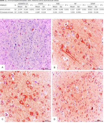
[image:4.612.90.526.76.591.2]setting identical to the image system. For exam -ining the sta-ining for each antibody, we used Table 1. Immunoperoxidase test results and statistical data
ANIMALS N ADAMTS-13 P < nNOS P < NSE P < NF P < GFAP P <
Mean Sd Mean Sd Mean Sd Mean Sd Mean Sd
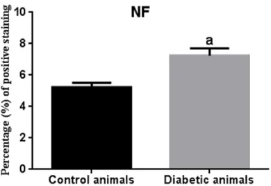
Control animals 10 2.074 0.141 0.001 1.540 0.074 0.001 2.298 0.146 0.001 5.227 0.287 0.001 4.912 0.125 0.001 STZ-treated animals 10 3.192 0.236 2.287 0.118 3.101 0.150 7.250 0.458 3.519 0.118
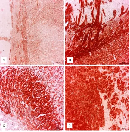
Figure 2. Control group; mild expression of ADAMTS-13 in neuronal and glial cells were detected. ABC technique
(anti-ADAMTS-13), Mayer’s hematoxylin counterstain, Bar, 100 μm (A) Diabetic group (B-D); Strong expression of ADAMTS-13 in neuronal and glial cells detected. ABC technique (anti-ADAMTS-13), Mayer’s hematoxylin counter
the same setting for all slides. Integrated opti-cal density of all the positive staining of
ADAMTS-13, nNOS, NSE, NF and GFAP in each photograph was measured. For the quantifica
-tion, mean was quantified as the ADAMTS-13, nNOS, NSE, and GFAP-positive area/total area
were measured and calculated by Leica Qwin Plus on the pictures. All images were collected under the same lighting conditions. To avoid
observer bias, all sections were quantified by
a blinded investigator. Data were statistically
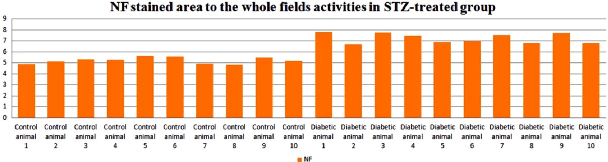
described in terms of mean and standard devi-ation (mean ± SD) for area %. After calculating the proportion (% pixels) of stained area to the
whole field, the mean (in % pixels) staining area for each slide was determined. For evaluating
the non-parametric data, Mann-Whitney U-test was performed to compare ADAMTS-13, nNOS,
NSE, NF and GFAP immunoreactive cells and
[image:5.612.91.522.73.503.2]Figure 4. Calculating the proportion (% pixels) of ADAMTS-13 stained area to the whole field activities in l STZ-induced diabetic animals.
[image:6.792.95.701.291.460.2]nificant. For the statistical analyses, MS-Excel
2003 and SPSS were used for Win. Ver.15.0 (SPSS Inc., Chicago IL, USA) programs. The data were presented as means ± SD. All statis-tical analyses and graphs were prepared using GraphPad Prism version 6.0 (Graph Pad Software, La Jolla California, USA). P < 0.05
was considered statistically significant.
Results
Histopathologic findings
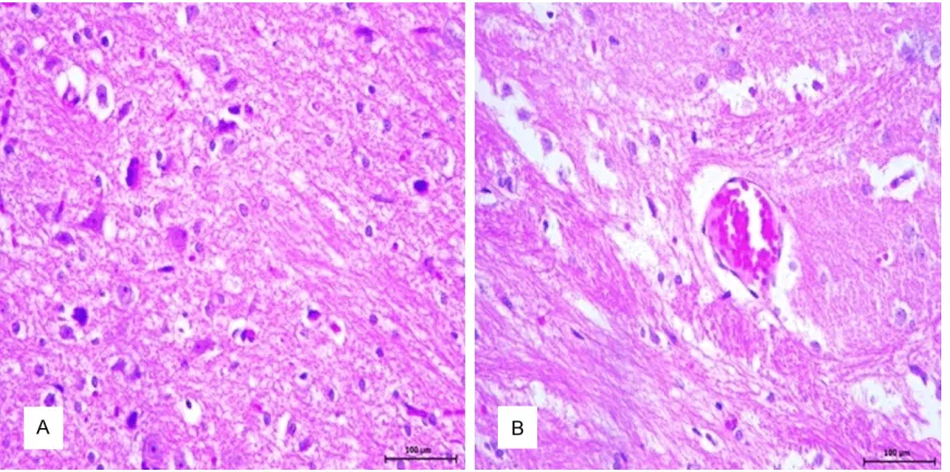
Microscopic lesions were observed in the brains of all STZ-induced diabetic animals. The normal laminar arrangement of neurons and glial cells were not observed. In addition,
hema-toxylin and eosin (H&E)-stained brain sections from healthy control animals exhibited norm- al architecture. Neurohistopathologic changes
were characterized by neuronal necrosis and
central chromatolysis (Figure 1A, 1B). Some degenerated neurons had cytoplasmic vacuo-
lization.
Immunoperoxidase findings
We analyzed protein expression levels of ADAMTS-13, nNOS, NSE, NF and GFAP in the
brain tissues from STZ-induced diabetic ani-mals and healthy control aniani-mals. Immunohis-
tochemical analysis showed significant up-reg -ulation of ADAMTS-13 (P < 0,001), nNOS (P < 0,001), and NSE (P < 0,001) expression and
significant down-regulation of GFAP (P < 0,001) expressions in the STZ-induced diabetic ani-mals in comparison to the control aniani-mals. Statistical analysis of the data on ADAMTS-13,
nNOS, NSE, NF and GFAP expressions in the
brain, measured by immunostaining in all the groups, are presented in Table 1.
A disintegrin and metalloprotease with throm-bospondin type I repeats-13 (ADAMTS-13) and neuronal nitric oxide synthase (nNOS) expres-sion
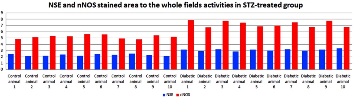
Fairly weak ADAMTS-13 (Figure 2A) and nNOS (Figure 3A) expression was observed in neu-rons and glial cells in healthy control group. We measured protein expression of ADAMTS-13 and nNOS in all parts of the brain. ADAMTS-13 (Figure 2C, 2D) and nNOS (Figure 3B, 3D)
expression increased significantly within and in
the periphery of the lesion in comparison to the healthy control groups. This assessment has also been interpreted quantitatively and
differ-ence was statistically significant [ADAMTS-13
(Figure 4) and nNOS, (Figure 5)].
ADAMTS-13 was expressed by some glial cells in the cerebral cortex but was predominantly expressed by the neurons (Figure 2B-D). The
most conspicuous finding of the present study
was that ADAMTS-13 and nNOS (Figure 3B-D) expression were markedly increased in the neurons (Figure 6). The number of nNOS-expressing neurons increased in all parts of the brain (Figure 7).
The results suggested that the increased expression of ADAMTS-13 and NO may play an
Figure 6.Statistical difference is indicated as letters. “a” represent values statistically higher than control group. Statistical analysis was performed according to Mann-Whitney U-test. The values represent means ± S.D. P < 0.05 was considered statistically signifi -cant.
important role in the regulation and protection of the CNS microenvironment in STZ-induced diabetic animals. Moreover, increased levels of NO may contribute to neuropathology related with STZ-induced diabetes.
STZ-induced diabetic animal brains showed enhanced levels of nNOS and ADAMTS-13, and prolonged release of NO, which may contribute to neurotoxicity and parenchyma degeneration. This may be responsible for the severity of the
disease and the permeability of the blood-brain barrier.
Neurofilament (NF), neuron-specific enolase (NSE) and glial fibrillary acidic protein (GFAP) expression
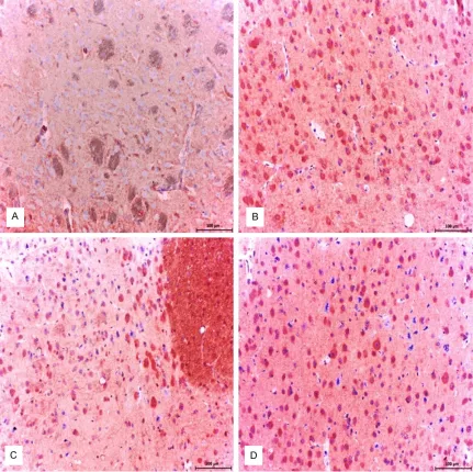
Fairly weak NF (Figure 8A) and NSE (Figure 9D) expressions were observed in neurons and glial cells in healthy control group. Strong/moderate
[image:8.612.92.524.72.509.2]GFAP expression was observed in glial cells in
Figure 8.Control group; normal accumulation of NF was noted. ABC technique (anti-NF), Mayer’s hematoxylin coun
-terstain, Bar, 200 μm (A). Diabetic group (B-D); Abnormal massive accumulation of NF was detected in the STZ-induced diabetic animal brains. ABC technique (anti-NF), Mayer’s hematoxylin counterstain, Bar, 200 μm. (B) Abnor
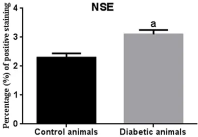
healthy control group (Figure 9A). Intense immunoreactivity for NSE expression was observed in cerebral sections (Figure 9E, 9F). NSE expression in the brain was higher in STZ-induced diabetic animals than in the control animals (Figure 5). The number of NSE-expressing neurons increased in all parts of the brain (Figure 10). Importantly, a significant decrease in the expression of GFAP was
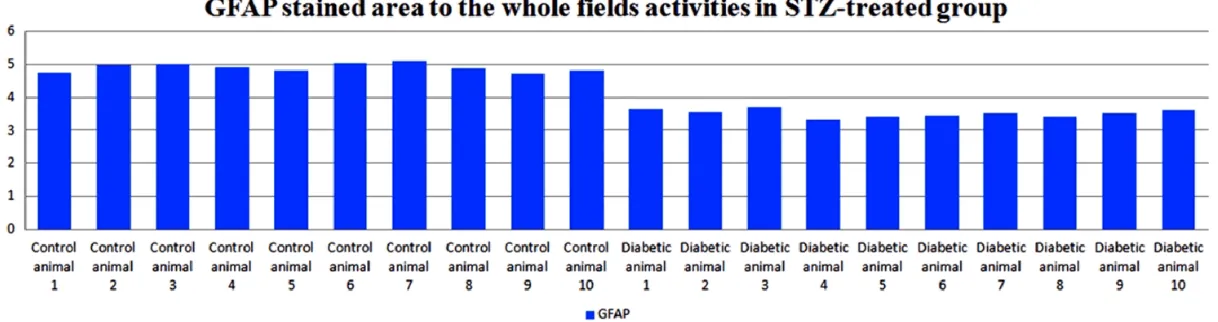
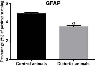
observed in the brain parenchyma in STZ-induced diabetic animals (Figures 9B, 9C, 11, 12).
Sites of enhanced NF immunoreactivity local
-ized to the necrotic lesions and this phenome
-non was significantly more pronounced in
STZ-induced diabetic animals than in the healthy control animals (Figures 8B-D, 13, 14).
Increased expression of NSE, NF and decreased expression of GFAP might give an idea of prog -ress of the disease and be critical for diagnosis of the disease.
Discussion
In the recent years, there have been studies on the role of ADAMTS-13 expression in the CNS,
but the function of this protein has not been
fully revealed [11, 14, 15, 47, 48]. For instance,
expression of ADAMTS-13 in mice brain tissue in experimental diabetes and its possible roles in the pathogenesis of the disease have not been studied before. In this study, we suggest that ADAMTS-13 may take an important role in
the inflammatory processes and the BBB per -meability. Moreover, we indicated that NO that is derived from nNOS causes degeneration of the CNS. nNOS is severely expressed in neu-rons and glial cells. In this study, the severity of the degeneration was indicated with NSE and
NF expressions.
Blood-brain barrier (BBB), which is the physical and metabolic barrier between the peripheral circulation and CNS, is involved the regulation and protection of the microenvironment in the
CNS [49]. Significant alteration in the level of
systemic glucose, as well as structural and functional disorders that occur in the BBB plays important roles in the pathogenesis of neuro-logical disorders that happen in DM [50-52]. In vivo and in vitro studies show that diabetes deteriorates the integrity of the BBB and increases the permeability of it [27, 28, 30, 31].
ADAMTS-13 closely associates with vWF and in mice inhibition of vWF induces very significant
increase in the BBB permeability [53]. Dincel and Kul, 2015, showed that ADAMTS-13 is highly expressed in the brains of small rumi-nants, which is infected with border disease virus and is suggested to decrease the degen-eration by reducing permeability of the BBB [11]. In a similar study with Toxoplasmic Ence- phalitis model, severe ADAMTS-13 expression in the brain of mice was suggested to protect the permeability of BBB and alleviate the sever-ity of infection [15]. In this research, we sug-gest that permeability of the BBB may be decreased by severe expression of ADAMTS-13 in order to suppress the negative effects of hyperglycemia on the BBB and protect its integ-rity. This situation indicates the tight connec-tion between ADAMTS-13 and the BBB perme-Figure 9.Control group; moderate expression of GFAP in astrocytes were noted. ABC technique (anti-GFAP), Mayer’s hematoxylin counterstain, Bar, 100 μm. (A) Diabetic group (B and C); Mild/no expression of GFAP in astrocytes were noted. ABC technique (anti-GFAP), Mayer’s hematoxylin counterstain, Bar, 100 μm. (B) Mild/no expression of GFAP in astrocytes were noted. ABC technique (anti-GFAP), Mayer’s hematoxylin counterstain, Bar, 100 μm (C) Control group; mild expression of NSE in neuronal cells were determined. ABC technique (anti- NSE), Mayer’s hematoxylin counterstain, Bar, 100 μm. (D) Diabetic group (E and F); Strong expression of NSE in neuronal cells were deter
-mined. ABC technique (anti- NSE), Mayer’s hematoxylin counterstain, Bar, 100 μm. (E) Strong expression of NSE in neuronal and glial cells were determined. ABC technique (anti- NSE), Mayer’s hematoxylin counterstain, Bar, 100 μm (F).
Figure 10.Statistical difference is indicated as let-ters. “a” represent values statistically higher than control group. Statistical analysis was performed according to Mann-Whitney U-test. The values repre-sent means ± S.D. P < 0.05 was considered
[image:10.612.91.287.183.319.2]ability. Therefore, discovering the effects of ADAMTS-13 on the BBB permeability may con-tribute to the understanding of the pathogene-sis of diabetic neuropathy and the control of the disease.
ADAMTS-13 contributes to the healing and remodelling process of the injured tissue [54]. In mice, inhibition of ADAMTS-13 induces a
severe inflammatory response and causes a
[image:12.612.93.289.73.208.2]rise in the infarct area in the heart [55-57] and brain [58, 59] depending on the ischemic/per-fusion injury. When the recombinant human ADAMTS-13 protein was injected to these mice, a decrease in the infarct area was detected
[58, 59]. These findings indicate that
ADAM-TS-13 acts as a systemic protection against myocardial and cerebral infarction. In this
study, we suggest that significantly high level of
ADAMTS-13 expression in brain tissue of dia-betic animals may help to alleviate the existing tissue degeneration and induce the healing process. Therefore, it is considered that under-standing the link between biosynthesis, expres-sion and inhibition of ADAMTS-13 with diabetic neuropathy and its complications will be crucial for revealing the pathogenesis of the disease and for the development of new treatment protocols.
Vascular complications and endothelial dys-functions related to Diabetes Mellitus are pathology that are widely studied and well
defined [60-62]. Injured or damaged endotheli -al cells lose some of the essenti-al molecules
that play role in homeostasis and blood flow.
Thus, in damaged endothelial cells there is an increase in the expression of adhesive mole-cules, procoagulant activity and predisposition to the development of thrombosis [63, 64]. Von
Willebrand Factor (VWF) plays a key role in
blood clotting and thrombus formation in severe vascular injury and damage [19, 20].
UL-VWF multimers that has high thrombotic activity is cleaved into less active VWF frag -ments by ADAMTS-13 [16, 65]. There are stud-ies showing that diabetes increases the ten-dency of the thrombosis and the risk of diabetes related stroke. However, in diabetes related stroke, complications that occur do not play an essential role. Dincel and Atmaca, 2015a found that ADAMTS-13 has a function in prevention of thrombus formation and may help for the inhibi-tion of a possible stroke [15]. The most striking
finding of this study was the absence of any
thrombus related pathology even though the veins that were damaged by hyperglycemia were a potential reason for microthrombus. On the other hand, in this study we suggest that increased ADAMTS-13 expression in endotheli-al cells has a role in prevention of a potentiendotheli-al microthrombus or pathology that are related to microthrombus. It is possible that the reason for why microthrombus and pathology related to microthrombus do not occur may be this severely expressed, protective protein, ADAM- TS-13.
Nitric oxide (NO) when produced at high levels, like other free radicals, induces neurotoxic effects [66]. There are studies showing that pathology occurring in the CNS may be based on severe level of NO [38, 67, 68]. In this study, it was suggested that NO produced in neurons may be responsible for degeneration and necrosis in the CNS seen in DM model. We think that NO expressed at pathological levels greatly contributes to degeneration and neuro-nal necrosis. It was described that NO at physi-ological levels inhibits apoptosis by blocking cytochrome c release [69]. Thus, it prevents the intrinsic apoptosis pathway. On the other hand, NO when produced above physiological limit, is known to induce apoptosis in CNS [37, 38, 40, 42]. It was shown that severe iNOS expression causes neurotoxicity in the hypo-thalamus and triggers neuronal apoptosis [70]. Along with this, severe iNOS expression was also shown to induce apoptosis [38, 71, 72]. Moreover, nNOS derived NO was shown to
Figure 12.Statistical difference is indicated as let-ters. “a” represent values statistically higher than control group. Statistical analysis was performed according to Mann-Whitney U-test. The values repre-sent means ± S.D. P < 0.05 was considered
cause degeneration by inducing apoptosis of neuronal and glial cells [11, 15]. In this study, nNOS derived NO expressed above the physio-logical limits in the CNS cells in diabetic ani-mals is considered as the reason of apoptosis that was previously observed in diabetes [1, 2, 43, 44].
In cerebral ischemia, NSE can be used as a marker to identify the severity of the neuronal
damage [73, 74]. A statistically significant
increase in the expression of NSE indicates severe degeneration of CNS. Therefore; in this study, NSE expression may help us to make comment on the severity of the neuronal degen-eration that occurs in the patients with diabe-tes. The reason for the severe reduction in the
expression of GFAP in DM model is thought to
be due to the apoptosis/degeneration of
astro-cytes. With the finding that there is a negative correlation between NSE and GFAP, this study
helps us to comment about the severity of the pathology that occur in CNS.
Neurofilaments are the most important compo -nents of the neuronal cytoskeleton and they are necessary for the maintenance of normal neuronal function [75, 76]. Abnormal
deposi-tion of NF is diagnostic indicator of parenchy -mal destruction in neurodegenerative and cerebral diseases [11, 46, 77, 78]. Thus, by
looking at the expression of NF in the cerebro
-spinal fluid, we may have an idea about the
severity of the degeneration [78]. In the
pres-ent study, significantly high-level of NF expres
-sion in the parenchymal tissue of diabetic
ani-mals was observed. A key finding of the present study was that the NF might contribute to the
diagnosis of the disease and provide insight about the severity of diabetes-related neuro- pathology.
Diabetes mellitus was defined to be associated
with demyelination in the brain and axonal inju-ry [25]. There are myelin sheath disorders and oligodendrocyte abnormalities in the STZ-induced diabetic animals. It was reported that the quality of myelin decreases [79] and the number of oligodendrocytes decreases in these animals [80]. It is announced that ADAMTS-13 may closely related to myelin production in the
border disease that is characterized by hypo -myelination [11]. Since we know that there is severe ADAMTS-13 expression and also major complications in myelin production in DM, we think that knowing the role of ADAMTS-13 in the hypomyelination and demyelination would support the understanding of the pathogenesis of diseases such as DM and other diseases associated with myelin damage.
It is thought that ADAMTS-13 acts a protective role in cerebral pathology, in prevention of the increase in BBB permeability or damage in endothelial cells that are induced by parasites, viruses or metabolic diseases. Considering this and other similar studies that we have done previously, we believe that ADAMTS-13 pro-tects CNS microenvironment and damaged endothelial cells and prevents neurons, endo-thelial and glial cells from an ischemic damage. It is clear that the knowing the correlation between diabetes and biosynthesis and expres-sion of ADAMTS-13 will help to understand pos-sible neuropathology and will help in the follow-up of the disease. In this study we show that there is severe level of neuronal degeneration and a decrease in the astrocytic activity that is far below the physiological level. This situation shows that astrocytes decrease in number when they are exposed to severe NO levels that results in apoptosis/degeneration or the dis-ruption in their functions. Subsequent to these
findings, it is believed that NF may be important
in identifying the severity and the disease
pro-gression during the follow-up, and NF levels may also be used clinically. The findings of this
research show the degeneration that occurs in the CNS may not only originate from
hypergly-Figure 14.Statistical difference is indicated as let-ters. “a” represent values statistically higher than control group. Statistical analysis was performed according to Mann-Whitney U-test. The values repre-sent means ± S.D. P < 0.05 was considered
[image:14.612.93.288.72.206.2]cemia, at the same time it may originate from nNOS derived NO expressed above on the phys-iological limits from neuronal and glial cells. The research also suggests that ADAMT-13 that is expressed severely in the brain tissue of dia-betic animals may be associated with myelin formation in addition to being closely associat-ed with BBB protection. ADAMTS-13 contribut-ed to the alleviation of degeneration in the brain and the healing process of the tissue is
also supported by the findings.
Acknowledgements
This work was funded and supported by the
Scientific Research Projects Commission of the Gümüşhane University, Gümüşhane, Turkey
(Project Code: 15.B0421.02.2). This study was presented as an oral presentation in the 32nd
World Veterinary Congress, 13-17 September 2015, Istanbul.
Disclosure of conflict of interest None.
Address correspondence to: Gungor Cagdas Dincel, Laboratory and Veterinary Health Program, Eskil Vocational School, Aksaray University Eskil Voca- tional High School Laboratory and Veterinary Health Program Eskil/Aksaray, Aksaray, Turkey. E-mail: gcdincel@yahoo.com.tr
References
[1] Li ZG, Zhang W, Grunberger G, Sima AA. Hip-pocampal neuronal apoptosis in type 1 diabe-tes. Brain Res 2002; 946: 221-231.
[2] Sima AAF, Kamiya H, Li ZG. Insulin, C-peptide,
hyperglycemia, and central nervous system complications in diabetes. Eur J Pharmacol 2004; 490: 187-197.
[3] Li PA, Siesjo BK. Role of hyperglycaemia-relat-ed acidosis in ischaemic brain damage. Acta Physiol Scand1997; 161: 567-580.
[4] Sridulyakul P, Wongeak-In N, Patumraj S. Cor-relations between endothelial functions and ROS detection in diabetic microvascular wall: Early and late ascorbic acid supplementation. Int J Vasc Med 2012; 2012: 709695.
[5] Yamamoto Y, Yamamoto H. RAGE-mediated
in-flammation, type 2 diabetes, and diabetic vas
-cular complication. Front Endocrinol (Laus -anne) 2013; 4: 105.
[6] Margineanu DG, Niespodziany I, Wülfert E. Hip
-pocampal slices from long-term streptozoto -cin-injected rats are prone to epileptiform re-sponses. Neurosci Lett 1998; 252: 183-6.
[7] Reijmer YD, Van Den Berg E, De Bresser J, Kes-sels RP, Kappelle LJ, Algra A, BiesKes-sels GJ. Ac-celerated cognitive decline in patients with type 2 diabetes: MRI correlates and risk fac-tors. Diabetes Metab Res Rev 2011; 27: 195-202.
[8] Garven A, Zaver S, Rincon N, Poliakov I,
Ro-sales-Hernandez A, Ayer A, Garven A, Zaver S,
Rincon N, Xu K, Tuor UI, Schmidt AM, Toth C. Differential impact of diabetes and hyperten-sion in the brain: adverse effects in white mat-ter. Neurobiol Dis 2011; 42: 446-458.
[9] Levy GG, Motto DG, Ginsburg D. ADAMTS13 turns 3. Blood 2005; 1: 11-7.
[10] Zhou W, Inada M, Lee TP, Benten D, Lyubsky S, Bouhassira EE, Gupta S, Tsai HM. ADAMTS13 is expressed in hepatic stellate cells. Lab In-vest 2005; 85: 780-8
[11] Dincel GC, Kul O. Increased Expressions of AD-AMTS-13, Neuronal Nitric Oxide Synthase, and
Neurofilament Correlate with Severity of Neu -ropathology in Border Disease Virus-Infected Small Ruminants. PLoS One 2015; 10: e0120005.
[12] Shang D, Zheng XW, Niiya M, Zheng XL. Apical sorting of ADAMTS13 in vascular endothelial cells and Madin-Darby canine kidney cells de-pends on the CUB domains and their associa-tion with lipid rafts. Blood 2006; 108: 2207-15.
[13] Shelat SG, Ai J, Zheng XL. Molecular biology of ADAMTS13 and diagnostic utility of ADAMTS13 proteolytic activity and inhibitor assays. Semin Thromb. Hemost 2005; 31: 659-72.
[14] Frentzou GA, Bradford C, Harkness KA, Had
-dock G, Woodroofe MN, Cross AK. IL-1β
down-regulates ADAMTS-13 mRNA expression in cells of the central nervous system. J Mol Neu-rosci 2012; 46: 343-51.
[15] Dincel GC, Atmaca HT. Increased expressions of ADAMTS-13 and apoptosis contribute to neuropathology during Toxoplasma gondii en-cephalitis in mice. Neuropathology 2015; [Epub ahead of print].
[16] Dong JF, Moake JL, Nolasco L, Bernardo A, Ar -ceneaux W, Shrimpton CN, Schade AJ, McIntire
LV, Fujikawa K, López JA. ADAMTS-13 rapidly
cleaves newly secreted ultralarge von Wille-brand factor multimers on the endothelial
sur-face under flowing conditions. Blood 2002;
100: 4033-9.
[17] Chauhan AK, Motto DG, Lamb CB, Bergmeier
W, Dockal M, Plaimauer B, Scheiflinger F, Gins -burg D, Wagner DD. Systemic antithrombotic effects of ADAMTS13. J Exp. Med 2006; 203: 767-76.
throm-botic thrombocytopenic purpura in genetically
susceptible ADAMTS13-deficient mice. J Clin
Invest 2005; 10: 2752-61.
[19] Savage B, Saldivar E, Ruggeri ZM. Initiation of
platelet adhesion by arrest onto fibrinogen or
translocation on von Willebrand factor. Cell 1996; 84: 289-97.
[20] Ruggeri ZM. Von Willebrand factor, platelets and endothelial cell interactions. J Thromb Haemost 2003; 7: 1335-42.
[21] Deng JT, Sutherland C, Brautigan DL, Eto M, Walsh MP. Phosphorylation of the myosin phosphatase inhibitors, CPI-17 and PHI-1, by integrin-linked kinase. Biochem J 2002; 367: 517-24.
[22] Reske-Nielsen E, Lundbaek K. Pathological changes in the central and peripheral nervous system of young long-term diabetics. II. The spinal cord and peripheral nerves. Diabetolo-gia 1968; 4: 34-43.
[23] Slager UT. Diabetic myelopathy. Arch. Pathol. Lab 1978; 102: 467-9.
[24] Slager UT, Webb AT. Pathologic findings in the
spinal cord. Arch Pathol 1973; 96: 388-94. [25] Huang M, Gao L, Yang L, Lin F, Lei H. Abnor
-malities in the brain of streptozotocin-induced
type 1 diabetic rats revealed by diffusion ten-sor imaging. Neuroimage Clin 2012; 14: 57-65.
[26] Jaffey PB, Gelman BB. Increased vulnerability
to demyelination in streptozotocin diabetic
rats. J Comp Neurol 1996; 373: 55-61 [27] Hawkins BT, Lundeen TF, Norwood KM, Brooks
HL, Egleton RD. Increased blood-brain barrier permeability and altered tight junctions in ex-perimental diabetes in the rat: contribution of hyperglycaemia and matrix metalloproteinas-es. Diabetologia 2007; 50: 202-211.
[28] Huber JD, VanGilder RL, Houser KA.
Strepto-zotocin-induced diabetes progressively in -creases blood-brain barrier permeability in
specific brain regions in rats. Am J Physiol.
Heart Circ. Physiol 2006; 291: 2660-2668. [29] Price TO, Eranki V, Banks WA, Ercal N, Shah
GN. Topiramate treatment protects blood-brain barrier pericytes from hyperglycemia-induced oxidative damage in diabetic mice. Endocrinol-ogy 2012; 153: 362-72.
[30] Starr JM, Wardlaw J, Ferguson K, Ercal N, Shah
GN. Increased blood-brain barrier permeability in type II diabetes demonstrated by gadolinium magnetic resonance imaging. J Neurol Neuro-surg Psychiatry 2003; 74: 70-76.
[31] Acharya NK, Levin EC, Clifford PM, Han M, Tourtellotte R, Chamberlain D, Pollaro M, Coretti NJ, Kosciuk MC, Nagele EP, Demarshall
C, Freeman T, Shi Y, Guan C, Macphee CH,
Wilensky RL, Nagele RG. Diabetes and hyper-cholesterolemia increase blood-brain barrier
permeability and brain amyloid deposition:
beneficial effects of the LpPLA2 inhibitor dara
-pladib. J Alzheimers Dis 2013; 35: 179-198.
[32] Zlokovic BV. The blood-brain barrier in health and chronic neurodegenerative disorders. Neuron 2008; 57: 178-201.
[33] Busija DW, Leffler CW. Dilator effects of amino
acid neurotransmitters on piglet pial arteri-oles. Am J Physiol 1989; 257: 1200-1203. [34] Faraci FM, Breese KR. Nitric oxide mediates
vasodilatation in response to activation of N-methyl-Daspartate receptors in brain. Circ Res 1993; 72: 476-480.
[35] Faraci FM, Brian JE. Nitric oxide and the cere -bral circulation. Stroke 1994; 25: 692-703. [36] Fergus A, Lee KS. Regulation of cerebral mi
-crovessels by glutamatergic mechanisms. Brain Res 1997; 754: 35-45.
[37] Bonfoco E, Krainc D, Ankarcrona M, Nicotera P, Lipton SA. Apoptosis and necrosis: Two distinct events induced, respectively, by mild and in-tense insults with N-methyl-D-aspartate or ni-tric oxide/superoxide in cortical cell cultures. Proc Natl Acad Sci U S A 1995; 92: 7162-7166. [38] Dincel GC, Kul O. eNOS and iNOS trigger apop-tosis in the brains of sheep and goats naturally infected with the border disease virus. Histol Histopathol 2015; 30: 1233-42.
[39] Brookes PS, Salinas EP, Darley-Usmar K,
Eis-erich JP, Freeman BA, Darley-Usmar VM, An -derson PG. Concentration-dependent effects of nitric oxide on mitochondrial permeability transition and cytochrome c release. J Biol Chem 2000; 275: 20474-20479.
[40] Heneka MT, Loschmann PA, Gleichmann M,
Weller M, Schulz JB, Wüllner U, Klockgether T.
Induction of nitric oxide synthase and nitric oxide-mediated apoptosis in neuronal PC12 cells after stimulation with tumor necrosis fac-tor-alpha/lipopolysaccharide. J Neurochem 1998; 71: 88-94.
[41] Hibbs JBJ, Taintor RR, Vavrin Z. Macrophage cytotoxicity: role for L-arginine deiminase and amino nitrogen oxidation to nitrite. Science 1987; 235: 473-476.
[42] Moriya R, Uehara T, Nomura Y. Mechanism of nitric oxide-induced apoptosis in human
neu-roblastoma SH-SY5Y cells. FEBS Lett 2000;
484: 253-260.
[43] Baraka A, Abdelgawad H. Targeting apoptosis
in the heart of streptozotocin-induced diabetic
rats. J Cardiovasc Pharmacol Ther 2010; 15: 175-181.
[44] Ma ZA, Zhao Z, Turk J. Mitochondrial dysfunc-tion and beta-cell failure in type 2 diabetes mellitus. Exp.Diabetes Res. 2012; Article ID 703538.
induced Maillard-type fluorescence and colla
-gen cross-linking in the heart of streptozotocin
diabetic rats. Pharmacol Res 2007; 55: 433-40.
[46] Dincel GC, Atmaca HT. Nitric oxide production increases during Toxoplasma gondii encephali-tis in mice. Exp Parasitol 2015; 156: 104-12. [47] Tauchi R, Imagama S, Ohgomori T, Natori T,
Shinjo R, Ishiguro N, Kadomatsu K. AD-AMTS-13 is produced by glial cells and upregu-lated after spinal cord injury. Neurosci. Lett 2012; 517: 1-6.
[48] Muroi C, Fujioka M, Mishima K, Irie K, Fujimura Y, Nakano T, Fandino J, Keller E, Iwasaki K, Fu -jiwara M. Effect of ADAMTS-13 on cerebrovas-cular microthrombosis and neuronal injury af-ter experimental subarachnoid haemorrhage. J. Thromb Haemost 2014; 12: 505-514. [49] Zlokovic BV. The blood-brain barrier in health
and chronic neurodegenerative disorders. Neuron 2008; 57: 178-201.
[50] Brownlee M. Biochemistry and molecular cell biology of diabetic complications. Nature 2001; 414: 813-20.
[51] Giacco F, Brownlee M. Oxidative stress and dia -betic complications. Circ Res 2010; 107: 1058-70.
[52] Mooradian AD, Thurman JE. Glucotoxicity: po-tential mechanisms. Clin Geriatr Med 1999; 15: 255.
[53] Noubade R, Del Rio R, McElvany B, Zachary JF,
Millward JM, Wagner DD, Offner H, Blanken-horn EP, Teuscher C. von-Willebrand factor
in-fluences blood brain barrier permeability and brain inflammation in experimental allergic
encephalomyelitis. Am J Pathol 2008; 173: 892-900.
[54] Niiya M, Uemura M, Zheng XW, Pollak ES,
Dockal M, Scheiflinger F, Wells RG, Zheng XL.
Increased ADAMTS-13 proteolytic activity in rat hepatic stellate cells upon activation in vitro and in vivo. J Thromb Haemost 2006; 4: 1063-70.
[55] Gandhi C, Motto DG, Jensen M, Lentz SR, Chauhan AK. ADAMTS13 deficiency exacer
-bates VWF-dependent acute myocardial isch -emia/reperfusion injury in mice. Blood 2012; 120: 5224-30.
[56] Doi M, Matsui H, Takeda H, Saito Y, Takeda M,
Matsunari Y, Nishio K, Shima M, Banno F, Aki -yama M, Kokame K, Miyata T, Sugimoto M. AD-AMTS13 safeguards the myocardium in a mouse model of acute myocardial infarction. Thromb Haemost 2012; 108: 1236-8.
[57] De Meyer SF, Savchenko AS, Haas MS, Schatz
-berg D, Carroll MC, Schiviz A, Dietrich B, Rot
-tensteiner H, Scheiflinger F, Wagner DD. Pro
-tective anti-inflammatory effect of ADAMTS13
on myocardial ischemia/reperfusion injury in mice. Blood 2012; 120: 5217-23
[58] Fujioka M, Hayakawa K, Mishima K, Kunizawa A, Irie K, Higuchi S, Nakano T, Muroi C, Fuku
-shima H, Sugimoto M, Banno F, Kokame K, Mi
-yata T, Fujiwara M, Okuchi K, Nishio K. AD -AMTS13 gene deletion aggravates ischemic brain damage: a possible neuroprotective role of ADAMTS13 by ameliorating postischemic hypoperfusion. Blood 2010; 115: 1650-3. [59] Zhao BQ, Chauhan AK, Canault M, Patten IS,
Yang JJ, Dockal M, Scheiflinger F, Wagner DD.
von Willebrand factor-cleaving protease AD-AMTS13 reduces ischemic brain injury in ex-perimental stroke. Blood 2009; 114: 3329-34. [60] Cade WT. Diabetes-related microvascular and
macrovascular diseases in the physical thera-py setting. Phys Ther 2008; 11: 1322-35. [61] Hadi HA, Suwaidi JA. Endothelial dysfunction in
diabetes mellitus. Vasc Health Risk Manag 2007; 6: 853-76.
[62] Tabit CE, Chung WB, Hamburg NM, Vita JA. En-dothelial dysfunction in diabetes mellitus: mo-lecular mechanisms and clinical implications. Rev Endocr Metab Disord 2010; 1: 61-74. [63] Wu KK, Thiagarajan P. Role of endothelium in
thrombosis and hemostasis. Annu RevMed 1996; 47: 315-31.
[64] Verhamme P, Hoylaerts MF. The pivotal role of
the endothelium in haemostasis and thrombo-sis. Acta Clin Belg 2006; 5: 213-9.
[65] Sadler JE. Von Willebrand factor, ADAMTS13, and thrombotic thrombocytopenic purpura. Blood 2008; 1: 11-8.
[66] Calabrese V, Mancuso C, Calvani M, Rizzarelli E, Butterfield DA, Stella AM. Nitric oxide in the
central nervous system: neuroprotection ver-sus neurotoxicity. Nat Rev Neurosci 2007; 8: 766-75.
[67] Boje KM, Aroa PK. Microglial-produced nitric oxide and reactive nitrogen oxides mediate neuronal cell death. Brain Res 1992; 587: 250-256.
[68] Dawson VL, Dawson TM, London ED, Bredt DS, Snyder SH. Nitric oxide mediates glutamate neurotoxicity in primary cultured cortical neu-rons. Proc Natl Acad Sci U S A 1991; 14: 6368-71.
[69] Natalie JT, Hajime H, Bronk S, Gores GJ. Nitric Oxide Inhibits Apoptosis 527 Downstream of Cytochrome c Release by Nitrosylating Cas-pase 9. Cancer Res 2002; 62: 1648-1653. [70] Wang C, Hikim AS, Ferrini M, Bonavera JJ,
Vernet D, Leung A, Lue YH, Gonzalez-Cadavid NF, Swerdloff RS. Male reproductive ageing:
Using the brown Norway 553 rat as a model for
man. Novartis Found Symp 2002; 242: 82-95.
[71] Pender MP, Rist JM. Apoptosis of inflammatory
cells in immune control of the 537 nervous system: role of glia. Glia 2001; 36: 137-144. [72] Brown GC. Nitric oxide and neuronal death.
[73] Hatfield RH, McKernan RM. CSF neuron-specif -ic enolase as a quantitative marker of neuro-nal damage in a rat stroke model. Brain Res 1992; 2: 249-52.
[74] Büttner T, Lack B, Jäger M, Wünsche W, Kuhn
W, Müller T, Przuntek H, Postert T. Serum levels of neuron-specific enolase and s-100 protein after single tonic-clonic seizures. J Neurol
1999; 6: 459-61.
[75] Barrya DM, Millecamps S, Julien JP, Garcia ML.
New movements in neurofilament transport,
turnover and disease. Exp Cell Res2007;313: 2110-2120.
[76] Yuan A, Rao MV, Veeranna Nixon RA. Neurofila -ments at a glance. J Cell Sci 2012; 125: 3257-3263.
[77] Liu Q, Xie F, Alvarado-Diaz A, Smith MA, Moreira PI, Zhu X, Perry G. Neurofilamentopathy in Neu -rodegenerative Diseases. Open Neurol J 2011; 5: 58-62.
[78] Norgren N, Rosengren L, Stigbrand T. Elevated
neurofilament levels in neurological diseases.
Brain Res 2003; 987: 25-31.
[79] Hernandez-Fonseca JP, Rincon J, Pedreanez A, Viera N, Viera N, Arcaya JL, Carrizo E, Mos -quera J. Structural and ultrastructural analysis of cerebral cortex, cerebellum, and hypothala-mus from diabetic rats. Exp Diabetes Res 2009; 329632: 1-12.
[80] Yang C, DeVisser A, Martinez JA, Poliakov I, Rosales-Hernandez A, Ayer A, Garven A, Zaver