Original Article
Methylation of DAPK, P16 and E-Cadherin gene in liver
cancer tissues and protein expression level
Hongmei Tang1, Xingguo Yao2, Chunlian Li3, Pingping Liu2, Bingqi Ma2
1Department of Outpatient, 2Department of Hepatobiliary Surgery, 3Department of Pediatrics, The Affiliated Hospi -tal of Weifang Medical University, Weifang, Shandong, China
Received November 16, 2015; Accepted January 12, 2016; Epub March 1, 2016; Published March 15, 2016
Abstract: As the most common mechanism of epigenetics, DNA methylation has not been fully illustrated in tumor disease. Hepatocellular carcinoma (HCC) is one common malignant tumor but with limited epigenetic knowledge about its molecular mechanism. This study thus mainly investigated the methylation status at promoter regions of death associated protein kinase (DAPK), poly tumor suppressor gene (P16) and E-Cadherin, along with protein expression levels. Paraffin sections of HCC or liver cirrhosis tissues were tested for promoter methylation condition of DAPK, P16 and E-cadherin genes using methylation specific PCR (MSP) approach. Immunohistochemistry (IHC) staining was applied to detect protein expression levels for statistical analysis. MSP assay showed methylation positive rates of DAPK, P16 and E-Cadherin genes at 69.23%, 79.49% and 58.97%, respectively, in HCC tissues. All of these rates were significantly higher than non-tumor tissues (28.21%, 2.56% and 25.64%, P<0.05). P16 gene methylation rate in cirrhosis tissues was lower than HCC group (P<0.01). While DAPK and E-Cadherin gene had not significantly different of methylation rate (P>0.05). IHC staining revealed significant correlation between methyla -tion and protein expression level of those genes (P<0.05), as positive methyla-tion HCC cells had lower protein levels than non-methylation group. Hyper-methylation existed in promoter of DAPK, P16 and E-Cadherin genes in HCC tis-sues, providing further evidences for early diagnosis of HCC.
Keywords: Hepatocellular carcinoma, death associated protein kinase, P16, E-Cadherin, gene methylation
Introduction
Hepatocellular carcinoma (HCC) is one com-mon malignant tumor worldwide, and is the third leading cause for death of tumors [1, 2]. Its detailed pathogenesis mechanism, howev-er, requires further study. Currently epigenetics has become one critical mechanism regulating tumor pathogenesis drawing research inter-ests. Recent studies recognized methylation of gene promoter region as one important reason for inactivation of tumor suppressor genes, for mediating cell apoptosis, cell cycle regulation,
DNA repair, message transduction and Wnt sig -naling pathway, in addition to close correlation between DNA methylation and tumor occur-rence or pathological types [3, 4]. DNA methyla-tion has been implicated in various cancers including colorectal cancer, esophagus carci-noma and pulmonary cancer [5-7]. Meanwhile, DNA methylation can work as potential tumor markers [8, 9] for studying related diseases.
The role of abnormal methylation of gene pro-moter in HCC, however, requires further
stud-ies. This study utilized methylation specific PCR
(MSP) technique, to detect methylation status of gene promoter regions of death associated protein kinase (DAPK), P16 and E-Cadherin genes in HCC and non-tumor liver tissues, in addition to protein expression level of those genes, in order to reveal the role of DNA meth-ylation in early diagnosis and treatment of HCC.
Materials and methods
General information
A total of 72 tissues samples were collected from liver cancer or liver cirrhosis patients between August 2014 and July 2015 in the
Affiliated Hospital of Weifang Medical University.
tissue samples were collected. Another 33 liver cirrhosis patients (20 males and 13 females, average age = 58.27 ± 6.81 years) were also
recruited. No significant difference has been
found regarding ages. All patients had
con-firmed diagnosis by pathological examinations,
and with complete medical history. This study has been approved by the ethical committee and is carried out under written consents from patients. All tissues were collected and kept in liquid nitrogen.
MSP primer
The methylation/un-methylation primer se- quence of DAPK, P16 and E-cadherin genes were designed as previously documented [10-12]. Sequences were shown in Table 1.
DNA extraction and bisulfite modification
Total DNA was extracted from tissues using
Wizard Genomic DNA Purification Kit (Promega, US) following manual instruction. DNA was quantified in UV spectrometer and was kept under -80°C. Bisulfite modification and purifi -cation was carried out using phenol and
sodi-um bisulfite (Sigma, US) as previously docu -mented [3].
Methylation specific PCR
MSP was carried out in a 30 μL system consist
-ing of 15 μL PCR mixture, 1 μL primers, 1~4 μL modified DNA and ddHO. PCR conditions were:
only positive reaction using UF/UF primers were
methylation negative. Immunohistochemistry
Tissue sections were baked in 70°C incubator
for 20~30 min, and were dewaxed by xylene
and gradient ethanol. After hydration, antigen retrieval was performed in heated citric acid buffer (pH 6.0) for 90 sec. Endogenous peroxi-dase activity was quenched by 3% H2O2 in methanol, followed by blocking in normal goat serum. Primary antibody was added for 4°C overnight incubation. Secondary antibody and development was performed using IHC test kit (Zhongshan, China). Cell nucleus was counter-stained by hematoxylin for 40 sec, followed by HCl-ethanol treatment, dehydration and
cover-slip mounting. Under a microscope, brown
granules in cytoplasm were deduced as the positive staining. A semi-quantitative analysis was performed as previously established [4]. High power (×200) images were captured, staining score was given by the sum of intensity score (0, no staining; 1, light yellow; 2, dark yel-low; 3, dark brown) and positive percentage
score (<5%, 0; 5%~25%, 1; 26%~50%, 2; 51%~75%, 3; >75%, 4). Total score was inter
-preted as negative (0), weak positive (1+, 1~2), positive (2+, 3~5) or strong positive (3+, 6~7).
Statistical analysis
[image:2.629.97.532.92.186.2]SPSS 16.0 software was used to process all collected data, of which measurement data Table 1. Methylation (M) and un-methylation (U) primer sequence
Target gene Sense strand (5’-3’) Anti-sense strand (5’-3’) Product size (bp)
DAPK (M) GGATAGTCGGATCGAGTTAACGTC TAACTAAAAATTCACCTACCGAC 98
DAPK (U) GGAGGATAGTTGGATTGAGTTAATGTT CACAACCAATCAACAACACA 106
P16 (M) TTATTAGAGGGTGGGGTGGATTGT CAACCCCAAACCACAACCATAA 150
P16 (U) TTATTAGAGGGTGGGGCGGATCGC GACCCCGAACCGCGACCGTAA 151
E-cadherin (M) TTAGGTTAGAGGGTTATCGCGT TAACTAAAAATTCACCTACCGAC 116
E-cadherin (U) TAATTTTAGGTTAGAGGGTTATTGT CACAACCAATCAACAACACA 97
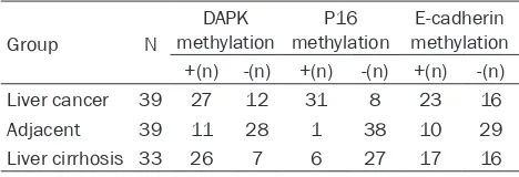
Table 2. Promoter methylation level of DAPK, P16 and E-cadherin genes
Group N
DAPK
methylation methylationP16 methylationE-cadherin ﹢(n) -(n) ﹢(n) -(n) ﹢(n) -(n)
Liver cancer 39 27 12 31 8 23 16
Adjacent 39 11 28 1 38 10 29
Liver cirrhosis 33 26 7 6 27 17 16
Note: n represents number of cases.
94°C pre-denature for 5 min, followed by 40 cycles each containing 94°C denature for 45 sec, 60°C annealing for 45 sec, and 72°C elongation for 45 sec, and ended with 72°C elongation for 7 min. Ampli-
fication products were separated by 2%
agarose gel electrophoresis and were imaged under an automatic gel imaging system. Those samples had positive
reac-tion using MF/MR specific primers were
[image:2.629.98.332.232.312.2]were presented as mean ± standard deviation (SD) while enumeration data were compared by
chi-square test. A statistical significance was defined as P<0.05.
Results
Methylation level of promoters
MSP method was used to detect the methyla-tion status of gene promoters of DAPK, P16
and E-cadherin genes. We found the methyla -tion positive percentage as 69.23% (27/39), 79.49% (31/39) and 58.97% (23/39) in liver cancer tissues for DAPK, P16 and E-cadherin
genes, respectively. Such figures in liver cirrho -sis tissues were 78.79% (26/33), 18.18% (6/33) and 51.52% (17/33), while in adjacent tissues were 28.21% (11/39), 2.56% (1/39) and 25.64% (10/39). Between-group compari-son revealed lower methylation level of P16 genes in cirrhosis group compared to liver
can-cer tissues (P<0.01), while no significant differ -ence existed between DAPK and E-cadherin
genes (P>0.05). The methylation level of all three genes was significantly higher in liver can -cer tissues compared to adjacent tissues (P<
0.01). Detailed results were shown in Table 2 and Figure 1. In addition, no significant differ -ence of methylation level has been discovered regarding clinical/pathological features.
Gene methylation status and protein expres-sion
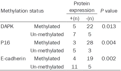
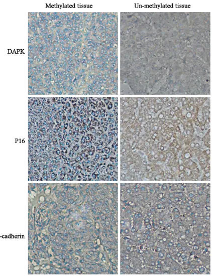
Based on the methylation status, we further analyzed protein expression level of DAPK, P16 and E-cadherin level in liver cancer tissues using IHC methods. As shown in Table 3, gene methylation level of DAPK, P16 and E-cadherin genes were negatively correlated with their
pro-tein expression levels (P<0.05). Propro-tein expres -sion level was decreased in hyper-methylation genes. Those liver cancer tissues with methyla-tion positive status had lower protein level than un-methylated counterparts (Figure 2).
Discussion
[image:3.629.102.535.80.180.2]Tumor pathogenesis is one progressive pro-cess in which both genetic and epigenetic mechanisms lead to the activation of onco-genes and inactivation of tumor suppressor genes, plus the stimulus from exogenous fac-tors [13]. Epigenetic is a type of gene expres-sion regulation in which DNA sequence keeps intact. In tumor patients, it is often observed that epigenetic gene silencing and consequent inactivation of tumor suppressor genes. The methylation of promoter region of tumor sup-pressor genes is thus believed as one impor-tant epigenetic mechanism underlying liver cancer, and may provide new insights for searching novel biomarkers of liver cancer diag-nosis [14, 15], has recently drawn lots of research interests. Currently the methylation of promoter region of DAPK, P16 and E-cadherin genes has been recognized, while further in-Figure 1. MSP bands showing promoter methylation of DAPK (upper), P16 (middle) and E-cadherin (lower) genes. M, methylated alleles; U, un-methylated alleles; Ca, liver cancer; N, non-tumor tissues; PC, methylated/un-methylated DNA positive control; NC, negative control.
Table 3. Methylation status of DAPK, P16 and E-cadherin gene and protein expression
Methylation status
Protein
expression P value ﹢(n) -(n)
DAPK Methylated 5 22 0.013
Un-methylated 7 5
P16 Methylated 3 28 0.004
Un-methylated 5 3
E-cadherin Methylated 4 19 0.002
Un-methylated 11 5
[image:3.629.100.297.268.384.2]depth studies are required. This study utilized MSP approach to detect the methylation status of DAPK, P16 and E-cadherin genes, in addition to analyzing protein expression levels of those
genes, in an attempt to find liver cancer related
methylation markers.
DAPK is one serine/threonine kinase partici-pating in various cellular processes including
cell apoptosis, autophage and inflammation
[image:4.629.112.534.78.628.2]ticipate in tumor cell metastasis for aggrava-tion of disease. Related studies have demon-strated the alternation of expression and function of DAPK in cancer patients [18, 19]. Its methylation phenomena are likely to be related with lymph node metastasis. Cell cyclin depen-dent kinase inhibitor P16, also named as mutli-tumor suppressor gene, belongs to INK4 family and participates in cell cycle control. It often display mutation, truncation, and abnormal methylation in tumor cells, leading to the inacti-vation of gene expression, further causing accelerated cell proliferation and shortening of cell cycles. It is the most common tumor-relat-ed abnormal methylation gene and can be detected even at early stage of tumors [20]. Studies believed that P16 might participate in tumor related pathways in conjunction with DAPK [21]. E-cadherin can regulate calcium-dependent cell adhesion in various epithelial tissues, as its malfunction could interrupt the interaction between cells. The mutation of E-cadherin in tumor patients may cause abnor-mal detachment and mobility of cells, further
strengthening cell invasion and infiltration/
metastasis potency [22]. Previous study has shown the down-regulation of E-cadherin gene in tumor tissues by promoter methylation, thus inducing unfavorable prognosis of patients [23, 24].
This study utilized a methylation specific PCR
approach, to detect methylation status of DAPK, P16 and E-cadherin gene promoters in liver cancer and non-tumor tissues. Results
showed that liver cancer tissues had signifi -cantly higher positive rate of gene methylation compared to adjacent non-tumor tissues, indi-cating the alternation of epigenetics in liver cancer. Such methylation may frequently occur in liver cancer pathogenesis. The methylation rate of P16 gene in liver cirrhosis was relatively lower than liver cancer group, while no
statisti-cally significant difference existed between
these two tissues regarding methylation rate of DAPK and E-cadherin gene, suggesting the pos-sible relationship between DAPK and E-cadherin with liver cirrhosis. In addition, IHC staining
revealed significant correlation between meth -ylation status of DAPK, P16 and E-cadherin genes and their protein levels. Those liver can-cer tissues with positive for methylation had lower protein expression level than un-methyl-ated groups. These results collectively
suggest-ed the participation abnormal methylation of promoter regions of those three genes in the pathogenesis of liver cancer via modulating protein expression, as consistent with previous
reports [25]. No significant correlation, howev -er, existed between gene methylation and clini-cal features, probably due to relatively small sample size in this study.
In summary, promote methylation of tumor related genes plays a crucial role during the occurrence and progression of cancers. This study indicated that abnormal methylation of DAPK, P16 and E-cadherin genes might be important molecular events during early stage liver cancer. These genes participate in occur-rence and progression of liver cancer. Abnormal methylation of promoters of these genes large-ly affects protein expression, providing new insights for investigating the mechanism of liver cancer pathogenesis. This study paved grounds for developing early molecular marker for diagnosis of liver cancer, although further large-sample and large-scale screening of gene promoter methylation are required.
Disclosure of conflict of interest None.
Address correspondence to: Dr. Xingguo Yao, De- partment of Hepatobiliary Surgery, The Affiliated Hospital of Weifang Medical University, 2428 Yuhe Road, Weifang 261031, Shandong, China. Tel: +86-536-3081329; Fax: +86-+86-536-3081329; E-mail: yap- xingguoo@sina.com
References
[1] Kao JH, Chen DS. Changing disease burden of hepatocellular carcinoma in the Far East and Southeast Asia. Liver Int 2005; 25: 696-703. [2] Song P, Tang W, Tamura S, Hasegawa K, Suga
-wara Y, Dong J, Kokudo N.The management of hepatocellular carcinoma in Asia: a guideline combining quantitative and qualitative evalua-tion. Biosci Trends 2010; 4: 283-7.
[3] Abdul-Kadir R, McLintock C, Ducloy AS, El-Re-faey H, England A, Federici AB, Grotegut CA, Halimeh S, Herman JH, Hofer S, James AH, Kouides PA, Paidas MJ, Peyvandi F, Winikoff R. Evaluation and management of postpartum hemorrhage: consensus from an international expert panel. Transfusion 2014; 54: 1756-68. [4] Liu S. Management of upper gastrointestinal
[5] Almeida FG, de Aquino PF, de Souza AD, de Souza AQ, do Carmo Vinhote S, Mac-Cormick TM, da Mota Silva MS, Chalub SR, de Saldanha da Gama Fischer J, Carvalho PC, da Gloria da Costa Carvalho M. Colorectal cancer DNA methylation patterns from patients in Manaus, Brazil. Biol Res 2015; 48: 50.
[6] Ling ZQ, Li P, Ge MH, Zhao X, Hu FJ, Fang XH, Dong ZM, Mao WM. Hypermethylation-modu -lated down-regulation of CDH1 expression con-tributes to the progression of esophageal can-cer. Int J Mol Med 2011; 27: 625-35.
[7] Xiao P, Chen JR, Zhou F, Lu CX, Yang Q, Tao GH, Tao YJ, Chen JL. Methylation of P16 in exhaled breath condensate for diagnosis of non-small cell lung cancer. Lung Cancer 2014; 83: 56-60.
[8] Henriksen SD, Madsen PH, Krarup H, Thorla-cius-Ussing O. DNA Hypermethylation as a Blood-Based Marker for Pancreatic Cancer: A Literature Review. Pancreas 2015; 44: 1036-45.
[9] Fang C, Jian ZY, Shen XF, Wei XM, Yu GZ, Zeng XT. Promoter Methylation of the Retinoic Acid Receptor Beta2 (RARbeta2) Is Associated with Increased Risk of Breast Cancer: A PRISMA Compliant Meta-Analysis. PLoS One 2015; 10: e0140329.
[10] Lewis R, Strachan A, Smith MM. Is high fidelity simulation the most effective method for the development of non-technical skills in nurs-ing? A review of the current evidence. Open Nurs J 2012; 6: 82-9.
[11] He D, Zhang YW, Zhang NN, Zhou L, Chen JN, Jiang Y, Shao CK. Aberrant gene promoter methylation of p16, FHIT, CRBP1, WWOX, and DLC-1 in Epstein-Barr virus-associated gastric carcinomas. Med Oncol 2015; 32: 92.
[12] Ribeiro-Filho LA, Franks J, Sasaki M, Shiina H, Li LC, Nojima D, Arap S, Carroll P, Enokida H, Nakagawa M, Yonezawa S, Dahiya R. CpG hy-permethylation of promoter region and inacti-vation of E-cadherin gene in human bladder cancer. Mol Carcinog 2002; 34: 187-98. [13] Sproul D, Meehan RR. Genomic insights into
cancer-associated aberrant CpG island hyper-methylation. Brief Funct Genomics 2013; 12: 174-90.
[14] Pogribny IP, Rusyn I. Role of epigenetic aberra-tions in the development and progression of human hepatocellular carcinoma. Cancer Lett 2014; 342: 223-30.
[15] Paziewska A, Dabrowska M, Goryca K, Antonie-wicz A, Dobruch J, Mikula M, Jarosz D, Zapala L, Borowka A, Ostrowski J. DNA methylation status is more reliable than gene expression at detecting cancer in prostate biopsy. Br J Can-cer 2014; 111: 781-9.
[16] Tang X, Wu W, Sun SY, Wistuba II, Hong WK, Mao L. Hypermethylation of the death-associ-ated protein kinase promoter attenuates the sensitivity to TRAIL-induced apoptosis in hu-man non-small cell lung cancer cells. Mol Can-cer Res 2004; 2: 685-91.
[17] Yu SB, Hu W, Zhao QY, Qin M, Huang H, Cui HY, Huang CX. Effect of anxiety and depression on the recurrence of persistent atrial fibrillation after circumferential pulmonary vein ablation. Chin Med J (Engl) 2012; 125: 4368-72. [18] Li Y, Zhu M, Zhang X, Cheng D, Ma X. Clinical
significance of DAPK promoter hypermethyl -ation in lung cancer: a meta-analysis. Drug Des Devel Ther 2015; 9: 1785-96.
[19] Lichtenstein IL, Shulman AG, Amid PK, Montl-lor MM. The tension-free hernioplasty. Am J Surg 1989; 157: 188-93.
[20] Jha AK, Nikbakht M, Jain V, Capalash N, Kaur J. p16(INK4a) and p15(INK4b) gene promoter methylation in cervical cancer patients. Oncol Lett 3: 1331-1335.
[21] Choudhury JH, Ghosh SK. Promoter Hyper-methylation Profiling Identifies Subtypes of Head and Neck Cancer with Distinct Viral, Envi -ronmental, Genetic and Survival Characteris-tics. PLoS One 2015; 10: e0129808.
[22] Du GS, Wang JM, Lu JX, Li Q, Ma CQ, Du JT, Zou SQ. Expression of P-aPKC-iota, E-cadherin, and beta-catenin related to invasion and metasta-sis in hepatocellular carcinoma. Ann Surg On-col 2009; 16: 1578-86.
[23] Zhai B, Yan HX, Liu SQ, Chen L, Wu MC, Wang HY. Reduced expression of E-cadherin/catenin complex in hepatocellular carcinomas. World J Gastroenterol 2008; 14: 5665-73.
[24] Tseng RC, Lee SH, Hsu HS, Chen BH, Tsai WC, Tzao C, Wang YC. SLIT2 attenuation during lung cancer progression deregulates beta-catenin and E-cadherin and associates with poor prognosis. Cancer Res 2010; 70: 543-51. [25] Harder J, Opitz OG, Brabender J, Olschewski M,