Original Article
The impact of storage time on HCV RNA quantitative
detection in serum samples by TaqMan real-time PCR
Xianjun Lao, Dongmei Yang, Minyan Liu, Wenjun Tang, Xue Qin, Shan Li
Department of Clinical Laboratory, First Affiliated Hospital of Guangxi Medical University, Nanning, Guangxi, China
Received October 28, 2015; Accepted December 25, 2015; Epub February 1, 2016; Published February 15, 2016
Abstract: Information on the concentration of hepatitis C virus (HCV) RNA is useful for patient management, in-cluding assessing treatment initiation and treatment response, and monitoring follow-up. Previous studies have
reported that poor sample processing and storage conditions might influence the stability of HCV RNA, and hence
its detectability. We analyzed 30 patients known to be positive with high/intermediate/low HCV RNA load, and kept their serum samples in storage at 4°C for 0, 1, 2, 3, 7, 14, 21 and 28 days. Quantitative detection of HCV RNA was performed by TaqMan real-time PCR. The results showed that no decrease in the concentration of HCV RNA was detected over the 14 days period at 4°C (P > 0.05) for the high and intermediate HCV RNA load groups. However,
after 21 and 28 days of storage, the mean viral loads in both groups differed significantly from those at Day 0 (P
> 0.005). For the low HCV RNA load group, an important loss of HCV RNA concentration was observed when the serums were stored at 4°C after 14 days, 21 days and 28 days (P < 0.001). These data demonstrated that serum samples with HCV RNA levels up to 104 IU/ml can be stored at 4°C for 14 days, and that lower than 104 IU/ml can
be performed for up to only 7 days when the samples are stored at 4°C.
Keywords: HCV RNA stability, serum sample storage, TaqMan real-time PCR
Introduction
Hepatitis C virus (HCV) infection is a serious public health issue in both developing and developed countries. According to available estimates, approximately 130-170 million peo-ple are currently infected with HCV and 3-4 mil-lion people are newly infected each year throughout the world [1, 2]. In China, HCV infec-tion is the fourth most common infectious dis-ease after hepatitis B, and it has become an emerging burden for society and health care systems [3]. In a clinical laboratory, the routine methods for diagnosis of HCV infection are the detection of circulating HCV antibodies (anti-HCV) and polymerase chain reaction (PCR) of HCV RNA [4]. Since quantitative detection of HCV RNA with TaqMan real-time reverse tran-scription polymerase chain reaction (TaqMan real-time PCR) is considered to be more precise and sensitive than traditional anti-HCV testing, it has become the most valuable tool for diag-nosing HCV infection. Furthermore, information on the concentration of HCV RNA has been found to be very important in guiding early
ther-apeutic intervention for patients during acute HCV infection and in monitoring patient follow-up [5].
In clinical practice, a serum or plasma speci-men may be stored for a period of time before an analysis can be conducted. While RNA is labile and RNAases are ubiquitous, one major point relative to the success of analytic meth-ods for HCV RNA detection is how the samples are stored after they have been collected. Suboptimal specimen processing and storage conditions may result in a significant decline in the concentration of HCV RNA or even in false-negative results, especially in low titer samples. To ensure the accuracy and reproducibility of the HCV viral load test results, it is necessary to define the optimal storage conditions of the clinical specimens when they are transported to the laboratory.
EDTA) at different storage temperatures (-80°C, -70°C, -20°C, 4°C, and room temperature) [6-15]. The results of these studies suggested that different storage conditions, such as short-term storage (≤ 5 days) at 4°C or long-short-term stor -age (up to 1 year) at -20°C or lower, had no effects on HCV viral load quantitation in serum or plasma samples. The effects that storage conditions of serum samples have been stud-ied for the cDNA polymerase chain reaction (cDNA-PCR), HCV PCR, competitive reverse transcription PCR (cRT-PCR), and branched DNA signal amplification assay; However, the effects of those storage conditions on HCV RNA load in serum samples using TaqMan real-time PCR have not yet been studied [11-13, 15]. The study reported here was designed to assess the influence on HCV RNA quantitation by TaqMan real-time PCR in serum stored at 4°C for 0, 1, 2, 3, 7, 14, 21 and 28 days. Materials and methods
Study population
Serum samples were obtained from 30 HCV RNA-positive patients at the Department of Clinical Laboratory, First Affiliated Hospital of Guangxi Medical University, Nanning, Guangxi China. The 30 patients were divided into three groups based on the HCV RNA loads: (i) 10 samples with high HCV RNA loads (106-107 IU/ ml); (ii) 10 samples with intermediate HCV RNA loads (104-106 IU/ml), and (iii) 10 samples with low HCV RNA loads (103-104 IU/ml). All of the participants involved in the study signed, a con-sent form and the study were approved by the Ethics Committee of the First Affiliated Hospital of Guangxi Medical University.
Serum sample processing
A whole blood sample was collected from each patient in 5.0 ml vacuum tubes (Shanghai Kehua Bio-Engineering, LLC, Shanghai, China) without using any anticoagulants. The serum
from the tubes was separated following a single centrifugation step (10 min at 3000 rpm), and then aliquoted in eight Eppendorf tubes. One of the sample tubes was immediately used for HCV RNA titer determination at Day 0. The seven remaining aliquots were stored at 4°C for 1, 2, 3, 7, 14, 21, and 28 days to evaluate the effect of storage time. All of the HCV RNA loads were investigated at the end of the aforemen-tioned periods of time.
TaqMan real-time PCR assay
Nucleic acid extraction and TaqMan RT-PCR were performed using the Hepatitis C virus RNA Quantification Kit (Shanghai Kehua Bio-Engineering, LLC, Shanghai, China), according to the manufacturer’s instructions. This meth-od had a detection limit of 250 IU/mL and lin-earity to 103-107 IU/mL. Intra-assay variability was defined by testing 10 replicates of each quantitation standard (104 IU/mL and 106 IU/ mL). The intra-assay coefficient of variability (CV) was ≤ 5% for both 104 IU/mL and 106 IU/ mL. The HCV RNA amplification and quantita -tive determination were carried out in a SLAN-96P real-time PCR system (Shanghai Hongshi Medical Technology Company, LTD, Shanghai, China). Cycling conditions were performed as follows: 25 min at 50°C, 2 min at 94°C, 10 sec at 94°C, 5 cycles of 10 sec at 94°C, 15 sec at 55°C, and then 15 sec at 72°C followed by 5 cycles of 10 sec at 94°C and 45 sec at 60°C. Each run contained a negative control, a posi-tive and a blank control for the absence of PCR inhibition and to avoid false-positive results. Moreover, internal standard RNA was added to each test specimen. The internal standard RNA was constructed to have the same primer-bind-ing site as the target viral RNA, which can serve as a non-competitive positive template [16]. Statistical analysis
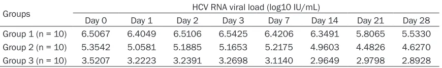
[image:2.612.90.525.85.152.2]The data were log10 transformed prior for anal-ysis and a pairwise comparison between each Table 1. Mean HCV RNA viral loads of three categories in different time points
Groups HCV RNA viral load (log10 IU/mL)
Day 0 Day 1 Day 2 Day 3 Day 7 Day 14 Day 21 Day 28 Group 1 (n = 10) 6.5067 6.4049 6.5106 6.5425 6.4206 6.3491 5.8065 5.5330 Group 2 (n = 10) 5.3542 5.0581 5.1885 5.1653 5.2175 4.9603 4.4826 4.6270 Group 3 (n = 10) 3.5207 3.2223 3.2391 3.2698 3.1140 2.9649 2.9798 2.8928
time point in the same group was conducted using repeated measures analysis. Test results with P values less than 0.05 were considered to be statistically significant. The statistical analysis was performed with SPSS 16.0 soft-ware (SPSS Chinese, China).
Results
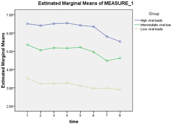
Thirty HCV RNA positive patients (16 males and 14 females; mean age, 45 years; range, 26-70 years) were enrolled in this study. The mean HCV RNA viral loads of all three sample catego-ries at 0, 1, 2, 3, 7, 14, 21 and 28 days are shown in Table 1 and Figure 1.
For the high HCV RNA group, the quantity of HCV RNA ranged from 6.18 log10 IU/ml to 6.94 log10 IU/ml, with a mean (± SD) of 6.50 ± 0.29 log10 IU/ml at Day 0. After 1, 2, 3, 7, and 14 days of storage at 4°C, the mean viral loads in the serum samples did not differ sig-nificantly from those taken at Day 0 (P > 0.05). However, when the serum samples stored at 4°C were checked after 21 days and 28 days of storage, the HCV RNA concentration was found to be significantly lower (0.70 log10, P = 0.011 and 0.97 log10, P = 0.005, respectively). For the intermediate HCV RNA group, the quan-tity of HCV RNA ranged from 4.48 log10 IU/ml to 5.95 log10 IU/ml, with a mean (± SD) of 5.35
ly significant decline in the mean viral loads was found on Day 14, Day 21, and Day 28 (0.56 log10, P < 0.001, 0.54 log10, P < 0.001 and 0.63 log10, P < 0.001, respectively).
Discussion
PCR detection of HCV RNA is a major technique that is used to screen and diagnose HCV infec-tion. Quantitation of HCV RNA has been described as a key predictor for patient man-agement, including assessing treatment initia-tion and treatment response, and monitoring follow-up. Previous studies have reported that different factors might have an effect on nucle-ic acid stability, such as shipping conditions, screening in blood banks, handling of samples, etc. To ensure optimal prognostic and thera-peutic value in monitoring chronically infected patients, it is critical to evaluate the effects of handling clinical specimens in routine clinical laboratory practice.
[image:3.612.91.369.72.277.2]In this study, 30 samples were included, cover-ing an approximately 3 log10 HCV RNA dynamic range, and these were divided based on HCV RNA loads, as follows: a high HCV RNA load group, an intermediate HCV RNA load group, and a low HCV RNA load group. The storage condition for all of the samples was 4°C for 0, 1, 2, 3, 7, 14, 21 and 28 days. The results showed high stability of HCV RNA over the
Figure 1. HCV RNA viral loads measurement in serum samples stored at 4°C after 0, 1, 2, 3, 7, 14, 21, and 28 days. High: high HCV-RNA loads group; Intermediate: intermediate HCV-RNA loads group; Low: low HCV-RNA loads group.
± 0.49 log10 IU/ml at Day 0. Similarly, the mean viral loads in serum samples over the 14-day period were found to be stable (0.39 log10, P = 0.116). However, the serum HCV RNA titer decreased sig-nificantly at 4°C after 21 days and 28 days of storage (0.87 log10, P = 0.003 and 0.73 log10, P = 0.005, respective- ly).
statistical-14-day period at 4°C in the high- and intermedi-ate HCV RNA load groups and no significant decrease in the HBV DNA titer was detected. However, for the samples stored for 21 and 28 days, the mean viral loads differed significantly from those at Day 0 for both of these days. For the low HCV RNA load group, an apparent decrease in the HCV RNA level was observed on Day 14. In our study, a 0.50 log10 or greater loss of HCV RNA was defined as the probability of specimen failure. Because it is generally accepted that a difference of less than 0.5 log10 IU/ml in viral loads detected using molec-ular assays should be considered the result of assay variance [17-19].Furthermore, with the exception of two samples with low viral loads, all the samples were found to be positive for HCV RNA at 28 days at 4°C storage. This is pos-sibly due to the fact that the particulate nature of the virus genome or the host’s properties may affect the continuation of HCV RNA positiv-ity [20]. Nevertheless, further studies are need-ed to more clearly address this issue.
Since Cuypers et al. [11] first examined the sta -bility of HCV RNA in serum stored at 4°C using the cDNA-PCR method in 1992, this subject has been studied extensively over the past sev-eral years. In one study, seven serum samples taken from patients with histologically-proven chronic hepatitis C was stored at 4°C until HCV RNA determination. The results showed that the HCV RNA levels were not significantly differ -ent from the levels at time point 0 after five days at 4°C, but the levels were loss completely after 6 months at 4°C [12]. In another study, the impact of storage time on HCV RNA detec-tion was analyzed in 11 serum samples sub-jected to 4°C storage for up to one month by using a Quantiplex branched-DNA assay. The authors reported that the estimated probability of specimen failure was 18%, which was lowest at 4°C [10]. Comparable results have also been presented in our studies. The probability of specimen failure was 14%, 14%, and 11% at Day 28 for the high, intermediate, and low HCV-RNA load groups, respectively. Moreover, Gessoni et al. [7] suggested that the standard procedures for storage schedules was 4°C for a maximum of 48 h without compromising the quality of the HCV RNA in whole blood samples. After examining the results of these studies, some differences were found in how the labora-tories handled the specimens and in the stan-dardizations they used for the nucleic acid test.
Therefore, evaluation of the optimal storage conditions for HCV RNA preservation should be conducted by individual laboratories based on their own technology.
Krajden et al. [10] the following elements are required to ensure an accurate assessment of the effect that storage at 4°C has on HCV RNA quantitative detection for clinical specimens: (1) sufficient samples to ensure statistical power; (2) a sensitive quantitative assay for viral load measurements; (3) an appropriate definition of a clinically relevant descriptive endpoint; and (4) reliable statistical analysis methods. In the present work, all of these crite-ria were applied. For clinical specimens, we included 30 HCV RNA-positive patients, and then these samples were equally divided into three groups in accordance with the HCV RNA loads. For the quantitative assay, a more direct and sensitive method, TaqMan real-time PCR, was used. This method had a detection limit of 250 IU/mL and linearity to 103-107 IU/mL. The intra-assay CV was ≤ 5% for both 104 IU/mL and 106 IU/mL. Moreover, in this assay, an internal standard RNA was added to each test specimen in order to avoid any false-negative results. For the descriptive endpoint, we defined the probability of specimen failure as a 0.50 log10 or greater loss of HCV RNA. For sta-tistical analysis, the repeated measures analy-sis method was performed with SPSS 16.0 software (SPSS Chinese, China). Test results with P values less than 0.05 were considered to be statistically significant.
In conclusion, our data demonstrated that serum samples with HCV RNA levels up to 104 IU/ml can only be stored at 4°C for 14 days, and those lower than 104 IU/ml can be per-formed for up to only 7 days when the samples are stored at 4°C.
Acknowledgements
We would like to thank Professor Zhiping Chen for the data analysis and Scribendi.com for its linguistic assistance during the preparation of this manuscript.
Disclosure of conflict of interest None.
Address correspondence to: Dr. Shan Li, Depart-
of Guangxi Medical University, Nanning 530021, Guangxi, China. Tel: 771-5356052; Fax: +86-0771-865353342; E-mail: [email protected]
References
[1] Global Burden Of Hepatitis C Working Group. Global burden of disease (GBD) for hepatitis C. J Clin Pharmacol 2004; 44: 20-29.
[2] Mohd Hanafiah K, Groeger J, Flaxman AD and
Wiersma ST. Global epidemiology of hepatitis
C virus infection: new estimates of age-specific
antibody to HCV seroprevalence. Hepatology 2013; 57: 1333-1342.
[3] Qin Q, Smith MK, Wang L, Su Y, Wang L, Guo W, Wang L, Cui Y and Wang N. Hepatitis C virus infection in China: an emerging public health issue. J Viral Hepat 2015; 22: 238-244. [4] Abe K and Konomi N. Hepatitis C virus RNA in
dried serum spotted onto filter paper is stable
at room temperature. J Clin Microbiol 1998; 36: 3070-3072.
[5] Comert F, Aktas E, Terzi HA, Kulah C, Ustundag Y, Kokturk F and Aydemir S. Evaluation of hep-atitis C virus RNA stability in room temperature and multiple freeze-thaw cycles by COBAS AmpliPrep/COBAS TaqMan HCV. Diagn Micro- biol Infect Dis 2013; 75: 81-85.
[6] Jose M, Curtu S, Gajardo R and Jorquera JI. The effect of storage at different temperatures on the stability of Hepatitis C virus RNA in plas-ma samples. Biologicals 2003; 31: 1-8. [7] Gessoni G, Barin P, Frigato A, Fezzi M, de Fusco
G, Arreghini N, Galli P and Marchiori G. The sta-bility of hepatitis C virus RNA after storage at +4 degrees C. J Viral Hepat 2000; 7: 283-286. [8] Damen M, Sillekens P, Sjerps M, Melsert R,
Frantzen I, Reesink HW, Lelie PN and Cuypers HT. Stability of hepatitis C virus RNA during specimen handling and storage prior to NASBA
amplification. J Virol Methods 1998; 72:
175-184.
[9] de Moreau de Gerbehaye AI, Bodeus M, Robert A, Horsmans Y and Goubau P. Stable hepatitis C virus RNA detection by RT-PCR during four days storage. BMC Infect Dis 2002; 2: 22. [10] Krajden M, Minor JM, Zhao J, Rifkin O and
Comanor L. Assessment of hepatitis C virus RNA stability in serum by the Quantiplex branched DNA assay. J Clin Virol 1999; 14: 137-143.
[11] Cuypers HT, Bresters D, Winkel IN, Reesink HW, Weiner AJ, Houghton M, van der Poel CL and Lelie PN. Storage conditions of blood sam-ples and primer selection affect the yield of cDNA polymerase chain reaction products of hepatitis C virus. J Clin Microbiol 1992; 30: 3220-3224.
[12] Halfon P, Khiri H, Gerolami V, Bourliere M, Feryn JM, Reynier P, Gauthier A and Cartouzou G. Impact of various handling and storage con-ditions on quantitative detection of hepatitis C virus RNA. J Hepatol 1996; 25: 307-311. [13] Quan CM, Krajden M, Zhao J and Chan AW.
High-performance liquid chromatography to assess the effect of serum storage conditions on the detection of hepatitis C virus by the polymerase chain reaction. J Virol Methods 1993; 43: 299-307.
[14] Cardoso MS, Koerner K, Hinz W, Lenz C, Schwandt A and Kubanek B. Hepatitis C virus stability: the issue! Vox. Sang 1999; 76: 124-127.
[15] Manzin A, Bagnarelli P, Menzo S, Giostra F, Brugia M, Francesconi R, Bianchi FB and Clementi M. Quantitation of hepatitis C virus genome molecules in plasma samples. J Clin Microbiol 1994; 32: 1939-1944.
[16] Kim K, Park J, Chung Y, Cheon D, Lee IB, Lee S, Yoon J, Cho H, Song C and Lee KH. Use of inter-nal standard RNA molecules for the RT-PCR
amplification of the faeces-borne RNA viruses.
J Virol Methods 2002; 104: 107-115.
[17] Miskovsky EP, Carrella AV, Gutekunst K, Sun CA, Quinn TC and Thomas DL. Clinical charac-terization of a competitive PCR assay for quan-titative testing of hepatitis C virus. J Clin Microbiol 1996; 34: 1975-1979.
[18] Kessler HH, Santner B, Umlauft F, Urbanek M, Kronawetter M, Pierer K, Stunzner D, Grunewald K and Marth E. Detection of hepati-tis C viral sequences in serum by ‘nested’ poly-merase chain reaction (PCR) and a commer-cial single-round PCR assay. Clin Diagn Virol 1995; 4: 239-250.
[19] Farma E, Boeri E, Bettini P, Repetto CM, McDermott J, Lillo FB and Varnier OE. Single-step PCR in molecular diagnosis of hepatitis C virus infection. J Clin Microbiol 1996; 34: 3171-3174.