Effect of Colchicine on Total Antioxidant Capacity,
Antioxidant Enzymes and Oxidative Stress Markers in
Patients with Knee Osteoarthritis
Metin Erden1*, Levent Ediz2, Özcan Hız2, Yasin Tuluce3, Halil Ozkol3, Murat Toprak1, Fahrettin Demirdag4
1Physical Medicine and Rehabilitation Department, District Education and Research Hospital, Van, Turkey; 2Physical Medicine and
Rehabilitation Department, Yuzuncu Yil University Medical Faculty, Van, Turkey; 3Medical Biology Department, Medical Faculty,
Yuzuncu Yil University, Van, Turkey;4Physical Medicine and Rehabilitation Department, District Education and Research Hospital, Elazig, Turke.
Email: *dr.metinerden@gmail.com
Received May 28th, 2012; revised June 30th, 2012; accepted July 15th, 2012
ABSTRACT
Background/Aim: We aimed to investigate the effects of colchicine on clinical recovery, as well as oxidative stress
markers and total antioxidant capacity (TAC) in whole blood of patients with knee osteoarthritis (OA). Materials and Methods: Sixty patients with grade 2 - 3 knee OA according to ACR knee OA criteria whom examination of the knee
joint synovial fluid by polarized light microscopy demonstrated CPDD crystals existence were included in the study. Fifty healthy subjects were included as a control group. Patients were divided randomly into two groups. The first group (paracetamol group) was given only paracetamol 3 gr daily p.o and the second group (colchicine and paracetamol group) was given colchicine 1.5 gr and paracetamol 3 gr daily p.o for six months. For outcome measures WOMAC and VAS were used. Superoxide dismutase (SOD), Catalase (CAT) ezyme activities and Glutathione (GSH) and Malon- dialdehyde (MDA) levels and TAC all were measured. Results: WOMAC scores were improved in both patient groups
compared with pre-treatment evaluation (p < 0.025). WOMAC morning stiffness scores were significantly more improved in colchicine group compared with paracetamol group (p > 0.05). TAC was signifcantly increased only in colchicine/paracetamol group. Oxidant parameter MDA levels were significantly decreased in both paracetamol group and colchicine/paracetamol group. CAT, SOD enzyme activities and GSH levels did not change before and after treatment protocols in both patient groups. Conclusion: Both paracetamol 3000 mg/day and 3000 mg paracetamol plus
1.5 gr/day colchicine is effective in the treatment of patients with knee osteoarthritis. But the addition of colchicine to paracetamol produced significantly greater symptomatic benefit than paracetamol alone. Our study also showed that colchicine lowers whole blood MDA which is a lipid peroxidation compound and elevates TAC levels in patients with knee OA. This may show probable disease modifying effect of colchicine in knee OA which require further long period laboratory and radiologic investigations.
Keywords: Knee Osteoarthritis; Colchicine; Paracetamol; Total Antioxidant Capacity; Malondialdehyde; Oxidative
Stres
1. Introduction
Osteoarthritis (OA) is a chronic degenerative disorder of multifactorial etiology characterized by loss of articular cartilage and changes in the underlying bone at the joint margins. OA is a leading cause of chronic joint pain and disability at older ages, largely due to knee and hip involvement. The etiology of OA is unclear. Mechanical, biochemical, metabolic, endocrine, genetic and environ- mental factors seem to play a role [1].
The incidence of osteoarthritis increases steeply after
50 years of age and the proportion of elderly people in the population continues to increase. More than 13% of Americans aged 55 to 64 years, and more than 17% of Americans aged 65 to 74 years, have pain and functional limitations related to knee OA [2].
Basic calcium phosphate (BCP) and calcium pyropho- sphate dihydrate (CPPD) crystals are commonly found in osteoarthritic joints. These crystals have been found in the synovial fluid of 60% of patients with knee OA and over 90% of a small group of patients with grade-4 OA [3,4].
Clinical and experimental data suggest a pathogenic
role for calcium-containing crystals in cartilage destruc- tion and OA development and progression [5].
Oxidative demage to collagen (oxidative DNA demage etc.) has also important role in etiopathogenesis of OA. Decreased joint fluid antioxidants and increased reactive oxygen species (ROS) have been shown in osteoarthritic joints compared to joints with macroscropically intact cartilage and subacute injury. ROS induced by IL-1β and TNF-α can directly cleave proteoglycans and collagen, modulate signaling pathways and activate MMPs, as well as alter chondrocyte apoptosis and cellular synthetic ac- tivity [4,6-24].
Recently there has been increase in trials on disease/ structure-modifying agents in OA (e.g. MMP inhibitors, glucosamine or chondritin sulphate, colchicine, strontium ranelate, biphosphonates and diacetylrhein/diacerein). This is an area of active research at present. Preclinical studies of potential disease modifying agents are promising. In clinical studies, generally, radiological joint space nar- rowing or width have been used as the structural outcome measure in these trials. More advanced compositional MRI techniques, and laboratory cartilage markers might enable to detect early cartilage changes before radio- graphic joint space narrowing becomes evident [25-27].
Furthermore there are studies in the literature, on slow acting disease/structure-modifying effects of colchicine in knee OA. The rationale using colchicine as disease/ structure-modifying drug to manage OA resides in the fact that calcium-containing crystals are frequently seen in OA (3, 4), and colchicine has been shown to be bene- ficial in preventing calcium crystal-induced inflammation (pseudogout). When used to treat gouty arthritis, colchi- cine is believed to work by inhibiting microtubule depen- dent cell infiltration and IL-1β production. However, in vitro and in vivo, colchicine also reduces monosodium urate (MSU)-induced ROS production by neutrophils [28-31].
Taking into account that increased incidence of cal- cium-containing crystals (from 60% upto 90%), decreased joint fluid antioxidants and increased reactive oxygen species (ROS) in osteoarthritic joints, we aimed to in- vestigate the effects of colchicine on clinical recovery, oxidative stress markers and total antioxidant capacity (TAC) in whole blood of patients with knee osteoar- thritis.
2. Materials and Methods
Sixty patients with grade 2 - 3 knee osteoarthritis accor- ding to ACR knee OA criteria whom examination of the knee joint synovial fluid by polarized light microscopy demonstrated CPDD crystals existence were included in the study. Fifty healthy subjects who have similar demo- graphic characteristics with patient group were included as a control group. The study protocol, consent form and
all recruitment materials were approved by the ethical Board of the University of Yuzuncu Yil University, Van, Turkey, and the study was conducted in accordance with the Declaration of Helsinki. Patients were divided ran- domly into two groups. The first group (paracetamol group) was given only paracetamol 3 gr daily p.o and the second group (colchicine and paracetamol group) was given colchicine 1.5 gr and paracetamol 3 gr daily p.o.
The patients with diabetes, gouty arthritis, secondary knee osteoarthritis due to inflammatory joint diseases, a history of intraarticular corticosteroid and/or hyaluronic acid injections, and a history of oral glucosamine, col- chicine use during the last 6 months, chronic alchohol consumption, chronic liver and kidney diseases, smokers, a history of antioxidant vitamin and/or mineral use during the last six months were excluded.
One of the primary clinical outcome measures was change in the WOMAC-Western Ontario and McMaster Universities Arthritis Index. The WOMAC has been used extensively in the quantitative assessment of knee OA, and has been proven to be effective in assessing func- tionality, pain in patients suffering from knee OA. Turkish WOMAC has been found valid and reliable [32].
Another clinical outcome measure was 100 mm VAS (visual analog scale) for pain measuremet.
Before drug treatment start and after the last treatment dose (6 months later), 7 mL sample of venous blood was taken in the morning before breakfast from each patient and control subjects. Serum samples were stored at
−20˚C until analysis.
2.1. Oxidant/Antioxidant Status Analysis
2.1.1. Superoxide Dismutase (SOD) Enzyme Analysis
SOD enzyme activity was measured using Genesys 10 UV-VIS Scanning spectrophotometer by Randox-Ransod enzyme kit at 505 nm and 37˚C.
2.1.2. Catalase (CAT) Enzyme Activity Determination
Catalase enzyme activity was measured by reading the absorbance with a spectrophotometer (Genesys 10 UV Scanning UV/VIS Spectrophotometer) set at 240 nm based on Aebi method.
2.1.3. Glutathion (GSH) Determination
GSH samples were measured by using a spectroflu- orimetry (Jasco 6000 USA) with excitation at 345 nm and emission at 425 nm.
2.1.4. Total Antioxidant Capacity (TAC)
Ready-kit was used according to the measurement me- thod.
2.1.5. Malondialdehyde (MDA) Determination
metabolism were analyzed spectrofluorimetrically, using the modified method by Hegde et al. [33]. The MDA was measured by a spectrofluorimetry (Jasco 6000, USA), with excitation at 520 nm and emission at 555 nm.
All outcome measures were performed before colchi- cine tretment and just after of colchicine treatmet finish (6 months later).
2.2. Statistical Analysis
Results were expressed as mean and standard deviation (SD). Statistical analysis was carried out using the SPSS program (version 11.5 software, SPSS Inc. Chicago, Illinois, USA). For the comparison of groups, indepen- dent student t test, ANOVA, Kruskal Wallis and Mann- Whitney U test were used. Categorical variables were evaluated with Pearson’s chi-square test. Within the same group comparisons in terms of the difference be- tween pretreatment and posttreatment measurements were investigated by the dependent t-test or Wilcoxon signed rank test. P values of less than 0.05 were regarded as significant.
3. Results
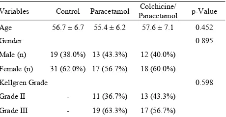
Demographic data of the groups were presented in Table
1. There was no significant difference between groups in
terms of demographic variables.
VAS scores according to patient and doctor evaluations of the tretment groups were presented in Table 2. Only
Colchicine group showed statistically significant change after the treatment in terms of VAS scores compared with pre-treatment evaluation.
Pre and post-treatment WOMAC scores of the patient groups were presented in Table 3. Post-treatment
[image:3.595.309.539.267.386.2]WOMAC scores were improved in both patient groups compared with pre-treatment evaluation (p < 0.025). WOMAC morning stiffness scores were significantly more
Table 1. Demographic properties of the groups. Variables Control Paracetamol Paracetamol Colchicine/ p-Value
Age 56.7 ± 6.7 55.4 ± 6.2 57.6 ± 7.1 0.452
Gender 0.895
Male (n) 19 (38.0%) 13 (43.3%) 12 (40.0%)
Female (n) 31 (62.0%) 17 (56.7%) 18 (60.0%)
Kellgren Grade 0.598
Grade II - 11 (36.7%) 13 (43.3%)
[image:3.595.56.539.415.494.2]Grade III - 19 (63.3%) 17 (56.7%)
Table 2. Pre-treatment and post-treatment VAS scores of the patient groups.
Variables Pre-treatment (mean(SD)) Post-treatment (mean(SD)) p-Valuea Change (mean(SD)) p-Valueb
VASpatient 0.645
Paracetamol 77.5 (17.25) 74.0 (17.25) 0.060 –4.0 (8.00)
Colchicine/Par 79.0 (11.50) 76.0 (14.00) 0.010 –4.0 (8.50)
VASdoctor 0.662
Paracetamol 75.5 (7.25) 74.0 (15.25) 0.043 –3.0 (9.25)
Colchicine/Par 73.0 (17.25) 68.0 (15.50) 0.033 –3.5 (12.00)
aComparisons between pre-treatment and post-treatment within the same groups, according to the Bonferroni correction p value < 0.025 was considered
statistically significant for the results; bComparisons between the groups in terms of treatment-related change, p-value < 0.05 was considered statistically
[image:3.595.56.540.548.709.2]significant for the results.
Table 3. Pre-teratment and post-treatment WOMAC scores of the patient groups.
Variables Pre-treatment (mean(SD)) Post-treatment (mean(SD)) p-Valuea Change (mean(SD)) p-Valueb
Pain 0.988
Paracetamol 11.5 (4.00) 11.0 (3.25) 0.008 –1.0 (2.25)
Colchicine/Par 11.0 (5.00) 9.5 (4.25) 0.011 –1.0 (3.00)
Morning stiffness 0.039
Paracetamol 1.0 (2.25) 1.0 (2.00) 0.206 0.0 (0.00)
Colchicine/Par 3.0 ( 4.25) 2.0 (3.25) 0.005 –1.0 (1.25)
Physical activity 0.755
Paracetamol 38.0 (11.00) 36.5 (14.25) 0.027 –2.5 (6.50)
Colchicine/Par 34.5 (17.00) 31.0 (13.25) 0.021 –2.0 (8.25)
Total 0.280
Paracetamol 12.2 (6.50) 11.5 (6.08) 0.002 –1.0 (1.91)
Colchicine/Par 13.3 (8.75) 11.9 (5.56) 0.002 –1.0 (2.85)
aComparisons between pre-treatment and post-treatment within the same groups, according to the Bonferroni correction p-value < 0.025 was considered
statistically significant for the results; bComparisons between the groups in terms of treatment-related change, p-value < 0.05 was considered statistically
improved in colchicine group compared with para- cetamol group (p > 0.05).
Baseline oxidant and anti-oxidant levels of the control group and the treatment groups were presented in Table 4. There were statistically significant differences between
control group and treatment groups in terms of baseline MDA levels (p < 0.001). Baseline MDA levels were higher in treatment groups than control group.
Comparisons of pre-treatment and post-treatment oxi- dant and antioxidant levels of the patient groups were presented in Table 5. Total antioxidant capacity (TAC)
was signifcantly increased only in colchicine/paraceta- mol group. Oxidant parameter MDA levels were signi- ficantly decreased in both paracetamol group and colchi- cine/paracetamol group. Catalase (CAT), superoxide dis- mutase (SOD) enzyme activities and glutathione (GSH) levels did not change before and after treatment protocols in both patient groups.
4. Discussion
In this study, the effects of colchicine on whole blood oxidant load, antioxidant capacity and clinical recovery in patients with OA were investigated. In the current study we found that baseline MDA levels were higher in patients with OA than healthy control group. Baseline SOD, CAT enzyme activities, GSH, and TAC levels were not differed between groups. When compared to baseline MDA levels, post-treatment MDA levels signi- ficantly decreased in only the group receiving colchicine/
paracetamol. Post-treatment TAC levels were also in- creased significantly in only the group receiving colchi- cine/paracetamol. No significant changes were observed in post-treatment SOD, CAT enzyme activities and GSH levels in both groups.
As well as colchicine treated group showed more improvement in clinical recovery parameters such as WOMAC and VAS scores than control and paracetamol treated groups. Our clinical recovery results are in accordance with previous studies by Ediz L. et al. and Das K. et al. [28,34].
[image:4.595.308.538.328.420.2]Basic calcium phosphate (BCP) and calcium pyropho- sphate dihydrate (CPPD) crystals are commonly found in osteoarthritic joints. These crystals have been found in the synovial fluid of 60% of patients with knee OA and over 90% of a small group of patients with grade-4 OA
Table 4. Pre-treatment (baseline) laboratory findings of control and treatment Groups.
Variables Control Paracetamol Paracetamol Colchicine/ p-Value
CAT 56.4 (18.30) 54.8 (13.02) 59.2 (18.97) 0.328
GSH 0.98 (0.09) 1.0 (0.17) 0.99 (0.06) 0.743
TAC 25.0 ± 5.78 24.0 ± 7.25 21.6 ± 5.31 0.081
MDA 5.7 (1.11)a,b 6.6 (1.32)a 6.1 (1.94)b <0.001
SOD 228.7 (72.52) 214.3 (90.97) 221.5 (124.23) 0.749
aStatistically significant difference between control group and paracetamol
group (p < 0.001); bStatistically significant difference between control group
[image:4.595.57.538.478.708.2]and clchicine group (p < 0.001).
Table 5. Comparison of pre-treatment and post-treatment oxidant and antioxidant levels of the patient groups.
Variables Pre-treatment (mean(SD)) Post-treatment (mean(SD)) p-Valuea Change (mean(SD)) p-Valueb
Catalase 0.738
Paracetamol 54.8 (13.02) 60.2 (16.52) 0.645 –0.09 (20.42)
Colchicine/Par 59.2 (18.97) 62.1 (16.62) 0.866 0.47 (20.32)
GSH 0.560
Paracetamol 1.0 (0.17) 1.0 (0.12) 0.949 0.01 (0.14)
Colchicine/Par 0.99 (0.06) 1.0 (0.07) 0.271 0.01 (0.11)
TAC 0.206
Paracetamol 24.0 ± 7.25 24.6 ± 7.10 0.754 0.6 ± 10.32
Colchicine/Par 21.6 ± 5.31 25.3 ± 5.55 0.018 3.7 ± 7.67
MDA 0.817
Paracetamol 6.6 (1.32) 5.6 (1.24) 0.002 –0.64 (1.87)
Colchicine/Par 6.1 (1.94) 5.6 (0.84) 0.014 –0.83 (2.22)
SOD 0.727
Paracetamol 214.3 (90.97) 228.7 (72.52) 0.610 20.4 (98.87)
Colchicine/Par 221.5 (124.23) 221.5 (95.15) 0.696 34.3 (109.37)
aComparisons between pre-treatment and post-treatment within the same groups, according to the Bonferroni correction p value < 0.025 was considered
statistically significant for the results; bComparisons between the groups in terms of treatment-related change, p value < 0.05 was considered statistically
[3,4].
These crystals induce and maintain inflammation and ROS production in osteoarthritic joint and hence degre- date more and more cartilage and produce pain and finally joint impairment occurs. To the best of our knowledge this is the first study in the literature evaluates colchicine effects on whole blood oxidant load and antioxidant levels in patients with knee OA. Our study showed that colchicine lowers whole blood MDA which is a lipid peroxidation compound and elevates TAC levels in patients with knee OA. Namely colchicine has antioxidant activity and also enhances total antioxiant capacity. However in this study, colchicine has no effect on whole blood SOD, CAT enzyme activities and GSH levels in patients with knee OA.
The current treatment of OA is primarily focused on relief of the symptoms by use of rapid action drugs (analgesics, cycloxygenase (COX-2) specific inhibitors and NSAIDS). These drugs do not effect the underlying pathogenesis of OA, thus have minimal role in modifying disease course and improving quality of life [35,36].
In conclusion both paracetamol 3000 mg/day and 3000 mg paracetamol plus 1.5 gr/day colchicine is effective in the treatment of patients with knee osteoarthritis. But the addition of colchicine to paracetamol produced signifi- cantly greater symptomatic benefit than paracetamol alone. Our study also showed that colchicine lowers whole blood MDA which is a lipid peroxidation com- pound and elevates TAC levels in patients with knee OA. This may show probable disease modifying effect of colchicine in knee osteoarthritis which require further long period laboratory and radiologic investigations.
5. Acknowledgements
This research was funded by Yuzuncu Yil University Science Researchs Supporting Agency.
REFERENCES
[1] M. B. Goldring and S. R. Goldring, “Osteoarthritis,”
Journal of Cellular Physiology, Vol. 213, No. 3, 2007, pp. 626-634. doi:10.1002/jcp.21258
[2] P. Sarzi-Puttini, M. A. Cimmino, R. Scarpa, R. Caporali, F. Parazzini, A. Zaninelli, F. Atzeni and B. Canesi, “Os- teoarthritis: An Overview of the Disease and İts Treat- ment Strategies,” Seminars in Arthritis and Rheumatism, Vol. 35, No. 1, 2005, pp. 1-10.
[3] Y. Z. Liu, A. P. Jackson and S. D. Cosgrove, “Contribution of Calcium-Containing Crystals to Cartilage Degradation and Synovial İnflammation in Osteoarthritis,” Osteoarth- ritis and Cartilage, Vol. 17, No. 10, 2009, pp. 1333-1340. [4] K. Jaovisidha and A. K. Rosenthal, “Calcium Crystals in
Osteoarthritis,” Current Opinion in Rheumatology, Vol. 14, No. 3, 2002, pp. 298-302.
[5] H. K. Ea and F. Liote, “Advances in Understanding Calcium-Containing Crystal Disease,” Current Opinion in Rheumatology, Vol. 21, No. 2, 2009, pp. 150-157. [6] E. A. Regan, R. P. Bowler and J. D. Crapo, “Joint Fluid
Antioxidants Are Decreased in Osteoarthritic Joints Com- pared to Joints with Macroscropically İntact Cartilage and Subacute İnjury,” Osteoarthritis Cartilage, Vol. 16, No. 4, 2008, pp. 515-521.
[7] O. Altindag, O. Erel, N. Aksoy, S. Selek, H. Celik and M. Karaoglanoglu, “Increased Oxidative Stress and Its Rela- tion with Collagen Metabolism in Knee Osteoarthritis,”
Rheumatology International, Vol. 27, No. 4, 2007, pp. 339-344.
[8] S. Sarban, A. Kocyigit, M. Yazar and U. E. Isıkan, “Plasma Total Antioxidant Capacity, Lipid Peroxidation, and Erythrocyte Antioxidant Enzyme Activities in Pati- ents with Rheumatoid Arthritis and Osteoarthritis,” Cli- nical Biochemistry, Vol. 38, No. 11, 2005, pp. 981-986. doi:10.1016/j.clinbiochem.2005.08.003
[9] A. F. Chen, C. M. Davies, M. De Lin and B. Fermor, “Oxidative DNA Damage in Osteoarthritic Porcine Arti- cular Cartilage,” Journal of Cellular Physiology, Vol. 217, No. 3, 2008, pp. 828-833.doi:10.1002/jcp.21562
[10] C. M. Davies, F. Guilak, J. B. Weinberg and B. Fermor, “Reactive Nitrogen and Oxygen Species in İnterleukin- 1-Mediated DNA Damage Associated with Osteoarthritis,”
Osteoarthritis Cartilage, Vol. 16, No. 5, 2008, pp. 624- 630. doi:10.1016/j.joca.2007.09.012
[11] Y. E. Henrotin and B. Kurz, “Oxygen and Reactive Oxy- gen Species in Cartilage Degradation: Friends or Foes?”
Osteoarthritis Cartilage, Vol. 13, No. 8, 2005, pp. 643- 654.
[12] J. C. Monboisse and J. P. Borel, “Oxidative Damage to Collagen,” EXS, Vol. 62, 1992, pp. 323-327.
[13] J. M. McCord, “Free Radicals and İnflammation: Protec- tion of Synovial Fluid by Superoxide Dismutase,” Sci- ence, Vol. 185, No. 150, 1974, pp. 529-531.
[14] A. Klamfeldt and S. Marklund, “Enhanced Breakdown in Vitro of Bovine Articular Cartilage Proteoglycans by Conditional Synovial Medium: The Effect of Superoxide Dismutase and Catalase,” Scandinavian Journal of Rheu- matology, Vol. 16, 1987, pp. 41-45.
[15] H. Burkhardt, M. Schwingel, H. Menninger, H. W. Macartney and H. Tschesche, “Oxygen Radicals as Ef- fectors of Cartilage Destruction: Direct Degradative Effect on Matrix Components and İndirect Action via Activation of Latent Collagenase from Polymorphonuclear Leukocytes,” Arthritis & Rheumatism, Vol. 29, 1986, pp. 379-387. doi:10.1002/art.1780290311
[16] H. M. Lander, A. J. Milbank, J. M. Tauras, D. P. Hajjar, B. L. Hempstead, G. D. Schwartz, et al., “Redox Regu- lation of Cell Signalling,” Nature, Vol. 381, 1996, pp. 380-381. doi:10.1038/381380a0
[18] F. J. Blanco, R. L. Ochs, H. Schwarz and M. Lotz, “Chondrocyte Apoptosis İnduced by Nitric Oxide,” Ame- rican Journal of Pathology, Vol. 146, No. 1, 1995, pp. 75-85.
[19] S. B. Abramson, M. Attur, A. R. Amin and R. Clancy, “Nitric Oxide and İnflammatory Mediators in the Per- petuation of Osteoarthritis,” Current Rheumatology Re- ports, Vol. 3, No. 6, 2001, pp. 535-541.
[20] D. Taskiran, M. Stefanovic-Racic, H. Georgescu and C. Evans, “Nitric Oxide Mediates Suppression of Cartilage Proteoglycan Synthesis by İnterleukin-1,” Biochemical and Biophysical Research Communications, Vol. 200, No. 1, 1994, pp. 142-148.
[21] Y. Hirai, K. Migita, S. Honda, Y. Ueki, S. Yamasaki and S. Urayama, et al., “Effects of Nitric Oxide on Matrix Metalloproteinase-2 Production by Rheumatoid Synovial Cells,” Life Sciences, Vol. 68, No. 8, 2001, pp. 913-920. doi:10.1016/S0024-3205(00)00998-X
[22] R. M. Clancy, S. B. Abramson, C. Kohne and J. Rediske, “Nitric Oxide Attenuates Cellular Hexose Monophosphate Shunt Response to Oxidants in Articular Chondrocytes and Acts to Promote Oxidant İnjury,” Journal of Cellular Physiology, Vol. 172, No. 2, 1997, pp. 183-191.
[23] N. Alaaeddine, J. A. Di Battista, J. P. Pelletier, J. M. Cloutier, K. Kiansa, et al., “Osteoarthritic Synovial Fib- roblasts Possess an İncreased Level of Tumor Necrosis Factor-Receptor 55 (TNF-R55) That Mediates Bio- logical Activation by TNF-Alpha,” Journal of Rheum- atology, Vol. 24, No. 10, 1997, pp. 1985-1994.
[24] M. Lotz, “The Role of Nitric Oxide in Articular Cartilage Damage,” Rheumatic Disease Clinics of North America, Vol. 25, No. 2, 1999, pp. 269-282.
[25] A. Anandacoomarasamy and L. March, “Current Evidence for Osteoarthritis Treatments,” Therapeutic Advances in Musculoskeletal Disease, Vol. 2, No. 1, 2010, pp. 17-28. [26] M. Fajardo and P. E. Di Cesare, “Disease-Modifying
Therapies for Osteoarthritis: Current Status,” Drugs Aging, Vol. 22, No. 2, 2005, pp. 141-161.
doi:10.2165/00002512-200522020-00005
[27] M. E. Zagaria, “Osteoarthritis in Seniors,” US Pharmacist, Vol. 31, No. 1, 2006, pp. 20-24.
[28] S. K. Das, S. Ramakrishnan, K. Mishra, et al., “A
Randomized Controlled Trial to Evaluate the Slow-Acting Symptom-Modifying Effects of Colchicine in Osteoarth- ritis of the Knee: A Preliminary Report,” Arthritis & Rheumatism, Vol. 47, No. 3, 2002, pp. 280-284.
doi:10.1002/art.10455
[29] M. Modriansky, Y. Y. Tyurina, V. A. Tyurin, T. Matsura, A. A. Shvedova, J. C. Yalowich and V. E. Kagan, “Anti-/ Pro-Oxidant Effects of Phenolic Compounds in Cells: Are Colchicine Metabolites Chain-Breaking Antioxidants?”
Toxicology, Vol. 177, No. 1, 2002, pp. 105-117. doi:10.1016/S0300-483X(02)00199-3
[30] D. Das, P. W. Pemberton, P. C. Burrows, C. Gordon, A. Smith, R. F. McMahon and T. W. Warnes, “Antioxidant Properties of Colchicine in Acute Carbon Tetrachloride İnduced Rat Liver İnjury and İts Role in the Resolution of Established Cirrhosis,” Biochimica et Biophysica Acta, Vol. 1502, 2000, pp. 351-362.
doi:10.1016/S0925-4439(00)00059-4
[31] E. W. Chia, R. Grainger and J. L. Harper, “Colchicine Suppresses Neutrophil Superoxide Production in a Mur- ine Model of Gouty Arthritis: A Rationale for Use of Low-Dose Colchicine,” British Journal of Pharmacology, Vol. 153, No. 6, 2008, pp. 1288-1295.
[32] E. H. Tüzün, L. Eker, A. Aytar, A. Daşkapan and M. Bayramoğlu, “Acceptability, Reliability, Validity and Responsiveness of the Turkish Version of WOMAC Osteoarthritis Index,” Osteoarthritis Cartilage, Vol. 13, No. 1, 2005, pp. 28-33. doi:10.1016/j.joca.2004.10.010 [33] K. R. Hegde, M. G. Henein and S. D. Varma, “Estab-
lishment of Mouse as an Animal Model for Study of Diabetic Cataracts: Biochemical Studies,” Diabetes, Ob- esity and Metabolism, Vol. 5, No. 2, 2003, pp. 113-119. [34] L. Ediz and I. Tekeoğlu, “Symptom Modifying Effect of
Colchicine in Patients with Knee Osteoarthritis,” Journal of Clinical and Analytical Medicine, Vol. 3, No. 1, 2012, pp. 63-67. doi:10.4328/JCAM.563
[35] A. Mahajan, S. Verma and V. Tandon, “Osteoarthritis,”
Journal of the Association of Physicians of India, Vol. 53, 2005, pp. 634-641.