1 1
C4 anatomy can evolve via a single developmental change
23
Authors: Marjorie R. Lundgren1,2 ([email protected]), Luke T. Dunning1 4
([email protected]), Jill K. Olofsson1 ([email protected]), Jose J. Moreno-5
Villena1 ([email protected]), Jacques W. Bouvier1 ([email protected]), 6
Tammy Sage3 ([email protected]), Roxana Khoshravesh3 ([email protected]), 7
Stefanie Sultmanis3 ([email protected]), Matt Stata3 8
([email protected]), Brad Ripley4 ([email protected]), Maria S. Vorontsova5 9
([email protected]), Guillaume Besnard6 ([email protected]), Claire Adams4 10
([email protected]), Nicholas Cuff7 ([email protected]), Anthony Mapaura8 11
([email protected]), Matheus Bianconi1 ([email protected]), Christine M. Long9 12
([email protected]), Pascal-Antoine Christin1 ([email protected]), Colin P. 13
Osborne1* ([email protected]) 14
2
1Department of Animal and Plant Sciences, University of Sheffield, Western Bank, Sheffield S10
16
2TN, UK 17
2Current address: Lancaster Environment Centre, Lancaster University, Lancaster, LA1 4YQ, UK
18
3Department of Ecology and Evolutionary Biology, University of Toronto, 25 Willcocks Street,
19
Toronto, Ontario M5S 3B2, Canada 20
4Botany Department, Rhodes University, Grahamstown 6139, South Africa
21
5Comparative Plant and Fungal Biology, Royal Botanic Gardens, Kew, Richmond, Surrey, TW9
22
3AB, UK 23
6Laboratoire Évolution & Diversité Biologique (EDB UMR5174), Université de Toulouse, CNRS,
24
ENSFEA, UPS, IRD, 118 route de Narbonne, 31062 Toulouse, France 25
7Northern Territory Herbarium, Department of Environment and Natural Resources. PO Box 496,
26
Palmerston, NT 0831, Australia 27
8National Herbarium and Botanic Garden, Harare, Zimbabwe
28
9Department of Primary Industry and Fisheries, Northern Territory Government, Darwin, NT 0801,
29
Australia 30
31
*Author for correspondence: Colin P. Osborne; [email protected]; Tel: +44-114-222-32
0146; Fax: +44-114-222-0002 33
34
Running title: One anatomical change key for C4 emergence 35
36
Keywords: Alloteropsis, bundle sheath, C3-C4 intermediate, C4 photosynthesis, evolution, grass, leaf 37
anatomy, mesophyll, vein density 38
39
Article type: Letter 40
3 Words in the abstract: 148
42
Words in the main text: 4999 43
Number of references: 57 44
Number of figures: 4 45
Number of tables: 2 46
Number of text boxes: 0 47
48
49
Statement of authorship: MRL, PAC and CPO designed the study. MRL produced and analyzed 50
the data, with the help of LTD, JJMV, and JWB. TS and RK performed the immunolocalisations and 51
TEM imaging. SS assisted with immunolocalisation sample preparation. MS assisted with tissue 52
fixation. MRL, LTD, JKO, BR, MSV, GB, CA, NC, AM, MB, CML, PAC, and CPO contributed 53
plant material. MRL, PAC, and CPO interpreted the results and wrote the paper, with the help of all 54
the authors. 55
56
Data accessibility statement: Should this manuscript be accepted, the data supporting the results 57
will be archived in the public repository Dryad and the data DOI will be included at the end of the 58
article. 59
60
Abbreviations: BS, bundle sheath; CCP, CO2 compensation point, IS, inner sheath; LDA, linear 61
4
Abstract
63
C4 photosynthesis is a complex trait that boosts productivity in warm environments. Paradoxically, it 64
evolved independently in numerous plant lineages, despite requiring specialized leaf anatomy. The 65
anatomical modifications underlying C4 evolution have previously been evaluated through 66
interspecific comparisons, which capture numerous changes besides those needed for C4 67
functionality. Here, we quantify the anatomical changes accompanying the transition between non-68
C4 and C4 phenotypes by sampling widely across the continuum of leaf anatomical traits in the grass 69
Alloteropsis semialata. Within this species, the only trait that is shared among and specific to C4 70
individuals is an increase in vein density, driven specifically by minor vein development that yields 71
multiple secondary effects facilitating C4 function. For species with the necessary anatomical 72
preconditions, developmental proliferation of veins can therefore be sufficient to produce a functional 73
C4 leaf anatomy, creating an evolutionary entry point to complex C4 syndromes that can become more 74
5
INTRODUCTION
76
The vast majority of plants use C3 photosynthesis, but some lineages evolved the C4 pathway to 77
overcome environmentally induced limitations on carbon fixation (Ehleringer et al. 1991; Sage et al. 78
2011). Net carbon fixation by C3 photosynthesis is decreased in warm, high light, arid, and saline 79
environments that lower CO2 concentrations within the leaf and increase photorespiration, the process 80
initiated when O2 instead of CO2 is fixed by the enzyme Rubisco (Chollet & Ogren 1975). To 81
circumvent the losses of carbon and energy caused by photorespiration, the C4 pathway spatially 82
separates the initial fixation of carbon and its assimilation by Rubisco across two leaf compartments, 83
thereby concentrating CO2 at the enzyme’s active site to promote CO2- rather than O2-fixation 84
(Downton & Tregunna 1968; Hatch 1976). A number of anatomical and biochemical functions must 85
work in concert to sustain the high fluxes of the C4 cycle, and comparisons of average C4 and C3 86
plants suggest that the evolution of the C4 phenotype required a large number and scale of changes 87
(Hattersley 1984). Despite this apparent complexity, the C4 trait evolved many times independently 88
(Sage et al. 2011). Resolving this paradox requires the quantitative distinction of changes that were 89
involved in the evolutionary transition from C3 to C4, from those that preceded or followed it. 90
In most C4 plants, carbon fixation within leaf mesophyll tissue (M) is used to concentrate CO2 91
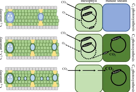
and boost Rubisco activity within bundle sheath tissue (BS), whereas Rubisco in C3 plants operates 92
within the M where it depends on atmospheric CO2 diffusion (Fig. 1; Brown 1975; Hattersley et al. 93
1977; Hatch 1987). Efficient C4 leaves require large BS volumes to accommodate the necessary 94
photosynthetic organelles, including chloroplasts containing abundant Rubisco, and a small distance 95
between M and BS compartments to allow the rapid transfer of metabolites (Fig. 1; Hattersley & 96
Watson 1975; Lundgren et al. 2014). These traits vary among C3 plant lineages and, in grasses, C4 97
photosynthesis evolved only within those groups with large fractions of BS (Christin et al. 2013; 98
Lundgren et al. 2014). Comparisons of multiple C4 lineages with their C3 relatives indicate that the 99
evolution of C4 leaf anatomy involved ultrastructural rearrangements and further decreases to the 100
6 Dengler 2007; Christin et al. 2013). These properties can be achieved via a variety of leaf structural 102
modifications, allowing C4 anatomy to be realized differently each time it evolved, in some cases 103
involving the use of different tissue types for the C4 BS function (Brown 1975; Soros and Dengler 104
2001; Christin et al. 2013; Freitag and Kadereit 2014; Lundgren et al. 2014). While the differences 105
between a diverse range of C3 and C4 species are well known, the minimum set of leaf anatomical 106
modifications required to carry out C4 photosynthesis remains to be established. 107
The grass Alloteropsis semialata provides an outstanding system to capture the early events 108
during C4 evolution because it includes genetically divergent C4 individuals, as well as a diversity of 109
non-C4 plants encompassing C3 and C3-C4 intermediate phenotypes (Ellis 1974; Lundgren et al. 110
2016), which emerged in the paleotropics (Lundgren et al. 2015). The inner sheath (i.e., the mestome 111
sheath), which is present in all C3 grasses, has been co-opted for the C4 BS function in A. semialata. 112
Previous studies have compared leaf properties among C4 and non-C4 leaves of a few A. semialata 113
accessions (Ellis 1974; Frean et al. 1983; Ueno & Sentoku 2006; Lundgren et al. 2016; Dunning et 114
al. 2017), but a broader sampling is required to establish which properties are unique to each 115
photosynthetic type. 116
The primary focus of this study is to compare leaf anatomy in accessions spanning the 117
diversity of each photosynthetic type to distinguish the structural diversifications that occurred before, 118
during, and after C4 emergence in this species. We hypothesize that the properties that predate C4 119
evolution will be shared by at least some of the non-C4 individuals, while those that happened after 120
C4 evolution in a phase of subsequent adaptation will be restricted to a subset of the C4 populations. 121
Properties unique to, and common among all, C4 accessions represent those that were involved in the 122
initial transition to a C4 physiology. We conducted a large scan of the diversity within the species 123
using traits linked to the number and size of different cell types, and used controlled growth 124
experiments to verify that anatomical differences are not environmentally induced. This evaluation 125
of the gross leaf morphology was accompanied by a focused study in some individuals to identify 126
7 shows that a complex trait of large ecological significance can evolve via a few key developmental 128
changes. 129
130
MATERIALS AND METHODS
131Characterizing photosynthetic types 132
Photosynthetic type was determined by a combination of stable isotope and CO2 compensation point 133
(CCP) data (Table S1; Dataset S1), as previously described (Lundgren et al. 2016). The carbon 134
isotope composition of plant tissues (δ13C) distinguishes photosynthetic types (von Caemmerer et al. 135
2014), such that plants with δ13C values higher than -17‰ were considered to have a fully functioning 136
C4 system, while those with values lower than this threshold were considered either C3 or C3-C4. 137
CCPs were used to distinguish C3-C4 from C3 plants, and to support the δ13C results. The CCP 138
indicates the CO2 concentration within the leaf at which CO2 assimilation via photosynthesis equals 139
CO2 loss via photorespiration and respiration. Because less CO2 is ultimately lost to photorespiration 140
in C3-C4 plants, they have very low CCPs compared to C3 plants. Thus, non-C4 plants with CCPs 141
greater than or equal to 35 μmol mol-1 were classified as C3, while those less than 35 μmol mol-1 were 142
classified as C3-C4. CCPs were calculated on 27 living accessions (6 C3, 4 C3-C4, and 17 C4), 143
following published protocols (Bellasio et al. 2016a,b; Lundgren et al. 2016). Non-C4 accessions for 144
which live material was unavailable were assumed to have the same photosynthetic type as their 145
closest relatives, as identified by phylogenetic relationships (Table S1). 146
147
Leaf samples 148
Fifty Alloteropsis semialata (R.Br.) Hitchc. accessions distributed across the species’ geographic 149
range, including 17 C3, 6 C3-C4, and 27 C4, were used to assess intraspecific anatomical variation. 150
Leaf samples from 44 of the 50 accessions were collected from their original field site and preserved 151
until embedding was possible. For the remaining six accessions, leaf samples were taken from plants 152
8 pieces 3-5 mm in length were embedded in methacrylate embedding resin (Technovit 7100, Heraeus 154
Kulzer GmbH, Wehrhein, Germany), sectioned 6-8 μm thick on a manual rotary microtome (Leica 155
Biosystems, Newcastle, UK), and stained with Toluidine Blue O (Sigma-Aldrich, St. Louis, MO, 156
USA). Stained leaf sections were imaged using microscopy-imaging software with a camera mounted 157
on a microscope (Cell A, Olympus DP71, and Olympus BX51, respectively; Olympus, Hamburg, 158
Germany) and the images were stitched together using DoubleTake (v2.2.9, Echo One, 159
Frederikssund, Denmark). 160
161
Leaf anatomy measurements 162
Anatomical traits were measured using ImageJ (Fig. S1; Schneider et al. 2012) from the cross-section 163
of a single leaf segment from the centre of the leaf blade, avoiding segments immediately adjacent to 164
the midrib and lateral edges of the cross-section. Vein orders were distinguished following Renvoize 165
(1987). A single segment was defined as the leaf area falling between two secondary veins, which are 166
large veins with metaxylem. Tertiary and minor veins (e.g., quaternary and quinary orders) lack 167
metaxylem. In this species, the extraxylary fibres that flank both the adaxial and abaxial edges of 168
tertiary veins distinguish them from higher order minor veins, which can be flanked by fibres on one 169
side only (Fig. S1). 170
The cross-sectional area of the whole segment, combining M, BS, epidermis and bulliform 171
cells, extraxylary fibres, and BS extensions, as well as any transverse veins or tear spaces was 172
measured. For all accessions, the total BS (i.e., the mestome sheath; the compartment used for the 173
Calvin cycle in C4 A. semialata), outer sheath, and vein areas were measured separately for secondary, 174
tertiary, and any minor veins. The area of M tissue was calculated as the total area remaining after 175
accounting for all other tissue types. In addition, the cross-sectional area of individual M and BS cells 176
(hereafter 'size') was measured (Fig. S1). Although the depth of individual cells can vary, it is their 177
cross-sectional areas, and not their three-dimensional volumes, that primarily influence the proportion 178
9 180
Linear discriminant analysis 181
We used a Linear Discriminant Analysis (LDA) to explain the variation between photosynthetic types 182
(i.e., the test maximizes between-group variance while minimising within-group variance). We 183
performed the LDA on the 50 accessions with leave-one-out cross-validation and then bootstrapping 184
over 100 runs, using the MASS package in R (Venables & Ripley 2002). Prior probabilities were 185
based on the relative sample size of the categorical variable (i.e., 0.34, 0.12, and 0.54 for C3, C3-C4, 186
and C4 groups, respectively). We chose predictor variables that were likely to influence the M:BS 187
ratio, including the number of M cells between major veins, average size of individual M cells, 188
number of minor veins per segment, leaf thickness, and the average size of BS cells on tertiary veins. 189
To test the generality of our findings from A. semialata, we carried out an equivalent LDA 190
for a larger sample of 157 grasses including one C3 and one C4 A. semialata and representing 17 191
independent C4 lineages. Predictor variables in this analysis were based on the anatomical 192
measurements of Christin et al. (2013) and chosen to best match the variables used in the A. semialata 193
LDA described above, including the number of mesophyll cells between veins, mesophyll cell width, 194
proportion of veins that are minor, leaf thickness, inner BS cell width, and outer BS cell width. The 195
species were grouped as C3, C4 species using the inner BS, and C4 species using the outer BS. 196
197
Vein order analysis 198
To determine whether the pattern of vein density observed in the main dataset was maintained across 199
a larger sample, we counted the total number of veins per segment and the presence or absence of 200
minor veins in 91 additional accessions consisting of herbarium specimens that had been rehydrated 201
in distilled water overnight at 4°C prior to embedding, sectioning, staining, and imaging as described 202
above. Together with the 50 previous samples, this larger dataset included a total of 72 C4 (i.e., δ13C > 203
-17‰) and 69 non-C4 (i.e., δ13C < -17‰) accessions distributed across the species’ geographic range. 204
10 Ultrastructure and immunohistochemistry
206
To investigate whether ultrastructural changes might also differ between photosynthetic types within 207
this species, we analysed the spatial distributions of organelles and enzymes in one population 208
representing each of the C3, C3-C4, and C4 types. Recently expanded mature leaf tissue was prepared 209
for transmission electron microscopy and processed for immunodetection of the large subunit of 210
Rubisco (RBCL) and glycine decarboxylase H subunit (GLDH) as previously described (see 211
Supporting Information Materials 1; Khoshravesh et al. 2017). 212
213
Leaf anatomy in a common environment 214
To determine the degree to which the various leaf anatomical phenotypes arose from plastic 215
development responses to their differing native growth environments, we compared field phenotypes 216
to those obtained from live tillers of 17 A. semialata accessions (5 C3, 4 C3-C4, and 8 C4) after growing 217
for a minimum of three months in a common growth chamber, with conditions as described in 218
Lundgren et al. 2016. The environmental conditions (Fick & Hijmans 2017) at the field collection 219
sites are detailed in Table S2. On both field and controlled environment samples, the number and 220
order of veins, minimum number of M cells separating veins, area of inner BS cells, segment length 221
and thickness, and IVD were determined on one segment per leaf. 222
223
Plasticity for leaf anatomy in response to low CO2
224
To further test whether C4-compatible phenotypes could emerge from plastic responses to the 225
environment, as previously suggested (Li et al., 2014), we carried out a CO2 manipulation experiment 226
designed to promote photorespiration. One C4 (MDG, South Africa) and one C3 (GMT, South Africa) 227
plant were initially grown from seed in a controlled environment chamber set as described in 228
Lundgren et al. 2016, but with 400 μmol mol-1 CO
2 concentration. Both plants were split into five 229
replicate cuttings and kept in the same growth chamber conditions to re-establish for four months. 230
11 ethanol:acetic acid solution. The growth chamber was then set to 180 μmol mol-1 CO
2 concentration 232
for the next four months to promote photorespiration, while maintaining the other environmental 233
conditions, and one new fully expanded leaf was again sampled and fixed. All leaf samples from the 234
400 (i.e., ambient) and 180 (i.e., low) CO2 treatments were embedded, sectioned, and imaged as 235
described above. To determine whether the plants used different photosynthetic pathways in the two 236
CO2 growth environments, we determined CCP and carboxylation efficiency, as described in 237
Lundgren et al. (2016). 238
239
240
RESULTS
241Alloteropsis semialata presents a continuum of leaf anatomy 242
The ratio of M to BS tissue, a trait known to differ among C3 and C4 species (Hattersley 1984), forms 243
a continuum within Alloteropsis semialata, along which photosynthetic types are sorted (Fig. 2). 244
Indeed, the smallest values are restricted to C4 accessions and the largest are found in C3 individuals. 245
When considering the area in cross-section between two secondary veins (i.e., a leaf segment from 246
here onward; Fig. S1), the M area is over ten times larger than the BS area in C3 accessions, but less 247
than five times larger in C4 accessions (Dataset S1). As expected, C3-C4 accessions are intermediate 248
in their overall leaf anatomy, with five to ten times more M than BS. These M:BS ranges are 249
consistent with those measured in other C3 and C4 grasses (Christin et al. 2013). 250
251
C3, C3-C4, and C4 Alloteropsis semialata have distinct leaf anatomy
252
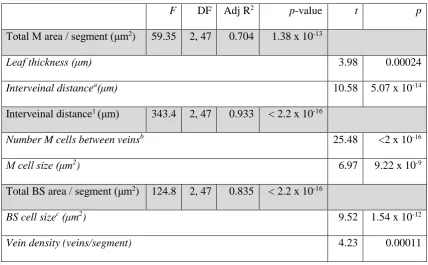
Variation in M:BS ratios can arise via changes to several underlying traits (Lundgren et al. 2014). 253
Our modelling shows that M area is the product of leaf thickness and interveinal distance (IVD; Table 254
1). The latter is predicted by the number and size of M cells between veins (Table 1). BS area is 255
explained by the number of BS units (i.e., the number of veins per segment) and the size of BS cells 256
12 between the three photosynthetic types is captured (Fig. 3a). In a bootstrapped sample, the mean 258
overall predictive accuracy is 0.986, which is statistically indistinguishable from 1.0 (95% CI = 0.966 259
– 1.000). The mean predictive accuracy for C4 (0.999, 95% CI = 0.995 – 1.000), C3 (0.976, 95% CI 260
= 0.918 – 1.000) and C3-C4 (0.926, 95% CI = 0.805 – 1.000) accessions are also statistically 261
indistinguishable from 1.0. The analysis therefore confirms that leaf anatomy varies among 262
photosynthetic types in a statistically predictable manner. 263
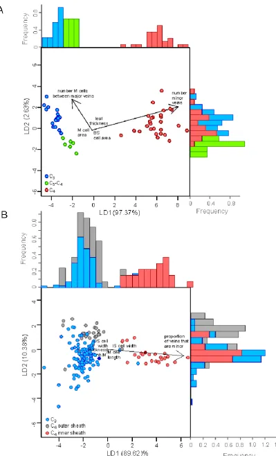
The first axis of the LDA explains 97.37% of the variance between photosynthetic types and 264
clearly distinguishes C4 from non-C4 accessions (Fig. 3a). This axis is most strongly associated with 265
the number of minor veins per segment, which were absent from all non-C4 accessions in this analysis 266
(Table 2). The second axis explains 2.63% of the variance between groups, clearly distinguishes C3 267
from C3-C4 plants, and is most strongly associated with the number of M cells between major veins 268
(i.e., secondary and tertiary order veins) and the number of minor veins (Fig. 3a; Table 2). Since 269
minor veins are restricted to C4 individuals, their contribution to LD2 is linked to diversity within the 270
C4 group. These results indicate that most of the variance in the dataset stems from the contrast 271
between C4 and non-C4 individuals, and is driven entirely by a single underlying trait, the presence 272
of minor veins. The phenotypic distance between C3 and C3-C4 individuals is very small, being 273
explained by the number of M cells between major veins. Conversely, leaf thickness and the cross-274
sectional areas of individual BS and M cells poorly distinguish photosynthetic types in this species. 275
The first two axes of an LDA of anatomical traits on the larger species dataset explain 89.62% 276
and 10.38% of the variation, respectively. The first axis clearly distinguishes C4 species that use the 277
inner bundle sheath from C4 species using the outer sheath and the C3 species (Fig. 3b), and is mostly 278
associated with the proportion of minor veins, while the remaining anatomical traits are weakly 279
correlated with both axes (Table 2). 280
281
Differences between C4 and non-C4 phenotypes arise from the development of minor veins
282
13 non-C4 accessions. When the M:BS ratio is calculated in the absence of minor veins, the clear 284
distinction between C3-C4 and C4 accessions disappears, with nearly half the C4 accessions 285
overlapping with C3-C4 plants (Fig. 2). This shows that the development of minor veins in C4 286
accessions reduces the M:BS ratio by increasing BS area and displacing M area. To confirm the 287
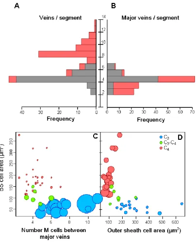
restriction of minor veins to C4 individuals, we screened vein architecture in a larger dataset (Fig. 288
4a,b; Dataset S2). Minor veins were present in all C4 accessions and absent in all but five non-C4 289
accessions. Four of these had only occasional and irregularly spaced minor veins, while the final 290
accession is an individual originating from a natural cross between C3-C4 and C4 individuals 291
(Olofsson et al. 2016). Our data therefore show that the presence of frequent and regularly spaced 292
minor veins is universally and uniquely associated with the C4 genomic background, captures nearly 293
all of the anatomical variation between C4 and non-C4 phenotypes, and explains overall differences 294
in relative M and BS areas. 295
The proliferation of minor veins explains a number of patterns associated with C4 anatomy. 296
As expected, the number of M cells between consecutive veins differs among photosynthetic types, 297
being the smallest in C4 accessions (1-3), compared to C3-C4 (3-6) and C3 (5-11) plants (Fig. S2). 298
However, the number of M cells between major veins overlaps between the C4 and non-C4 groups 299
(Fig. 4c), which indicates that the reduced distance between any pair of M and BS cells in C4 300
accessions is caused by the differentiation of ground meristem cells into minor veins rather than a 301
reduced proliferation of M cells. The high vein density of C4 plants following the development of 302
minor veins is accompanied by more than a twofold increase in extraxylary fibres (i.e., tissue area per 303
segment length) than is found in non-C4 accessions (Fig. S3). Because the area of extraxylary fibres 304
per vein does not differ between the photosynthetic types (Fig. S3), the increased fibre area in C4 305
plants derives entirely from their greater vein density. 306
307
Other anatomical changes happened before or after the transition from C3-C4 to C4 physiology
308
14 accessions, and is therefore linked to the emergence of a fully functioning C4 physiology from a C3 -310
C4 intermediate state. Evolutionary changes that happened once this C4 physiology was in place 311
would be restricted to some, but not all, C4 individuals. In our dataset, such changes include further 312
reductions to the M:BS ratio, potentially achieved via contractions to M airspace (Byott 1976) or 313
increases in BS cell size. Indeed, although BS cell sizes of different photosynthetic types overlap, 314
large increases to BS cell size characterize some African C4 accessions (Figs S4-S5). The BS cell 315
enlargement was therefore involved in the adaptation of C4 physiology after it had emerged, possibly 316
to accommodate more or larger organelles for a more efficient C4 cycle, rather than being involved 317
in its origin. Occasional hybridization between C4 and non-C4 individuals could affect the distribution 318
of trait values, however non-C4 A. semialata individuals are restricted to Africa, so that hybridization 319
outside of Africa is unlikely. Yet, Asia and Australian accessions exhibit some of the smallest BS 320
cells among C4 accessions (Fig. S5). 321
Some characters observed in C4 accessions are also present in C3-C4 individuals, but not C3 322
ones, indicating that they are not associated with the transition to fully functional C4 physiology, but 323
might have facilitated it. These include a small increase in BS cell sizes in C3-C4 compared with C3 324
plants and a decrease in outer sheath cell size, with C3-C4 accessions bridging the anatomical gap 325
between C3 and C4 outer sheath cell sizes (Fig. 4c-d). This reduced outer sheath in C3-C4 and C4 A. 326
semialata likely facilitates metabolite exchanges between M and BS cells. 327
328
Differences between C4 and non-C4 leaves are not environmentally induced
329
Alloteropsis semialata plants grow naturally in diverse environments, depending on their 330
photosynthetic background and evolutionary history (Lundgren et al. 2015). To verify that the 331
differences we observe among photosynthetic types are not induced by environmental variations, we 332
compared the leaves of field-collected plants after transplanting and growing them in a common 333
controlled environment growth chamber for at least three months (Dataset S3; Fig. S6). Compared to 334
15 environment. Moreover, C3-C4 plants produced thicker leaves (p = 0.040), such that leaf thickness of 336
the three photosynthetic types converged in the common environment, which is likely a result of the 337
non-limiting light, nutrients, and water available in these conditions. However, the other traits were 338
not influenced by growth conditions and leaf anatomy of the three photosynthetic types remained 339
distinct when grown in the common environment. 340
We further verified that historical changes in atmosphere composition did not influence the 341
leaf phenotype by comparing C3 and C4 A. semialata under current ambient (400 ppm) and the 342
Pleistocene minimum (180 ppm) CO2 concentrations. Plants grown under the low CO2 concentration 343
experience elevated photorespiration rates, which might have induced a more C4-like anatomy. 344
However, we found that plants did not shift photosynthetic state under the differing CO2 conditions 345
(i.e., mean CCPs in ambient/low CO2 for C3 = 49.8/53.1 and C4 = 4.6/8.1 µmol mol-¹; Dataset S4). 346
Both C3 and C4 plants produced thinner leaves in the low CO2 environment (p = 0.0049 C3 / 0.0065 347
C4), and C4 plants developed smaller BS cells (p = 0.011; Fig. S6), probably because the lower carbon 348
supply restricted development (Ripley et al. 2013). Importantly, the C3 plants did not produce more 349
veins (or any minor veins), larger BS cells, or fewer M cells between veins when grown under this 350
high photorespiration condition. These results show that, even when photorespiration is high, a C4 -351
like phenotype is not plastically induced in C3 A. semialata. 352
353
354
DISCUSSION
355Photosynthetic types form a continuum, along which multiple biochemical, anatomical, and 356
ultrastructural alterations increase the proportion of CO2 fixed via the C4 cycle. The emerging model 357
of C4 evolution involves gradual and overlapping phenotypic changes (Heckmann et al., 2013; Sage 358
et al., 2014; Bräutigam & Gowik, 2016; Schlüter & Weber, 2016; Dunning et al., 2017), with traits 359
acquired in differing orders among C4 lineages (Williams et al., 2013). Different traits may be 360
16 of that phenotype (Christin & Osborne, 2014; Watcharamongkol et al., 2018). Within the grass 362
Alloteropsis semialata, we have shown that the only gross leaf property distinguishing all C4 from all 363
non-C4 phenotypes is the development of frequent minor veins. The presence of these minor veins 364
has multiple consequences, including an overall increase of vein density, enlargement of the total 365
volumes of BS tissue, and a displacement of M tissue. These anatomical changes combine to facilitate 366
C4 cycle activity, as demonstrated by a strong correlation between leaf vein frequency and carbon 367
isotope composition observed for this species (Lundgren et al., 2016). Our analyses of leaf 368
ultrastructure indicate that the evolution of C4 photosynthesis in A. semialata may have involved 369
additional changes in organelle distribution among cell types (Supporting Information Materials 1; 370
Figs S7-9), although the small sample of populations prevents us from differentiating ultrastructural 371
changes linked to the transition to C4 from those that happened later. 372
The change in venation inferred during the evolution of C4 photosynthesis in A. semialata 373
may have a number of physiological and ecological consequences. First, the increase in vein 374
frequency is accompanied by an enhancement of unpigmented extraxylary fibres, which improves 375
light transmission to the BS, and thus ATP production in these cells, facilitating photosynthetic 376
carbon reduction (Bellasio & Lundgren 2016). Enhanced fibre density may also increase leaf 377
toughness, reduce digestibility, and consequently deter herbivores (Caswell et al. 1973; Wilson et al. 378
1983). Secondly, the insertion of additional veins may influence leaf hydraulics. Model simulations 379
for other plant species demonstrate that an increase in minor vein density can lead to greater leaf 380
hydraulic conductance (McKown et al., 2010). However, empirical studies show that this is unlikely 381
to improve drought tolerance, since the decline in hydraulic conductance during drought arises 382
primarily outside veins (Scoffoni & Sack, 2017; Scoffoni et al., 2017a), while embolisms arise first 383
in the midrib, not minor veins (Scoffoni et al., 2017b). 384
Our results complement those from previous comparisons among species, which show that an 385
additional order of minor veins develops during the evolutionary transition from non-C4 to C4 forms 386
17 (Kümpers et al., 2017). Our further analysis of leaf gross anatomy across multiple grass species shows 388
that the insertion of additional minor veins is a frequent developmental mechanism for decreasing the 389
M:BS ratio in those C4 grasses that primarily localise Rubisco within the mestome sheath. The 390
insertion of minor veins could occur via relatively few developmental changes, likely underpinned 391
by changes to auxin, brassinosteroids, SHORTROOT/SCARECROW, and/or INDETERMINATE 392
DOMAIN transcription factors (Kumar & Kellogg 2018; Sedelnikova et al. 2018). In grasses, vein 393
orders develop sequentially as leaves grow wider, such that minor veins are initiated considerably 394
later than other vein orders, usually once the leaf ceases to widen (Nelson & Langdale 1989; 395
Sedelnikova et al. 2018). Thus, the development of functional minor veins likely arises via the 396
heterochronic regulation of the existing machinery for vein formation, sustaining vein differentiation 397
beyond that of non-C4 plants (Nelson 2011; Sedelnikova et al. 2018), probably through the prolonged 398
production of auxin during later phases of leaf elongation (Scarpella et al. 2010). Alternatively, minor 399
veins may also result from a heterotopic specialization of auxin maxima that permits them to form 400
closer together (Kumar & Kellogg 2018). 401
The possibility that a transition from non-C4 to C4 states can be caused by a single 402
developmental alteration is a plausible explanation for the recurrent origins of C4 leaf anatomy, and 403
helps to resolve the paradox of how this complex trait emerged so many times. We also show that 404
organelle number and size differs among photosynthetic types of A. semialata, but, here too, recent 405
work indicates that one gene can control multiple ultrastructural modifications (Wang et al. 2017). 406
Finally, transcriptome comparisons show that few genes encoding enzymes are upregulated during 407
the transition from non-C4 to C4 in A. semialata (Dunning et al. 2017). We therefore conclude that 408
the overall transition from a non-C4 state to the form of C4 photosynthesis observed in A. semialata 409
involved relatively few genetic mutations. 410
The limited number of changes involved in the emergence of C4 anatomy in A. semialata is 411
partially explained by the presence of relatively enlarged BS in the C3-C4 accessions, since C3 A. 412
18 Dunning et al. 2017). The C3 A. semialata phenotype might represent an evolutionary reversal from 414
a C3-C4 state (Dunning et al. 2017), such that its leaf anatomy derives from an ancestral C3-C4 415
intermediate form. C3-C4 A. semialata are characterised by fewer M cell compared to C3 accessions, 416
and higher BS organelle abundance (Figs 3a, 4c and S7-9). These properties that had been selected 417
for the C3-C4 physiology eased the subsequent transition to a full C4 state, but it is important to note 418
that the physiology and anatomy of C3-C4 A. semialata are typical for C3-C4 plants in general 419
(Lundgren et al., 2016), and their anatomical characteristics can be found among C3 grasses 420
(Hattersley 1984; Christin et al. 2013; Lundgren et al. 2014). The background against which C4 421
anatomy evolved in A. semialata is therefore not exceptional. 422
Our conclusion that C4 leaf anatomy can arise from one key developmental modification is 423
apparently incompatible with the great anatomical specialization of other C4 lineages, as well as the 424
large phenotypic gaps separating them from their closest C3 relatives (Dengler et al. 1994; Christin 425
et al. 2013). However, most C3 and C4 sister lineages are separated by long periods of evolution, and 426
comparing these groups therefore captures all of the changes that happened after the origin of C4 427
photosynthesis to improve the efficiency of C4 physiology and adapt it to various organismal and 428
ecological contexts (Christin & Osborne 2014). Indeed, photosynthetic efficiency may be 429
significantly lower in C4 A. semialata than in species from some older C4 lineages (Lundgren et al., 430
2016; Bräutigam et al., 2018). This suggests that C4 photosynthesis in A. semialata may represent a 431
rudimentary version of the physiological trait (Ueno & Sentoku 2006). The biochemical 432
characteristics of the C4 cycle in A. semialata may be one reason for this (Bräutigam et al., 2018), 433
and the presence of Rubisco protein in M could be another (Ueno & Sentoku, 2006). Anatomical 434
diversity may also explain some of the variation in physiological efficiency among A. semialata 435
populations (Lundgren et al. 2016). Indeed, in A. semialata, enlargements of the BS cells beyond 436
those seen in non-C4 individuals are restricted to a subset of C4 populations (Fig. 2) and thus happened 437
after the emergence of C4 physiology. Over time, accumulated modifications will move C4 leaf 438
19 C4 phenotype and the associated physiology can be accessed via a single modification likely placed 440
multiple groups on a selective highway to highly specialized and successful variants of the C4 441
syndrome. 442
443
ACKNOWLEDGEMENTS 444
This work was funded by a University of Sheffield Prize Scholarship to MRL, an ERC grant (grant 445
number ERC-2014-STG-638333) and a Royal Society Research Grant (grant number RG130448). 446
LTD and JKO are supported by a NERC grant (grant number NE/M00208X/1) and PAC is 447
supported by a Royal Society University Research Fellowship (grant number URF120119). JWB 448
was supported by 301 and Think Ahead Sheffield Undergraduate Research Experience grants to 449
MRL. The work on ultrastructure was supported by a Natural Sciences and Engineering Research 450
Council of Canada grant (no 2015-04878) to TLS. The authors thank Peter Westhoff, Stefanie 451
Schulze, and Udo Gowik for use of their GLDH antibody, Susanne von Caemmerer for advice 452
about outer bundle sheath cell resistance, Paul Hattersley for leaf samples and 13C isotope data, 453
John Thompson for field assistance and sample collection, Emma Jardine for discussion of linear 454
discriminant analysis, Heather Walker for mass spectrometry assistance, Gareth Fraser for the use 455
of his vibratome, and Emanuela Samaritani for histology assistance. Herbarium leaf samples were 456
obtained from Kew Herbarium at the Royal Botanic Garden, the National Herbarium of South 457
Africa in Pretoria, and National Museums of Kenya in Nairobi, and the National Botanic Garden of 458
Belgium, Brussels, with the assistance of Martin Xanthos, Lyn Fish, Caroline Mashau, and Itambo 459
Malombe. 460
461
462
20
REFERENCES
464
Bellasio, C. & Lundgren, M.R. (2016). Anatomical constraints to C4 evolution: light harvesting 465
capacity in the bundle sheath. New Phytol., 212, 485-496. 466
467
Bellasio, C., Beerling, D.J. & Griffiths, H. (2016a). An Excel tool for deriving key photosynthetic 468
parameters from combined gas exchange and chlorophyll fluorescence: theory and practice. Plant 469
Cell Environ., 39, 1180-1197. 470
471
Bellasio, C., Beerling, D.J. & Griffiths, H. (2016b). Deriving C4 photosynthetic parameters from 472
combined gas exchange and chlorophyll fluorescence using an Excel tool: theory and practice. Plant 473
Cell Environ., 39, 1164-1179. 474
475
Bräutigam, A. & Gowik, U. (2016) Photorespiration connects C3 and C4 photosynthesis. J. Exp. Bot., 476
67, 2953-2962. 477
478
Brautigam, A., Schluter, U., Lundgren, M.R., Flachbart, S., Ebenhoh, O., Schonknecht, G., Christin, 479
P.A., Bleuler, S., Droz, J.M., Osborne, C. & Weber, A. (2018). Biochemical mechanisms driving 480
rapid fluxes in C4 photosynthesis. bioRxiv, p.387431. 481
482
Brown, W.V. (1975). Variations in anatomy, associations, and origins of Kranz tissue. Am. J. Bot., 483
62, 395-402. 484
485
Byott, G.S. (1976). Leaf air space systems in C3 and C4 species. New Phytol., 76, 295-299. 486
487
Caswell, H., Reed F, Stephenson S. N. & Werner P.A. (1973). Photosynthetic pathways and selective 488
21 490
Christin, P.A. & Osborne, C.P. (2014). The evolutionary ecology of C4 plants. New Phytologist, 204, 491
765-781. 492
493
Christin, P.A., Osborne, C.P., Chatelet, D.S., Columbus, J.T., Besnard, G., Hodkinson, T.R., et al. 494
(2013). Anatomical enablers and the evolution of C4 photosynthesis in grasses. Proc. Natl. Acad. 495
Sci., 110, 1381-1386. 496
497
Chollet, R. & Ogren W.L. (1975). Regulation of photorespiration in C3 and C4 species. Bot. Rev., 41, 498
137-179. 499
500
Dengler, N.G., Dengler, R.E., Donnelly, P.M. & Hattersley, P.W. (1994). Quantitative leaf anatomy 501
of C3 and C4 grasses (Poaceae): bundle sheath and mesophyll surface area relationships. Ann. Bot., 502
73, 241-255. 503
504
Downton, W.J.S. & Tregunna, E.B. (1968). Carbon dioxide compensation-its relation to 505
photosynthetic carboxylation reactions, systematics of the Gramineae, and leaf anatomy. Can. J. Bot., 506
46, 207-215. 507
508
Dunning, L.T., Lundgren M.R., Moreno‐ Villena J.J., Namaganda M., Edwards E.J., Nosil P., et al. 509
(2017). Introgression and repeated co-option facilitated the recurrent emergence of C4 photosynthesis 510
among close relatives. Evolution 71, 1541-1555. 511
512
Ehleringer, J.R., Sage, R.F., Flanagan, L.B. & Pearcy R.W. (1991). Climate change and the evolution 513
of C4 photosynthesis. Trends Ecol. Evol., 6, 95-99. 514
22 Ellis, R.P. (1974). The significance of the occurrence of both Kranz and non-Kranz leaf anatomy in 516
the grass species Alloteropsis semialata. S. Afr. J. Sci., 70, 169-173. 517
518
Fick, S.E. & R.J. Hijmans. (2017). Worldclim 2: new 1-km spatial resolution climate surfaces for 519
global land areas. Int. J. Climatol.,37, 4302-4315. 520
521
Frean, M.L., Ariovich, D. & Cresswell, C.F. (1983). C3 and C4 Photosynthetic and anatomical forms 522
of Alloteropsis semialata (R. Br.) Hitchcock: 2. A comparative investigation of leaf ultrastructure and 523
distribution of chlorenchyma in the two forms. Ann. Bot., 51, 811-821. 524
525
Freitag, H. & Kadereit, G. (2014). C3 and C4 leaf anatomy types in Camphorosmeae 526
(Camphorosmoideae, Chenopodiaceae). Plant Systematics and Evolution, 300, 665-687. 527
528
Hatch, M.D. (1976). Photosynthesis: the path of carbon. In: Plant Biochemistry, {eds. Bonner J. & 529
Varner J.} Academic Press, New York. USA. pp. 797-844. 530
531
Hatch, M.D. (1987). C4 photosynthesis: a unique blend of modified biochemistry, anatomy and 532
ultrastructure. Biochim. Biophys. Acta, 895, 81-106. 533
534
Hattersley, P.W. (1984). Characterization of C4 type leaf anatomy in grasses (Poaceae). M:BS area 535
ratios. Ann. Bot., 53, 163-180. 536
537
Hattersley, P.W. & Watson, L. (1975). Anatomical parameters for predicting photosynthetic 538
pathways of grass leaves: the 'maximum lateral cell count' and the 'maximum cells distant count’. 539
Phytomorphology, 25, 325-333. 540
23 Hattersley, P.W., Watson, L. & Osmond C.B. (1977). In situ immunofluorescent labelling of ribulose-542
1, 5-bisphosphate Carboxylase in leaves of C3 and C4 plants. Aust. J. Plant Physiol., 4, 523-539. 543
544
Heckmann, D., Schulze, S., Denton, A., Gowik, U., Westhoff, P., Weber, A.P.M., Lercher, M.J. 545
(2013) Predicting C4 photosynthesis evolution: modular, individually adaptive steps on a Mount Fuji 546
fitness landscape. Cell 153, 1579-1588. 547
548
Khoshravesh, R., Lundsgaard-Nielsen. V., Sultmanis, S. & Sage, T.L. (2017). Light Microscopy, 549
Transmission Electron Microscopy, and Immunohistochemistry Protocols for Studying 550
Photorespiration. Photorespiration. Humana Press, New York, USA. pp. 243-270. 551
552
Kumar, D. & Kellogg, E. A. (2018). Getting closer: vein density in C4 leaves. New Phytologist 553
(https://doi.org/10.1111/nph.15491) 554
555
Kümpers, M.C., Burgess, S.J., Reyna-Llorens, I., Smith-Unna, R., Boursnell, R., Hibberd, J.M. 556
(2017). Shared characteristics underpinning C4 leaf maturation derived from analysis of multiple C3 557
and C4 species of Flaveria. J. Exp. Bot. 68, 177-189. 558
559
Li, Y., Xu, J., Haq, N.U., Zhang, H., Zhu, X.G. (2014) Was low CO2 a driving force of C4 evolution: 560
Arabidopsis responses to long-term low CO2 stress. J. Exp. Bot., 65, 3657-3667. 561
562
Lundgren, M.R., Osborne, C.P. & Christin, P.A. (2014). Deconstructing Kranz anatomy to 563
understand C4 evolution. J. Exp. Bot., 65, 3357-3369. 564
565
Lundgren, M.R. Besnard, G., Ripley, B.S., Lehmann, C.E.R., Chatelet, D.S., Kynast R.G., et al. 566
24 1029.
568
569
Lundgren, M.R., Christin P.A., Gonzalez Escobar, E., Ripley, B.S., Besnard, G., Long, C.M., et al. 570
(2016). Evolutionary implications of C3-C4 intermediates in the grass Alloteropsis semialata. Plant 571
Cell Environ., 39, 1974-1885. 572
573
McKown, A.D. & Dengler, N.G. (2007). Key innovations in the evolution of Kranz anatomy and C4 574
vein pattern in Flaveria (Asteraceae). Am. J. Bot., 94, 382-399. 575
576
McKown, A.D. & Dengler, N.G. (2009). Shifts in leaf vein density through accelerated vein 577
formation in C4 Flaveria (Asteraceae). Ann. Bot., 104, 1085-1098. 578
579
McKown, A.D., Cochard, H., Sack, L. (2010) Decoding leaf hydraulics with a spatially explicit 580
model: principles of venation architecture and implications for its evolution. Am. Nat., 175, 447-460. 581
582
Nelson, T. (2011). The grass leaf developmental gradient as a platform for a systems understanding 583
of the anatomical specialization of C4 leaves. J. Exp. Bot., 62, 3039-3048. 584
585
Nelson, T. & Langdale, J.A. (1989). Patterns of leaf development in C4 plants. Plant Cell, 1, 3-13. 586
587
Olofsson, J.K. Bianconi, M., Besnard, G., Dunning, L.T., Lundgren, M.R., Holota, H., et al. (2016). 588
Genome biogeography reveals the intraspecific spread of adaptive mutations for a complex trait. Mol. 589
Ecol., 25, 6107-6123. 590
591
Renvoize, S.A. (1987). A survey of leaf-blade anatomy in grasses XI. Paniceae. Kew Bull., 42, 739-592
25 594
Ripley, B.S., Cunniff, J. & Osborne, C.P. (2013). Photosynthetic acclimation and resource use by the 595
C3 and C4 subspecies of Alloteropsis semialata in low CO2 atmospheres. Glob. Change Biol., 19, 900-596
910. 597
598
Sedelnikova, O.V., Hughes, T.E. & Langdale, J.A. (2018). Understanding the genetic basis of C4 599
Kranz anatomy with a view to engineering C3 crops. Annual Review of Genetics, 52 600
(https://doi.org/10.1146/annurev-genet-120417-031217).
601
602
Sage, R. F., Christin P.A., & Edwards E.J. (2011). The C4 plant lineages of planet Earth. J. Exp. 603
Bot., 62, 3155-3169. 604
605
Sage, R.F., Khoshravesh, R., & Sage, T.L. (2014). From proto-Kranz to C4 Kranz: building the bridge 606
to C4 photosynthesis. J. Exp. Bot., 65, 3341-3356. 607
608
Scarpella, E., Barkoulas, M. & Tsiantis, M. (2010). Control of leaf and vein development by 609
auxin. CSH Perspect. Biol., 2, p.a001511. 610
611
Schlüter, U. & Weber, A.P. (2016). The road to C4 photosynthesis: evolution of a complex trait via 612
intermediary states. Plant and Cell Physiology, 57, 881-889. 613
614
Schneider, C.A., Rasband, W.S. & Eliceiri, K.W. (2012). NIH Image to ImageJ: 25 years of image 615
analysis. Nat. Methods, 9, 671-675. 616
617
Scoffoni C. & Sack L. (2017) The causes and consequences of leaf hydraulic decline with 618
26 620
Scoffoni, C., Albuquerque, C., Broderson, C.R., Townes, S.V., John, G.P., Bartlett, M.K., Buckley, 621
T.N., McElrone, A.J., Sack, L. (2017a) Outside-xylem vulnerability, not xylem embolism, controls 622
leaf hydraulic decline during dehydration. Plant Physiol., 173, 1197-1210. 623
624
Scoffoni, C., Albuquerque, C., Broderson, C.R., Townes, S.V., John, G.P., Cochard, H., Buckley, 625
T.N., McElrone, A.J., Sack, L. (2017b) Leaf vein xylem conduit diameter influences susceptibility to 626
embolism and hydraulic decline. New Phytol. 213, 1076-1092. 627
628
Soros, C.L. & Dengler, N.G. (2001). Ontogenetic derivation and cell differentiation in photosynthetic 629
tissues of C3 and C4 Cyperaceae. American Journal of Botany, 88, 992-1005.
630
631
Ueno, O. & Sentoku, N. (2006). Comparison of leaf structure and photosynthetic characteristics of 632
C3 and C4 Alloteropsis semialata subspecies. Plant Cell Environ., 29, 257-268. 633
634
Venables, W.N. & Ripley, B.D. (2002). Modern Applied Statistics with S. 4th edition. Springer, New 635
York, USA. 636
637
von Caemmerer, S., Ghannoum, O., Pengelly, J.J. & Cousins, A.B. (2014). Carbon isotope 638
discrimination as a tool to explore C4 photosynthesis. J. Exp. Bot., 65, 3459-3470. 639
640
Wang, P., Khoshravesh, R., Karki, S., Tapia, R., Balahadia, C.P., Bandyopadhyay, A., et al. (2017). 641
Re-creation of a key step in the evolutionary switch from C3 to C4 leaf anatomy. Curr. Biol., 27, 3278-642
3287. 643
644
27 climates but promoted migration to cooler ones. Ecology letters, 21, 376-383.
646
647
Williams, B.P., Johnston, I.G., Covshoff, S., Hibberd, J.M. (2013) Phenotypic landscape inference 648
reveals multiple evolutionary paths to C4 photosynthesis. eLife 2, e00961. 649
650
Wilson, J.T.R., Brown, R.H. & Windham, W.R. (1983). Influence of leaf anatomy on the dry matter 651
28
TABLES
653
[image:28.595.58.487.166.431.2]654
Table 1. Results of linear regression analyses on leaf components underlying M:BS in Alloteropsis 655
semialata.
656
F DF Adj R2 p-value t p
Total M area / segment (μm2) 59.35 2, 47 0.704 1.38 x 10⁻¹³
Leaf thickness (μm) 3.98 0.00024
Interveinal distancea(μm) 10.58 5.07 x 10⁻¹⁴
Interveinal distance1 (μm) 343.4 2, 47 0.933 < 2.2 x 10⁻¹⁶
Number M cells between veinsb 25.48 <2 x 10-16
M cell size (μm2
) 6.97 9.22 x 10⁻⁹
Total BS area / segment (μm2) 124.8 2, 47 0.835 < 2.2 x 10⁻¹⁶
BS cell sizec (μm2) 9.52 1.54 x 10⁻¹²
Vein density (veins/segment) 4.23 0.00011
657
aAverage distance between all veins; bNumber of mesophyll (M) cells between all veins; cCross-sectionalarea
658
of inner bundle sheath (BS) cells on tertiary order veins.
659
29 Table 2. Coefficients of linear discriminants in a linear discriminant analysis on (top) five leaf anatomical
661
traits expected to drive overall mesophyll to bundle sheath area ratios in Alloteropsis semialata and on
662
(bottom) six leaf anatomical traits in 157 grass species, grouped as C3 species, C4 species using the inner
663
sheath, and C4 species using the outer sheath.
664
665
LDA on Alloteropsis semialata accessions
Trait LD1 LD2
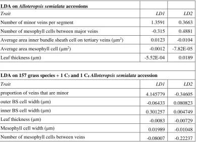
Number of minor veins per segment 1.3591 0.3663
Number of mesophyll cells between major veins -0.315 0.4881
Average area inner bundle sheath cell on tertiary veins (μm2) 0.0123 -0.0104
Average area mesophyll cell (μm2) -0.0012 -7.82E-05
Leaf thickness (μm) -5.52E-04 0.0189
LDA on 157 grass species + 1 C3 and 1 C4 Alloteropsis semialata accession
Trait LD1 LD2
proportion of veins that are minor 4.145779 -0.34605
outer BS cell width (μm) -0.06433 0.080823
inner BS cell width (μm) 0.301257 0.004749
Leaf thickness (μm) -0.0083 -0.00729
Mesophyll cell width (μm) 0.01989 -0.01048
Number of mesophyll cells between veins -0.08007 -0.22237
666
30
FIGURE LEGENDS
668
Figure 1. Schematic of leaf anatomy and photosynthetic pathway in C3, C3-C4, and C4 grasses. In C3
669
plants, CO2 assimilation via the Calvin-Benson cycle (solid black circle) and CO2 release via photorespiration
670
(dashed black circle) both occur in mesophyll cells (light green). C3 leaves consequently have larger areas
671
of mesophyll tissue than bundle sheath tissue, where no photosynthetic activity occurs. C3-C4 plants use an
672
intermediate physiology called C2 photosynthesis, where the Calvin cycle occurs in mesophyll cells, like in C3
673
plants. However, because glycine decarboxylase (GDC) is specifically localized to bundle sheath cells, the
674
photorespiratory cycle is split across these two cell types, creating a weak CO2-concentrating mechanism,
675
where CO2 is released in the bundle sheath and can be reassimilated via the Calvin cycle. C2 photosynthesis
676
therefore requires large areas of mesophyll for photosynthesis via an initial Calvin cycle, but also close contact
677
between mesophyll and bundle sheath cells for the photorespiratory CO2 pump. C4 plants have a strong CO2
678
concentrating mechanism whereby CO2 is biochemically shuttled from the mesophyll into the bundle sheath.
679
The high CO2 concentration in the bundle sheath largely avoids oxygenation and thus, photorespiration.
680
Photosynthesis via the C4 cycle therefore requires large areas of bundle sheath tissue, but less mesophyll, which
681
can be achieved via the insertion of minor veins. Dark blue, bundle sheath lacking chloroplasts; dark green,
682
bundle sheaths with chloroplasts; light green, mesophyll cells; yellow, extraxylary fibers/bundle sheath
683
extensions; grey, epidermal cells; light blue, veins; white, metaxylem.
684
685
Figure 2. Continuous variation in Alloteropsis semialata leaf anatomy, but distinct division among C3,
686
C3-C4, and C4 types. Ratios of mesophyll (M) to bundle sheath (BS) area of individual accessions of C3 (blue
687
circles), C3-C4 (green circles), and C4 (solid red circles) plants, ranked by M:BS value. n = 50. Lines delineating
688
M:BS ratios that distinguish C3 from C3-C4 (green) and C3-C4 from C4 (red) are shown. For C4 individuals,
689
M:BS ratios are also calculated in the absence of minor veins (open red circles).
690
691
Figure 3. Linear discriminant analysis of leaf anatomical traits. The first (LD1) and second (LD2)
692
dimensions of the LDA are plotted against each other with histograms of each dimension shown on the
693
opposing axis for (A) the LDA on C3, C3-C4, and C4 Alloteropsis semialata accessions and (B) the LDA on
694
157 additional C3, C4 inner sheath, and C4 outer sheath grass species. One C3 and one C4A. semialata accession
31
were included this larger LDA and denoted by solid blue and red circles, respectively. Loading plots are
696
overlaid via black arrows. M, mesophyll, IS, inner sheath; OS, outer sheath, nb.M, number of mesophyll cells
697
between veins.
698
699
Figure 4. Diversity of intraspecific anatomical components. Histograms of (A) vein density (i.e., the total
700
number of veins per segment) and (B) the number of major veins per segment in C4 (red; n=72) and non-C4
701
(grey, n= 69) accessions.Scatter plots show (C) the average number of mesophyll (M) cells between major
702
veins versus the average area of individual bundle sheath (BS) cells, with dot size scaled to the M:BS ratio,
703
and (D) BS cell area versus outer sheath cell area, with dot size scaled to the number of veins per segment.
704
Colors indicate photosynthetic type with C3 (blue; n=17), C3-C4 (green; n=6), and C4 (red; n=27).
705
32
FIGURES
707 708 709 710 711 712
FIGURE 1
713 C3 an at o m y C3 -C4 an at o m y C4 an at o m y C 3 p h o to sy n th es is C 2 p h o to sy n th es is C 4 p h o to sy n th es ismesophyll bundle sheath
CO2
O2
CO2
O2
CO2
CO
233 714
FIGURE 2
34 716
FIGURE 3
[image:34.595.84.484.64.723.2]35 718
719
720
FIGURE 4
721
[image:35.595.101.506.57.571.2]36
SUPPORTING INFORMATION
723
Supporting Information Materials 1. Ultrastructural characterization of C3, C3-C4 and C4 accessions
724
725
Dataset S1. Comprehensive anatomical measurements for all accessions
726
Dataset S2. Larger vein density dataset
727
Dataset S3. Field v controlled environment plasticity dataset
728
Dataset S4. Growth CO2 concentration plasticity dataset
729
730
Figure S1. Example leaf anatomy measurement methods
731
Figure S2. Relationships between anatomical traits
732
Figure S3. Comparison of extraxylary fibre area in C3, C3-C4 and C4 accessions
733
Figure S4. Relationships between inner BS cell size and overall BS areas
734
Figure S5. Comparison of bundle sheath size in African and non-African C4 accessions.
735
Figure S6. Plasticity for leaf anatomical components by photosynthetic type
736
Figure S7. Organelle abundance differs between photosynthetic types.
737
Figure S8. Immunodetection of GLDH
738
Figure S9. Immunodetection of Rubisco large subunit
739
740
Table S1. Details used to determine photosynthetic pathway for accessions
741
Table S2. Details of the accessions used in the plasticity dataset
742