AUTHOR QUERY FORM
Book: Applications of
Advanced Omics
Technologies:
From Genes to
Metabolites, 64
Chapter: 2
Please e-mail your responses
and any corrections to:
E-mail:
[email protected]
Dear Author,
Any queries or remarks that have arisen during the processing of your manuscript are listed below and are highlighted by flags in the proof. (AU indicates author queries; ED indicates editor queries; and TS/TY indicates typesetter queries.) Please check your proof carefully and answer all AU queries. Mark all corrections and query answers at the appropriate place in the proof (e.g., by using on-screen annotation in the PDF file http://www.elsevier.com/book-authors/science-and-technology-book-publishing/overview-of-the-publishing-process) or compile them in a separate list, and tick off below to indicate that you have answered the query.
Please return your input as instructed by the project manager.
Location in Chapter Query / remark
AU:1, page42 Please provide the page number for this
reference.
AU:2, page42 Please check the changes made in the page
number for this reference.
AU:3, page43 Please check the inserted volume and page
numbers for this reference.
AU:4, page23 Please provide the significance of“*”in
Chapter 2
C0010
microRNA Profiling: An
Overview of Current
Technologies and Applications
Sinead M. Smith*and David W. Murray{
*Department of Clinical Medicine, School of Pharmacy and Pharmaceutical Sciences, Trinity College Dublin, Dublin 2, Ireland
{Department of Physiology and Medical Physics, Royal College of Surgeons in Ireland,
Dublin 2, Ireland
Chapter Outline
1. Introduction 21 2. miRNA Biogenesis and
Nomenclature 23 3. Considerations for miRNA
Profiling 24
4. miRNA Extraction 26 5. qPCR Analysis of miRNAs 28 6. Microarray Profiling of
miRNAs 30
7. Small RNA-seq 31
8. In SilicomiRNA Analysis
Resources 33
9. miRNA Functional Analysis 35 10. Recent Developments in
the Application of miRNA Profiling to Cancer
Research 36
11. Conclusions 38
References 39
s0005
1 INTRODUCTION
ncRNAs may be subdivided according to their size. First, long ncRNAs (lncRNAs) range in size from a few hundred nucleotides (nt) to multiple kilobases in length represent the largest class of ncRNAs and account for much of the transcribed genome (6). In contrast to lncRNAs, small ncRNAs are less than 300 nt in length and include microRNAs (miRNAs), piwi-interacting RNAs, small-interfering RNAs, and small nuclear RNAs among others (7). The miRNAs (18–25 nt) are the most widely studied family of the small ncRNAs. Since their discovery in 1993 (8,9), the diversity and significance of this class of regulatory molecule has become increasingly appreciated. miRNAs posttranscriptionally decrease the expression of thousands of target genes by binding to specific messenger RNA (mRNA) tar-gets and promoting their degradation and/or inhibiting their translation (10–12). Although a relatively limited number of miRNAs (approximately 1000) have been identified in humans compared with the number of mRNAs and proteins (approximately 30,000), a single miRNA may regulate hundreds of mRNAs and thus has the potential to greatly impact gene-expression networks(10).
s0010
2 miRNA BIOGENESIS AND NOMENCLATURE
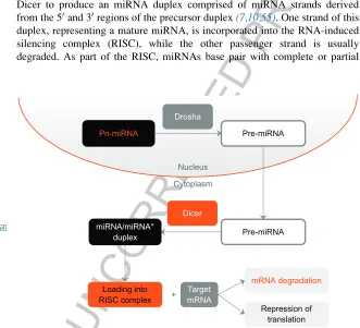
p0015 An understanding of the multistep processes involved in miRNA processing and biogenesis is required for the design and application of analytical techni-ques for miRNA detection and quantitation. Primary miRNA (pri-miRNA) transcripts are either transcribed by RNA polymerase II from independent genes or represent introns of protein-coding genes (54–56). A pri-miRNA transcript can contain a single miRNA or multiple miRNAs that are processed from the same transcript(25). The pri-miRNA folds into a hairpin structure, which acts as a substrate for cleavage by the endonuclease Drosha resulting in an approximately 70–100 nt long precursor miRNA (pre-miRNA) (Figure 1). Following the Drosha cleavage, the pre-miRNA is exported to the cytoplasm by Exportin, where it is further processed by the endonuclease Dicer to produce an miRNA duplex comprised of miRNA strands derived from the 50and 30regions of the precursor duplex(7,10,55). One strand of this duplex, representing a mature miRNA, is incorporated into the RNA-induced silencing complex (RISC), while the other passenger strand is usually degraded. As part of the RISC, miRNAs base pair with complete or partial
Pri-miRNA Pre-miRNA
Drosha
Nucleus
Cytoplasm
Pre-miRNA miRNA/miRNA*
duplex
Dicer
Loading into RISC complex
Target mRNA +
mRNA degradation
[image:4.504.81.412.265.566.2]Repression of translation
FIGURE 1
f0005 miRNA Biogenesis. In the nucleus, pri-miRNA transcripts are either transcribed by RNA polymerase II from independent genes or represent introns of protein-coding genes. Drosha mediates the processing of a pri-miRNA to a miRNA. Following Drosha cleavage, the pre-miRNA is transported to the cytoplasm. Once in the cytoplasm, Dicer processes the pre-pre-miRNA to produce a miRNA duplex derived from the 50and 30regions of the precursor. Mature miRNAs are loaded into the RISC complex to target specific mRNA molecules for degradation or inhibi-tion of translainhibi-tion.
complementarity to sequences in the 30untranslated region (30-UTR) of target mRNAs and induce mRNA translational repression or instability by deadeny-lation and degradation(55).
p0020 Different mature miRNA species can be produced from a single pre-miRNA molecule, as distinct pre-miRNAs are generated from the 30 and 50arms of the pre-miRNA duplex. In addition, a given mature miRNA may comprise a distribution of sizes centered around 22 nt rather than a discrete length. The variation in mature miRNA length is due to 30 or 50 end posttranscriptional modifications that include addition or deletion of nucleotides (10,57,58), which have been shown to affect miRNA stability and function. Addition or deletion of nucleotides to the 50 end of the mature miRNA can have signifi-cant effects on miRNA function by shifting the sequence of the seed region, which is the +2 to +8 nucleotide position from the 50 end of the miRNA that determines the mRNA target within the RISC (59). Profiling approaches therefore need to distinguish among pri-miRNAs, pre-miRNAs, and mature miRNAs and take into account mature miRNA sequence variations.
p0025 miRNAs are usually designated with a three letter species prefix (e.g., hsa-miR-X for Homo sapiens; mmu-miR-X for Mus musculus) and a number that designates the specific miRNA (56). Prefixes may also be added to reflect the stage of miRNA biogenesis of a given transcript (e.g., pri-mir-X for a pri-miRNA; pre-mir-X for a precursory miRNA) (10). Furthermore, suffixes are also added to indicate whether a mature miRNA arose from the 30(miR-X-3p) or 50arm (miR-X-5p) of the pre-miRNA hairpin, and a cap-ital “R” is used in mature miRNA nomenclature. miRNAs with related sequences belonging to the same family often have lower case letters follow-ing the name (e.g., miR-Xa; miR-Xb, miR-Xc) (56). Mature miRNAs with identical sequences that are derived from transcripts encoded by multiple loci are differentiated by a numerical suffix (e.g., miR-X-1, miR-X-2, miR-X-3) (10). miRNA nomenclature allows for distinction between the two single-stranded mature sequences that originate from the two strands of the double-stranded miRNA molecule, for example, miR-X and miR-X*, which indicate the major and minor strands, respectively (10,56). Since recent NGS analysis has shown that the relative quantities of miRNA strands can vary in different cell types and tissues, this form of strand differentiation may soon be replaced by solely describing the strands using the -5p and -3p suffixes.
s0015
3 CONSIDERATIONS FOR miRNA PROFILING
for use as a universal primer binding site or in selective enrichment processes. This has implications, given that miRNAs must be selectively detected in a background of diverse RNA molecules, including pri-miRNAs and pre-miRNAs that contain the same sequence as the mature miRNA (10,58). miRNAs are heterogeneous in their GC content, resulting in variations in their melting temperatures (Tm). miRNAs within a family can differ by a single nucleotide. Additionally, as mentioned inSection 2, posttranscriptional mod-ifications can result in mature miRNAs of different lengths centered around 22 nt(10,57,58).
s0020
4 miRNA EXTRACTION
[image:7.504.85.407.105.595.2]p0040 High-quality miRNA isolation is a fundamental step in the analytical process. miRNAs can be successfully extracted and purified from cell lines and a vari-ety of tissue specimens and biofluids, including blood products and urine
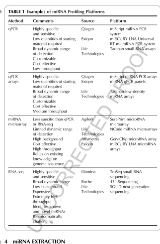
TABLE 1
t0005 Examples of miRNA Profiling Platforms
Method Comments Source Platform
qPCR Highly specific and sensitive
Low quantities of starting material required Broad dynamic range of detection Customizable Cost effective Low throughput Qiagen Exiqon Life Technologies
miScript miRNA PCR system
miRCURY LNA Universal RT microRNA PCR system Taqman small RNA assays
qPCR arrays
Highly specific
Low quantities of starting material required Broad dynamic range of detection Customizable Cost effective Medium throughput Qiagen Exiqon Life Technologies
miScript miRNA PCR arrays miRNA qPCR panels
Taqman low-density miRNA arrays
miRNA microarray
Less specific than qPCR or RNA-seq
Limited dynamic range of detection
High background Cost effective High throughput Relies on existing knowledge on genome sequence Agilent Life Technologies Affymetrix Exiqon SurePrint microRNA microarray
NCode miRNA microarrays
GeneChip microRNA array miRCURY LNA microRNA arrays
RNA-seq Highly specific and sensitive Broad dynamic range Low background Expensive Extremely high throughput Identifies known and novel miRNAs Bioinformatically challenging
Illumina TruSeq small RNA sequencing Roche 454 Sequencing Life
Technologies
(62,63). Cell lines and tissue samples yield far greater amounts of miRNA than plasma or urine, which contain high levels of endogenous ribonuclease (RNase) enzymes that degrade RNA molecules even in very small quantities (10,64). Generally speaking, the principles for miRNA isolation are similar to those for total RNA isolation, except that miRNA purification protocols are modified to retain or enrich the fraction of small RNA molecules less than 200 nt. As with extracting total RNA, extreme care must be taken to avoid degradation of the RNA sample by exposure to RNase enzymes(65). Samples for RNA processing should be harvested as rapidly as possible to protect against RNase activity and to prevent unwanted changes in gene expression. Samples should be frozen immediately at80C, placed in a suitable RNA stabilization solution such as RNAlater for tissue samples (Qiagen, Life Tech-nologies, and Sigma Aldrich) and RNAprotect Cell Reagent for cultured cells (Qiagen), or lysed and homogenized immediately upon harvesting in the pres-ence of RNase-denaturing buffers. RNA work should be performed in a desig-nated area of the laboratory using dedicated equipment and pipettors with nuclease-free aerosol-resistant tips. RNase decontamination solutions, such as RNase Zap (Life Technologies) or RNaseKiller (Qiagen) should be used to remove RNase contamination from bench surfaces and laboratory equip-ment. Aseptic technique is recommended when working with RNA samples, and powder-free latex or vinyl gloves should be worn and changed frequently to prevent the introduction of RNase contamination. Samples and reagents should be prepared on ice to inhibit RNase activity and sterile, disposable, certified RNase-free plasticware should be used.
p0045 A variety of isolation kits are commercially available that involve solvent-based miRNA extraction, followed by solid-phase purification on columns (62). The miRVana miRNA Isolation kit (Life Technologies) utilizes an organic extraction procedure followed by purification on a glass fiber filter to isolate total RNA ranging from 10 nt to multiple kilobases from cells and fresh or frozen tissue samples. The miRNeasy range of miRNA isolation kits (Qiagen) employ a combined phenol/guanidine-based lysis procedure, fol-lowed by a silica-membrane-based purification of total RNA from 18 nt upward. The miRNeasy mini Kit isolates total RNA including the small RNA fraction from cultured cells, laser capture microdissected specimens, and fresh or frozen tissue samples, while the miRNeasy Serum/Plasma Kit copurifies total RNA and small RNAs from serum/plasma or urine samples. The lysis buffer provided with these kits enables sample lysis, RNase activity inhibition, and cellular DNA and protein removal by organic extraction. p0050 Histopathology archives of FFPE tissue samples are valuable resources for
to miRNA isolation using the miRNeasy FFPE kit, the FFPE tissue specimens are treated with a solution provided with the kit that reverses formaldehyde modification as much as possible without further RNA degradation. Proteinase K is contained within the lysis buffer to release RNA from the tissue sections. A short high-temperature incubation step partially reverses formalin cross-linking of the released nucleic acids and is followed by a DNase treatment to eliminate genomic DNA, and subsequent ethanol precipitation and purification on columns. Using either the miRVana miRNA Isolation kit or the miRNeasy range, further enrichment of the small RNA species (<200 nt) may be per-formed to increase the sensitivity of downstream analyses.
p0055 The concentration of the RNA sample is determined by measuring the absorbance at 260 nm in a spectrophotometer. Because larger RNA species will dominate the absorbance measurement, it is not recommended to quantify purified small RNA fractions by spectrophotometry. RNA concentration and quality should be assessed using the total RNA preparation before enriching for the small RNA fraction. Based on the fact that a 40-mg/mL sample of pure RNA has an absorbance of 1 at 260 nm, the concentration of the sample can be calculated(65). The ratio of the absorbance at 260 and 280 nm is used to determine the quality of the preparation. A ratio of between 1.8 and 2.1 is indicative of high-quality RNA(65). Recently developed NanoDrop spectro-photometers (Thermo Scientific) are useful for the measurement of limited sample preparations as they accurately determine RNA concentration and quality in samples volumes as small as 0.5mL. The integrity and size distribu-tion of total RNA can be determined using the Bioanalyzer 2100 (Agilent Technologies) or by agarose gel electrophoresis and ethidium bromide stain-ing. In a high-quality RNA sample, the 28s ribosomal RNA (rRNA) and 18s rRNA bands should appear as sharp peaks on the Bioanalyzer or sharp bands on a gel. The desirable ratio of 28s rRNA to 18s rRNA is approximately 2:1. The Bioanalyser 2100 also provides an RNA Integrity Number that should be close to 10, especially if the RNA samples are to be considered for use in downstream applications such as microarrays or RNA-seq.
s0025
5 qPCR ANALYSIS OF miRNAs
certain considerations need to be taken into account for primer design for mature miRNA analysis by qPCR. One method involves tailing of miRNAs with a common sequence and performing RT using a universal primer. One such approach involves the addition of a poly(A) tail to the mature miRNA molecule, for example, Qiagen’s miScript PCR System(66). Total RNA con-taining the small RNA fraction is used as a starting material. Mature miRNAs are polyadenylated by poly(A) polymerase and reverse transcribed into cDNA in a first-strand cDNA synthesis reaction using oligo-dT primers containing a universal tag on the 50 end. miRNA abundance is then detected by SYBR green qPCR using a miRNA-specific forward primer and a universal reverse primer. As the oligo-dT primer detects all poly(A)-tailed miRNA molecules during the RT reaction, this assay enables detection of multiple miRNAs within a single cDNA preparation. The miRCURY LNA Universal RT micro-RNA PCR system (Exiqon) also employs a universal RT approach involving the addition of a 50 universal tagged poly(A) tail, followed by qPCR with miRNA-specific locked nucleic acid (LNA)-enhanced forward and reverse primers. LNA is a synthetic RNA/DNA analog characterized by increased thermostability of nucleic acid duplexes. As each incorporated LNA monomer increases the Tm of a given primer (58), this technology overcomes PCR specificity and sensitivity issues due to Tm variations resulting from miRNAs that are heterogeneous in their GC content.
p0065 Another approach for detecting miRNA levels by qPCR is through the use of stem-loop RT primers that are specific for the 30end of miRNAs (67). Taqman Small RNA assays (Life Technologies) are based on this approach. Following first-strand cDNA synthesis using a stem-loop primer, the RT cDNA product is quantified by Taqman PCR using an miRNA-sequence-specific forward primer, a specific reverse primer, and a Taqman probe. Because this approach utilizes a sequence-specific stem-loop RT primer, a separate RT reac-tion is required for quantitareac-tion of each miRNA of interest. However, there are advantages to using the stem-loop RT primer. By annealing a short RT priming sequence to the 30 end of the miRNA, better specificity for discriminating similar mature miRNA sequences is achieved (58,67,68). In addition, the double-stranded stem-loop structure inhibits hybridization of the RT primer to pre-miRNAs and other long RNAs. Further, the base stacking of the stem-loop enhances the stability of miRNA and DNA duplexes, improving the efficiency of the RT reaction. Lastly, the stem-loop structure when unfolded adds sequence downstream of the miRNA after RT, resulting in a longer RT template more suitable for TaqMan assay design(68).
starting total RNA. Following RT, the resulting cDNA templates are mixed with a PCR master mix and added to the microfluidic array cards or multiwell plates prior to standard qPCR amplification and analysis.
p0075 In order to control for variations in RNA input or RT efficiency when performing qPCR using either individual miRNA assays or PCR-based arrays, normalization to endogenous control genes is recommended. Inclusion of con-trols enables normalization of qPCR results for relative quantitation analysis. An ideal control demonstrates gene expression that is highly abundant and constant across cell types and tissues. Endogenous control assays may be cus-tom designed or are available predesigned commercially and include members of the snRNA (e.g., U6 snRNA(26,69)) and snoRNA (e.g., snoRNA202(70)) families. In addition to low-medium throughput miRNA profiling, qPCR is usually the method of choice for validating results from microarray analysis (Section 6) or RNA-seq profiling (Section 7) or miRNAs.
s0030
6 MICROARRAY PROFILING OF miRNAs
p0080 miRNA microarray technology is based on complementary hybridization of labeled miRNAs to an array of immobilized synthetic oligonucleotide probes on a gene chip. This technology allows for high-throughput analysis of miRNA expression and is cheaper to perform than RNA-seq. However, it is not as sensitive as qPCR or RNA-seq approaches. As it is based on current knowledge on genome sequence, miRNA microarray profiling does not per-mit the identification of novel previously uncharacterized miRNAs. Various commercial platforms are available for miRNA microarray profiling (Table 1). These platforms employ different approaches to label miRNAs for subsequent hybridization to the DNA probes on the microarray chip. One approach involves the enzymatically catalyzed ligation of a fluorophore-conjugated oligonucleotide to the 30end of the miRNA molecule using T4 RNA ligase prior to hybridization (SurePrint microRNA microarray; Agilent). Because Dicer processing during miRNA biogenesis results in the exposure of a 50phosphate on the mature miRNA that could lead to circulari-zation during the ligation reaction, procedures involving T4-mediated ligation are preceded with a dephosphorylation step to remove the 50 phosphate. p0085 Alternative microarray approaches are available that involve the 30tailing
As a result, it may be difficult to use a temperature that suits the annealing of all the probes to their target miRNAs during hybridization. To overcome the issue of diminished specificity, LNA probes may be used to increase the Tm value (miRCURY LNA microRNA arrays, Exiqon).
s0035
7 SMALL RNA-SEQ
p0090 Next-generation RNA-seq provides a more precise measurement of levels of transcripts and their isoforms than other methods and is not limited by varia-bility in Tm, small variations in miRNA sequences among families or post-transcriptional modifications. NGS technologies rely on a combination of procedures involving template preparation, sequencing, and imaging, followed by sequence alignment to a reference genome and data analysis(71). In the case of miRNAs, adapter sequences are firstly ligated to the 50 and 30 ends of the mature miRNA molecule, taking advantage of the natural structure common to most mature miRNA molecules of a 30-hydroxyl group and 50-phosphate that result from cleavage by the enzyme Dicer(56). Adapter ligation is followed by an RT reaction, a PCR amplification, and a purification step. Sample quality should be assessed on the Agilent Bioanalyzer 2100 at each step of the cDNA library preparation to ensure that the integrity of the samples is sufficient for sequencing. The high-quality cDNA template preparation is attached to a solid support surface and this immobilization of spatially separated template sites allows for thousands of sequencing reactions to be performed simultaneously in order to obtain millions of short-sequence reads(60,71). Both Illumina and Roche platforms (Table 1) sequence by synthesis. The Illumina platform sup-ports massively parallel sequencing using a reversible terminator-based cyclic method that comprises nucleotide incorporation, fluorescence imaging, and cleavage. A fluorescently labeled terminator is imaged as each dNTP is added and then removed to allow incorporation of the next base. Since all four revers-ible terminator-bound dNTPs are present during each sequencing cycle, natural competition minimizes incorporation bias. By contrast, Roche sequencing tech-nology involves pyrosequencing, where each nucleotide addition results in the activation of luciferase activity that produces light that is detected during the imaging process. As this technique does not require terminator removal at each step, increased sequencing speed can be achieved (56). The SoLID platform sequences by ligation. Primers hybridize to the adapter sequences on template DNA and a set of four fluorescently labeled probes compete for ligation to the sequencing primer.
quality control assessment are available from web sites including Galaxy(72), FASTX-Toolkit, and FastQC. These web sites provide useful tools that deter-mine whether there are any problems with the datasets before commencing with further downstream analysis. In addition, FASTX-Toolkit enables file preprocessing, including shortening of reads to remove barcodes or noise (FASTQ/A Trimmer), conversion of sequence reads to the reverse com-plement (FASTQ/A Reverse-Comcom-plement), or removal of adapter sequences
TABLE 2
t0010 Analysis Software Available for Small RNA-seq Data Analysis
Resource URL Tools
Galaxy https://usegalaxy.org/ Sequencing data quality assessment
FASTX-Toolkit http://hannonlab.cshl.edu/ fastx_toolkit/index.html
Sequencing data quality assessment and preprocessing, for example, adapter sequence removal, sequence trimming, and conversion of sequence reads to reverse complement
FastQC http://www.bioinformatics. babraham.ac.uk/projects/fastqc/
Sequence data quality assessment
BWA http://bio-bwa.sourceforge.net/ Alignment of sequence reads to a reference genome
Bowtie http://bowtie-bio.sourceforge.net/ index.shtml
Alignment of sequence reads to a reference genome
TopHat http://tophat.cbcb.umd.edu/ Alignment of sequence reads to a reference genome
UCSC genome browser
http://genome.ucsc.edu/cgi-bin/ hgGateway
Visualization of aligned sequence reads in the genome of interest
Integrative Genomics Viewer
http://www.broadinstitute.org/igv/ Visualization of aligned sequence reads in the genome of interest
Integration with clinical and phenotypic data
Gene Expression Omnibus
http://www.ncbi.nlm.nih.gov/geo/ Publicly available functional genomics repository
Cufflinks http://cufflinks.cbcb.umd.edu/ index.html
Differential expression analysis
Gobyweb http://campagnelab.org/software/ gobyweb/
from the sequence reads (FASTQ/A Clipper). Following quality control, sequence reads are then aligned to a reference genome to produce a genome-wide transcription map using bioinformatics tools such as BWA(73), Bowtie (74), or TopHat (75). Aligned files may be visualized using the UCSC genome browser (76,77) or the Integrative Genomics Viewer (IGV) (78). The UCSC genome browser provides a web-based tool for the display of any requested portion of the genome at the desired scale. Multiple annotation tracks of sequence data are publicly available on the web site. Alternatively, users may add their own NGS files to the browser as custom tracks for visu-alization, or custom tracks of interest that have been published in the literature and made available through resources such as the NCBI Gene Expression Omnibus database(79). The IGV is freely available for download to personal computers as a desktop application, and also enables visualization of genomic data from public sources as well as visualization of users own datasets. In addition, the IGV allows for integration of clinical and phenotypic data. Finally, relative quantitation of differential miRNA expression in terms of the number of sequence reads for a particular miRNA relative to the total reads in a sample can be determined using differential expression platforms such as Cufflinks(80)and Gobyweb (81). Following differential expression analysis, differentially regulated miRNAs identified by RNA-seq analysis are usually validated by qPCR approaches.
p0100 Bioinformatics analysis of the sequence reads identifies both known and novel miRNAs and provides precise sequence information, which is important to distinguish between miRNA molecules that differ by as little as a single nucleotide. Not all novel miRNAs identified by small RNA-seq are genuine functional mature miRNAs. A number of criteria have been suggested before a novel miRNA may be considered genuine and entered into the miRBase miRNA repository, including a sequence length of approximately 22 nt, conservation across species, a genomic origin that predicts a hairpin pre-miRNA sequence and identification in the data reads that correspond to both the -5p -3p arms of the pre-miRNA hairpin(82,83).
s0040
8
IN SILICO
miRNA ANALYSIS RESOURCES
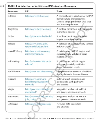
RNA-seq datasets deposited in the NCBI Gene Expression Omnibus reposi-tory(10,54,79). Target prediction tools involve mRNA target prediction based on an inputted miRNA sequence or accession number(85). Numerous target prediction resources are available. TargetScan identifies mRNA targets in many species by detecting the presence of conserved 7mer and 8mer sites that match the seed region of each miRNA(86). Similar toTargetScan, PicTar uses genomewide alignment to predict target mRNAs in different species(87). The false-positive discovery rates for PicTar and TargetScan have been reported to be between 20% and 30% with between 80% and 90% overlap in target predictions between the two for human targets(88).
TABLE 3
t0015 A Selection ofIn SilicomiRNA Analysis Resources
Resource URL Tools
miRBase http://www.mirbase.org A comprehensive database of miRNA nomenclature and sequences Links to target prediction web sites and RNA-seq datasets.
TargetScan http://www.targetscan.org/ A tool for prediction miRNA targets in multiple species
PicTar http://pictar.mdc-berlin.de/ A tool for prediction of miRNA targets in multiple species
Tarbase http://www.diana.pcbi. upenn.edu/tarbase.html
A database of experimentally verified miRNA targets
microRNA.org http://www.microrna.org/ microrna/home.do
A database of miRNA targets and experimentally validated tissue expression levels
miRNAMap http://mirnamap.mbc.nctu. edu.tw/
A database of miRNA targets and experimentally validated tissue expression levels
mir2Disease http://www.mir2disease.org/ Comprehensive resource of miRNA dysregulation in human disease
mirWalk http://www.umm.uni-heidelberg.de/apps/zmf/ mirwalk/
miRNA target prediction and associations with pathways and disease
Magia http://gencomp.bio.unipd.it/ magia/start/
Integrative analysis of miRNA and gene-expression networks
mirConnex http://mirconnx.csb.pitt.edu/ job_config
Integrative analysis of miRNA and gene-expression networks
p0110 Many online resources collect experimentally confirmed miRNA–mRNA interactions. One of these is Tarbase(89), which collects interactions reported in the literature from a variety of experimental techniques. Numerous other online resources exist that contain information on experimentally validated interactions and expression patterns including microRNA.org (90) and miRNAMap(91). Online tools can be used to study a disease state of interest including miR2disease(92), which is a curated database for microRNA dereg-ulation in human disease. miR2disease is updated frequently and contains information on miRNA–disease relationships, including expression patterns of specific miRNAs in specific disease states as well as the method used for detection of miRNA expression. miR2disease also contains information on experimentally verified miRNA target genes as well as links to other miRNA databases. miRwalk(93)is another comprehensive database of experimentally verified miRNA interactions associated with pathways and diseases. Informa-tion is provided on the chromosomal locaInforma-tion of the miRNA as well as links to the original published study. Integrative analysis of miRNA expression profiles with large-scale mRNA expression datasets may also be performed usingin silicotools such as MAGIA(94)and mirConnX(95), which enable target prediction, integrated analysis of expression profiles, posttranscriptional regulatory network browsing, functional annotation, and enrichment analysis in combination with the network visualization tool Cytoscape(96).
s0045
9 miRNA FUNCTIONAL ANALYSIS
in which the 30UTR of the target mRNA has been subcloned, can be tested. Decreased target protein expression may be validated by Western blot analy-sis. Such experiments provide insight into the functional consequences of altered miRNA expression.
s0050
10 RECENT DEVELOPMENTS IN THE APPLICATION
OF miRNA PROFILING TO CANCER RESEARCH
p0120 The role of miRNAs in cancer development and progression has been widely reported (11,97–99). Generally speaking, miRNA profiling and functional studies in cancer research can be divided into (i) those that focus on decipher-ing the mechanistic role of miRNA dysregulation in the tumor phenotype with the ultimate aim of developing therapeutic strategies to target these mechan-isms and (ii) those that involve biomarker discovery to identify miRNA pro-files associated with disease type or predicted response to therapy. Various forms of the miRNA profiling methods outlined above have been used to describe miRNAs whose expression is altered in cancer cell lines, tumor tis-sue, and plasma or serum samples from cancer patients. Using a combination of qPCR and miRNA microarrays, numerous studies have delineated miRNAs that are differentially expressed in cancer of the breast (34,37), lung (42,43,46,47), pancreas (100), liver (40,41), and in B-cell lymphoma(101). Additionally, dysregulated miRNAs have been associated with treatment outcome. By combining miRNA microarray analysis with stem-loop qPCR validation, Yang et al.described significantly decreased let-7i expression in chemotherapy-resistant epithelial ovarian cancer (102). Tomimura et al.
showed that increased miR-21 expression in hepatocellular carcinoma leads to increased resistance to the antitumor effect of combination therapy involv-ing interferon-aand 5-fluorouracil(40). Increased miR-21 expression has also been reported in colon adenocarcinoma and is associated with poor survival and therapeutic outcome(39).
differences in miRNA expression in lung cancer tissue compared with adja-cent normal tissue in mouse models of lung carcinoma. miR-31 was shown to be significantly increased in the murine lung cancer tissue and the finding was confirmed using human tissue samples. Functional analysis indi-cated that miR-31 inhibition decreased lung cancer cell growth and tumorige-nicity. Using the bioinformatics resources TargetScan, Pictar, and miRanda, Liuet al.also identified the tumor suppressor genes, large tumor suppressor 2 (LATS2) and the PP2A regulatory subunit B alpha isoform (PPP2R2A) as targets of miR-31, and validated these targets using 30-UTR luciferase-binding assays and by demonstrating inverse expression of miR-31 and LATS2 or PPP2R2A in mouse and human lung cancers(43). In another study, Volinia
et al. used microarrays to identify dysregulated miRNAs in samples from six solid tumor types, namely lung, breast, stomach, prostate, colon, and pancreatic (35). They defined a solid tumor signature of overexpression of miRNAs, including miR-17-5p, miR-20a, miR-21, miR-92, miR106a, and miR-155. Using TargetScan, the retinoblastoma 1 tumor suppressor gene and transforming growth factor, beta receptor II were identified as predicted targets. The mRNA targets were confirmed experimentally using 30-UTR luciferase assays and by monitoring target protein expression to support a functional role for the role of miRNAs in solid cancer pathogenesis. Using a similar approach involving combined miRNA profiling, target identi-fication, and functional analysis, decreased expression of the miRNA let-7a has been suggested to contribute to the development of prostate cancer (103). More recently, RNA-seq analysis has shed further light on how miRNA expression contributes to cancer progression, in particular, in the lung. Using a mouse model of lung adenocarcinoma, Valdmaniset al.utilized small RNA-seq to show increased expression of a cluster of miRNAs at the Dlk1-Dio3 locus, some of which are known to be involved in key cancer-associated pathways (46). Perdomo et al. identified an association of miR-4423 with lung cancer in primates by utilizing a small RNA-seq approach(47).
levels that endured beyond the end of active was reported (107). Although not yet in clinical trials, select miRNAs have emerged as potential clinical targets for cancer treatment. miR-34 has a well-defined role as a tumor sup-pressor and miRNA replacement therapy by means of miRNA mimics has shown promise in animal studies(108). Like all therapeutics, the major bar-riers to targeting miRNAs in humans are ensuring high bioavailability, achieving target specificity, and reducing toxicity. The use of nanoparticles to target miRNA is a potential option for the treatment of cancers. Recently, an miR-155-targeting nanoparticle containing miR-155 antisense nucleotides, was shown to reduce the growth of B-cell lymphoma in a murine model of disease (109), suggesting a promising therapeutic option for lymphoma and leukemia.
p0135 In relation to biomarker discovery, many studies have focused on identify-ing cancer-associated biomarkers in plasma or serum as a noninvasive means for diagnosis or predictive of response to therapy. A good example of this is miR-22, the plasma levels of which were used to distinguish lung cancer patients from healthy individuals by qPCR, thereby supporting its potential role as a diagnostic biomarker that could be further evaluated in clinical trials (44). In prostate cancer, circulating levels of miR-375 and miR-141 have been reported to correlate with disease progression and lymph node metastasis(53). miR-1, mir-92a, miR-133a, and miR-133b were identified as the most impor-tant diagnostic markers for breast cancer detection in a recent study by Chan
et al.(51). In terms of utilizing miRNAs as predictive biomarkers for response to therapy, qPCR on circulating levels of miR-125b are predictive of the chemotherapy response in breast cancer(36). The translation of these findings into either noninvasive rapid diagnostic tools or as viable therapeutic targets depends on additional carefully designed and executed profiling studies.
s0055
11 CONCLUSIONS
previously unidentified miRNAs. As the cost of these techniques is falling, RNA-seq will become a more accessible platform for rapid and large-scale miRNA profiling, offering the potential to provide invaluable insight into the role of these key regulatory molecules a variety of settings. It is clear that the elucidation of the biological, pathological, and clinical roles of miRNA regulation, expression, and functional properties has greatly contributed to our understanding of pathogenesis. Continued investigation involving both miRNA discovery as well as miRNA targeting will be of great importance to the translation of miRNA biology into clinical applications for diagnosis of disease and novel therapeutics.
REFERENCES
1. Derrien, T.; Johnson, R.; Bussotti, G.; Tanzer, A.; Djebali, S.; Tilgner, H.; Guernec, G.; Martin, D.; Merkel, A.; Knowles, D. G.; Lagarde, J.; Veeravalli, L.; Ruan, X.; Ruan, Y.; Lassmann, T.; Carninci, P.; Brown, J. B.; Lipovich, L.; Gonzalez, J. M.; Thomas, M.; Davis, C. A.; Shiekhattar, R.; Gingeras, T. R.; Hubbard, T. J.; Notredame, C.; Harrow, J.; Guigo, R.Genome Res.2012,22, 1775–1789.
2. Djebali, S.; Davis, C. A.; Merkel, A.; Dobin, A.; Lassmann, T.; Mortazavi, A.; Tanzer, A.; Lagarde, J.; Lin, W.; Schlesinger, F.; Xue, C.; Marinov, G. K.; Khatun, J.; Williams, B. A.; Zaleski, C.; Rozowsky, J.; Roder, M.; Kokocinski, F.; Abdelhamid, R. F.; Alioto, T.; Antoshechkin, I.; Baer, M. T.; Bar, N. S.; Batut, P.; Bell, K.; Bell, I.; Chakrabortty, S.; Chen, X.; Chrast, J.; Curado, J.; Derrien, T.; Drenkow, J.; Dumais, E.; Dumais, J.; Duttagupta, R.; Falconnet, E.; Fastuca, M.; Fejes-Toth, K.; Ferreira, P.; Foissac, S.; Fullwood, M. J.; Gao, H.; Gonzalez, D.; Gordon, A.; Gunawardena, H.; Howald, C.; Jha, S.; Johnson, R.; Kapranov, P.; King, B.; Kingswood, C.; Luo, O. J.; Park, E.; Persaud, K.; Preall, J. B.; Ribeca, P.; Risk, B.; Robyr, D.; Sammeth, M.; Schaffer, L.; See, L. H.; Shahab, A.; Skancke, J.; Suzuki, A. M.; Takahashi, H.; Tilgner, H.; Trout, D.; Walters, N.; Wang, H.; Wrobel, J.; Yu, Y.; Ruan, X.; Hayashizaki, Y.; Harrow, J.; Gerstein, M.; Hubbard, T.; Reymond, A.; Antonarakis, S. E.; Hannon, G.; Giddings, M. C.; Ruan, Y.; Wold, B.; Carninci, P.; Guigo, R.; Gingeras, T. R.Nature2012,489, 101–108.
Liu, J.; Liuni, S.; McWilliam, S.; Madan Babu, M.; Madera, M.; Marchionni, L.; Matsuda, H.; Matsuzawa, S.; Miki, H.; Mignone, F.; Miyake, S.; Morris, K.; Mottagui-Tabar, S.; Mulder, N.; Nakano, N.; Nakauchi, H.; Ng, P.; Nilsson, R.; Nishiguchi, S.; Nishikawa, S.; Nori, F.; Ohara, O.; Okazaki, Y.; Orlando, V.; Pang, K. C.; Pavan, W. J.; Pavesi, G.; Pesole, G.; Petrovsky, N.; Piazza, S.; Reed, J.; Reid, J. F.; Ring, B. Z.; Ringwald, M.; Rost, B.; Ruan, Y.; Salzberg, S. L.; Sandelin, A.; Schneider, C.; Schonbach, C.; Sekiguchi, K.; Semple, C. A.; Seno, S.; Sessa, L.; Sheng, Y.; Shibata, Y.; Shimada, H.; Shimada, K.; Silva, D.; Sinclair, B.; Sperling, S.; Stupka, E.; Sugiura, K.; Sultana, R.; Takenaka, Y.; Taki, K.; Tammoja, K.; Tan, S. L.; Tang, S.; Taylor, M. S.; Tegner, J.; Teichmann, S. A.; Ueda, H. R.; van Nimwegen, E.; Verardo, R.; Wei, C. L.; Yagi, K.; Yamanishi, H.; Zabarovsky, E.; Zhu, S.; Zimmer, A.; Hide, W.; Bult, C.; Grimmond, S. M.; Teasdale, R. D.; Liu, E. T.; Brusic, V.; Quackenbush, J.; Wahlestedt, C.; Mattick, J. S.; Hume, D. A.; Kai, C.; Sasaki, D.; Tomaru, Y.; Fukuda, S.; Kanamori-Katayama, M.; Suzuki, M.; Aoki, J.; Arakawa, T.; Iida, J.; Imamura, K.; Itoh, M.; Kato, T.; Kawaji, H.; Kawagashira, N.; Kawashima, T.; Kojima, M.; Kondo, S.; Konno, H.; Nakano, K.; Ninomiya, N.; Nishio, T.; Okada, M.; Plessy, C.; Shibata, K.; Shiraki, T.; Suzuki, S.; Tagami, M.; Waki, K.; Watahiki, A.; Okamura-Oho, Y.; Suzuki, H.; Kawai, J.; Hayashizaki, Y.; The FANTOM Consortium, RIKEN Genome Exploration Research Group and Genome Science Group.Science2005,309, 1559–1563.
5. Consortium, E. P.; Bernstein, B. E.; Birney, E.; Dunham, I.; Green, E. D.; Gunter, C.; Snyder, M.Nature2012,489, 57–74.
6. Wahlestedt, C.Nat. Rev. Drug Discov.2013,12, 433–446.
7. Pagani, M.; Rossetti, G.; Panzeri, I.; de Candia, P.; Bonnal, R. J.; Rossi, R. L.; Geginat, J.; Abrignani, S.Immunol. Rev.2013,253, 82–96.
8. Lee, R. C.; Feinbaum, R. L.; Ambros, V.Cell1993,75, 843–854. 9. Wightman, B.; Ha, I.; Ruvkun, G.Cell1993,75, 855–862.
10. Pritchard, C. C.; Cheng, H. H.; Tewari, M.Nat. Rev. Genet.2012,13, 358–369.
11. Lu, J.; Getz, G.; Miska, E. A.; Alvarez-Saavedra, E.; Lamb, J.; Peck, D.; Sweet-Cordero, A.; Ebert, B. L.; Mak, R. H.; Ferrando, A. A.; Downing, J. R.; Jacks, T.; Horvitz, H. R.; Golub, T. R.Nature2005,435, 834–838.
12. Djuranovic, S.; Nahvi, A.; Green, R.Science2012,336, 237–240.
13. Lim, L. P.; Glasner, M. E.; Yekta, S.; Burge, C. B.; Bartel, D. P.Science2003,299, 1540. 14. Reinhart, B. J.; Slack, F. J.; Basson, M.; Pasquinelli, A. E.; Bettinger, J. C.; Rougvie, A. E.;
Horvitz, H. R.; Ruvkun, G.Nature2000,403, 901–906. 15. Chen, X.Science2004,303, 2022–2025.
16. Houbaviy, H. B.; Murray, M. F.; Sharp, P. A.Dev. Cell2003,5, 351–358.
17. Brennecke, J.; Hipfner, D. R.; Stark, A.; Russell, R. B.; Cohen, S. M.Cell2003,113, 25–36. 18. Xu, P.; Vernooy, S. Y.; Guo, M.; Hay, B. A.Curr. Biol.2003,13, 790–795.
19. O’Carroll, D.; Mecklenbrauker, I.; Das, P. P.; Santana, A.; Koenig, U.; Enright, A. J.; Miska, E. A.; Tarakhovsky, A.Genes Dev.2007,21, 1999–2004.
20. Cobb, B. S.; Nesterova, T. B.; Thompson, E.; Hertweck, A.; O’Connor, E.; Godwin, J.; Wilson, C. B.; Brockdorff, N.; Fisher, A. G.; Smale, S. T.; Merkenschlager, M. J. Exp. Med.2005,201, 1367–1373.
21. Li, Q. J.; Chau, J.; Ebert, P. J.; Sylvester, G.; Min, H.; Liu, G.; Braich, R.; Manoharan, M.; Soutschek, J.; Skare, P.; Klein, L. O.; Davis, M. M.; Chen, C. Z.Cell2007,129, 147–161. 22. Rodriguez, A.; Vigorito, E.; Clare, S.; Warren, M. V.; Couttet, P.; Soond, D. R.; van
23. Thai, T. H.; Calado, D. P.; Casola, S.; Ansel, K. M.; Xiao, C.; Xue, Y.; Murphy, A.; Frendewey, D.; Valenzuela, D.; Kutok, J. L.; Schmidt-Supprian, M.; Rajewsky, N.; Yancopoulos, G.; Rao, A.; Rajewsky, K.Science2007,316, 604–608.
24. O’Neill, L. A.; Sheedy, F. J.; McCoy, C. E.Nat. Rev. Immunol.2011,11, 163–175. 25. O’Connell, R. M.; Rao, D. S.; Baltimore, D.Annu. Rev. Immunol.2012,30, 295–312. 26. O’Connell, R. M.; Taganov, K. D.; Boldin, M. P.; Cheng, G.; Baltimore, D.Proc. Natl. Acad.
Sci. U. S. A.2007,104, 1604–1609.
27. Taganov, K. D.; Boldin, M. P.; Chang, K. J.; Baltimore, D.Proc. Natl. Acad. Sci. U. S. A. 2006,103, 12481–12486.
28. Junker, A.FEBS Lett.2011,585, 3738–3746.
29. Kurowska-Stolarska, M.; Alivernini, S.; Ballantine, L. E.; Asquith, D. L.; Millar, N. L.; Gilchrist, D. S.; Reilly, J.; Ierna, M.; Fraser, A. R.; Stolarski, B.; McSharry, C.; Hueber, A. J.; Baxter, D.; Hunter, J.; Gay, S.; Liew, F. Y.; McInnes, I. B.Proc. Natl. Acad. Sci. U. S. A.2011,108, 11193–11198.
30. Nakasa, T.; Miyaki, S.; Okubo, A.; Hashimoto, M.; Nishida, K.; Ochi, M.; Asahara, H.
Arthritis Rheum.2008,58, 1284–1292.
31. Luo, X.; Yang, W.; Ye, D. Q.; Cui, H.; Zhang, Y.; Hirankarn, N.; Qian, X.; Tang, Y.; Lau, Y. L.; de Vries, N.; Tak, P. P.; Tsao, B. P.; Shen, N.PLoS Genet.2011,7, e1002128. 32. Dai, R.; Zhang, Y.; Khan, D.; Heid, B.; Caudell, D.; Crasta, O.; Ahmed, S. A.PLoS One
2010,5, e14302.
33. Calin, G. A.; Dumitru, C. D.; Shimizu, M.; Bichi, R.; Zupo, S.; Noch, E.; Aldler, H.; Rattan, S.; Keating, M.; Rai, K.; Rassenti, L.; Kipps, T.; Negrini, M.; Bullrich, F.; Croce, C. M.Proc. Natl. Acad. Sci. U. S. A.2002,99, 15524–15529.
34. Iorio, M. V.; Ferracin, M.; Liu, C. G.; Veronese, A.; Spizzo, R.; Sabbioni, S.; Magri, E.; Pedriali, M.; Fabbri, M.; Campiglio, M.; Menard, S.; Palazzo, J. P.; Rosenberg, A.; Musiani, P.; Volinia, S.; Nenci, I.; Calin, G. A.; Querzoli, P.; Negrini, M.; Croce, C. M.
Cancer Res.2005,65, 7065–7070.
35. Volinia, S.; Calin, G. A.; Liu, C. G.; Ambs, S.; Cimmino, A.; Petrocca, F.; Visone, R.; Iorio, M.; Roldo, C.; Ferracin, M.; Prueitt, R. L.; Yanaihara, N.; Lanza, G.; Scarpa, A.; Vecchione, A.; Negrini, M.; Harris, C. C.; Croce, C. M.Proc. Natl. Acad. Sci. U. S. A. 2006,103, 2257–2261.
36. Wang, H.; Tan, G.; Dong, L.; Cheng, L.; Li, K.; Wang, Z.; Luo, H.PLoS One2012,7, e34210. 37. Tavazoie, S. F.; Alarcon, C.; Oskarsson, T.; Padua, D.; Wang, Q.; Bos, P. D.; Gerald, W. L.;
Massague, J.Nature2008,451, 147–152.
38. Michael, M. Z.; O’Connor, S. M.; van Holst Pellekaan, N. G.; Young, G. P.; James, R. J.Mol. Cancer Res.2003,1, 882–891.
39. Schetter, A. J.; Leung, S. Y.; Sohn, J. J.; Zanetti, K. A.; Bowman, E. D.; Yanaihara, N.; Yuen, S. T.; Chan, T. L.; Kwong, D. L.; Au, G. K.; Liu, C. G.; Calin, G. A.; Croce, C. M.; Harris, C. C.JAMA2008,299, 425–436.
40. Tomimaru, Y.; Eguchi, H.; Nagano, H.; Wada, H.; Tomokuni, A.; Kobayashi, S.; Marubashi, S.; Takeda, Y.; Tanemura, M.; Umeshita, K.; Doki, Y.; Mori, M.Br. J. Cancer 2010,103, 1617–1626.
41. Ji, J.; Yamashita, T.; Budhu, A.; Forgues, M.; Jia, H. L.; Li, C.; Deng, C.; Wauthier, E.; Reid, L. M.; Ye, Q. H.; Qin, L. X.; Yang, W.; Wang, H. Y.; Tang, Z. Y.; Croce, C. M.; Wang, X. W.Hepatology2009,50, 472–480.
43. Liu, X.; Sempere, L. F.; Ouyang, H.; Memoli, V. A.; Andrew, A. S.; Luo, Y.; Demidenko, E.; Korc, M.; Shi, W.; Preis, M.; Dragnev, K. H.; Li, H.; Direnzo, J.; Bak, M.; Freemantle, S. J.; Kauppinen, S.; Dmitrovsky, E.J. Clin. Invest.2010,120, 1298–1309.
44. Shen, J.; Liu, Z.; Todd, N. W.; Zhang, H.; Liao, J.; Yu, L.; Guarnera, M. A.; Li, R.; Cai, L.; Zhan, M.; Jiang, F.BMC Cancer2011,11, 374.
45. Rani, S.; Gately, K.; Crown, J.; O’Byrne, K.; O’Driscoll, L.Cancer Biol. Ther.2013,14. Au1 46. Valdmanis, P. N.; Roy-Chaudhuri, B.; Kim, H. K.; Sayles, L. C.; Zheng, Y.; Chuang, C. H.;
Caswell, D. R.; Chu, K.; Zhang, Y.; Winslow, M. M.; Sweet-Cordero, E. A.; Kay, M. A.
Oncogene2013.
47. Perdomo, C.; Campbell, J. D.; Gerrein, J.; Tellez, C. S.; Garrison, C. B.; Walser, T. C.; Drizik, E.; Si, H.; Gower, A. C.; Vick, J.; Anderlind, C.; Jackson, G. R.; Mankus, C.; Schembri, F.; O’Hara, C.; Gomperts, B. N.; Dubinett, S. M.; Hayden, P.; Belinsky, S. A.; Lenburg, M. E.; Spira, A.Proc. Natl. Acad. Sci. U. S. A.2013,110, 18946–18951. 48. Nicoloso, M. S.; Spizzo, R.; Shimizu, M.; Rossi, S.; Calin, G. A.Nat. Rev. Cancer2009,9,
293–302.
49. Ryan, B. M.; Robles, A. I.; Harris, C. C.Nat. Rev. Cancer2010,10, 389–402.
50. Xie, Y.; Todd, N. W.; Liu, Z.; Zhan, M.; Fang, H.; Peng, H.; Alattar, M.; Deepak, J.; Stass, S. A.; Jiang, F.Lung Cancer2010,67, 170–176.
51. Chan, M.; Liaw, C. S.; Ji, S. M.; Tan, H. H.; Wong, C. Y.; Thike, A. A.; Tan, P. H.; Ho, G. H.; Lee, A. S.Clin. Cancer Res.2013,19, 4477–4487.
52. Corcoran, C.; Friel, A. M.; Duffy, M. J.; Crown, J.; O’Driscoll, L.Clin. Chem.2011,57, 18–32. 53. Mahn, R.; Heukamp, L. C.; Rogenhofer, S.; von Ruecker, A.; Muller, S. C.; Ellinger, J.Urology Au2
2011,77, 1265.e9–1265.e16.
54. Ghildiyal, M.; Zamore, P. D.Nat. Rev. Genet.2009,10, 94–108. 55. Krol, J.; Loedige, I.; Filipowicz, W.Nat. Rev. Genet.2010,11, 597–610.
56. Guerau-de-Arellano, M.; Alder, H.; Ozer, H. G.; Lovett-Racke, A.; Racke, M. K.
J. Neuroimmunol.2012,248, 32–39.
57. Git, A.; Dvinge, H.; Salmon-Divon, M.; Osborne, M.; Kutter, C.; Hadfield, J.; Bertone, P.; Caldas, C.RNA2010,16, 991–1006.
58. Benes, V.; Castoldi, M.Methods2010,50, 244–249.
59. Tan Gana, N. H.; Victoriano, A. F.; Okamoto, T.Genes Cells2012,17, 11–27. 60. Wang, Z.; Gerstein, M.; Snyder, M.Nat. Rev. Genet.2009,10, 57–63. 61. Ozsolak, F.; Milos, P. M.Nat. Rev. Genet.2011,12, 87–98.
62. Accerbi, M.; Schmidt, S. A.; De Paoli, E.; Park, S.; Jeong, D. H.; Green, P. J.Methods Mol. Biol.2010,592, 31–50.
63. Doleshal, M.; Magotra, A. A.; Choudhury, B.; Cannon, B. D.; Labourier, E.; Szafranska, A. E.J. Mol. Diagn.2008,10, 203–211.
64. Jensen, S. G.; Lamy, P.; Rasmussen, M. H.; Ostenfeld, M. S.; Dyrskjot, L.; Orntoft, T. F.; Andersen, C. L.BMC Genomics2011,12, 435.
65. Smith, S. M.; Murray, D. W.Methods Mol. Biol.2012,823, 119–138. 66. Ach, R. A.; Wang, H.; Curry, B.BMC Biotechnol.2008,8, 69.
67. Chen, C.; Ridzon, D. A.; Broomer, A. J.; Zhou, Z.; Lee, D. H.; Nguyen, J. T.; Barbisin, M.; Xu, N. L.; Mahuvakar, V. R.; Andersen, M. R.; Lao, K. Q.; Livak, K. J.; Guegler, K. J.
Nucleic Acids Res.2005,33, e179.
68. Schmittgen, T. D.; Lee, E. J.; Jiang, J.; Sarkar, A.; Yang, L.; Elton, T. S.; Chen, C.Methods 2008,44, 31–38.
69. Cheng, Y.; Kuang, W.; Hao, Y.; Zhang, D.; Lei, M.; Du, L.; Jiao, H.; Zhang, X.; Wang, F.
70. Yang, L.; Boldin, M. P.; Yu, Y.; Liu, C. S.; Ea, C. K.; Ramakrishnan, P.; Taganov, K. D.; Zhao, J. L.; Baltimore, D.J. Exp. Med.2012,209, 1655–1670.
71. Metzker, M. L.Nat. Rev. Genet.2010,11, 31–46.
72. Goecks, J.; Nekrutenko, A.; Taylor, J.; Galaxy, T.Genome Biol.2010,11, R86. 73. Li, H.; Durbin, R.Bioinformatics2009,25, 1754–1760.
74. Langmead, B.; Trapnell, C.; Pop, M.; Salzberg, S. L.Genome Biol.2009,10, R25. 75. Kim, D.; Salzberg, S. L.Genome Biol.2011,12, R72.
76. Kent, W. J.; Sugnet, C. W.; Furey, T. S.; Roskin, K. M.; Pringle, T. H.; Zahler, A. M.; Haussler, D.Genome Res.2002,12, 996–1006.
77. Karolchik, D.; Barber, G. P.; Casper, J.; Clawson, H.; Cline, M. S.; Diekhans, M.; Au3 Dreszer, T. R.; Fujita, P. A.; Guruvadoo, L.; Haeussler, M.; Harte, R. A.; Heitner, S.; Hinrichs, A. S.; Learned, K.; Lee, B. T.; Li, C. H.; Raney, B. J.; Rhead, B.; Rosenbloom, K. R.; Sloan, C. A.; Speir, M. L.; Zweig, A. S.; Haussler, D.; Kuhn, R. M.; Kent, W. J.Nucleic Acids Res.2013,1, D764–D770.
78. Thorvaldsdottir, H.; Robinson, J. T.; Mesirov, J. P.Brief. Bioinform.2013,14, 178–192. 79. Barrett, T.; Wilhite, S. E.; Ledoux, P.; Evangelista, C.; Kim, I. F.; Tomashevsky, M.;
Marshall, K. A.; Phillippy, K. H.; Sherman, P. M.; Holko, M.; Yefanov, A.; Lee, H.; Zhang, N.; Robertson, C. L.; Serova, N.; Davis, S.; Soboleva, A.Nucleic Acids Res.2013,
41, D991–D995.
80. Trapnell, C.; Williams, B. A.; Pertea, G.; Mortazavi, A.; Kwan, G.; van Baren, M. J.; Salzberg, S. L.; Wold, B. J.; Pachter, L.Nat. Biotechnol.2010,28, 511–515.
81. Dorff, K. C.; Chambwe, N.; Zeno, Z.; Simi, M.; Shaknovich, R.; Campagne, F.PLoS One 2013,8, e69666.
82. Ambros, V.; Bartel, B.; Bartel, D. P.; Burge, C. B.; Carrington, J. C.; Chen, X.; Dreyfuss, G.; Eddy, S. R.; Griffiths-Jones, S.; Marshall, M.; Matzke, M.; Ruvkun, G.; Tuschl, T.RNA2003,
9, 277–279.
83. Chiang, H. R.; Schoenfeld, L. W.; Ruby, J. G.; Auyeung, V. C.; Spies, N.; Baek, D.; Johnston, W. K.; Russ, C.; Luo, S.; Babiarz, J. E.; Blelloch, R.; Schroth, G. P.; Nusbaum, C.; Bartel, D. P.Genes Dev.2010,24, 992–1009.
84. Kozomara, A.; Griffiths-Jones, S.Nucleic Acids Res.2011,39, D152–D157. 85. Watanabe, Y.; Tomita, M.; Kanai, A.Methods Enzymol.2007,427, 65–86. 86. Lewis, B. P.; Burge, C. B.; Bartel, D. P.Cell2005,120, 15–20.
87. Krek, A.; Grun, D.; Poy, M. N.; Wolf, R.; Rosenberg, L.; Epstein, E. J.; MacMenamin, P.; da Piedade, I.; Gunsalus, K. C.; Stoffel, M.; Rajewsky, N.Nat. Genet.2005,37, 495–500. 88. Rajewsky, N.Nat. Genet.2006,38 Suppl, S8–S13.
89. Vergoulis, T.; Vlachos, I. S.; Alexiou, P.; Georgakilas, G.; Maragkakis, M.; Reczko, M.; Gerangelos, S.; Koziris, N.; Dalamagas, T.; Hatzigeorgiou, A. G.Nucleic Acids Res.2012,
40, D222–D229.
90. Betel, D.; Wilson, M.; Gabow, A.; Marks, D. S.; Sander, C.Nucleic Acids Res.2008,36, D149–D153.
91. Hsu, S. D.; Chu, C. H.; Tsou, A. P.; Chen, S. J.; Chen, H. C.; Hsu, P. W.; Wong, Y. H.; Chen, Y. H.; Chen, G. H.; Huang, H. D.Nucleic Acids Res.2008,36, D165–D169. 92. Jiang, Q.; Wang, Y.; Hao, Y.; Juan, L.; Teng, M.; Zhang, X.; Li, M.; Wang, G.; Liu, Y.
Nucleic Acids Res.2009,37, D98–D104.
93. Dweep, H.; Sticht, C.; Pandey, P.; Gretz, N.J. Biomed. Inform.2011,44, 839–847. 94. Sales, G.; Coppe, A.; Bisognin, A.; Biasiolo, M.; Bortoluzzi, S.; Romualdi, C.Nucleic Acids
Res.2010,38, W352–W359.
96. Smoot, M. E.; Ono, K.; Ruscheinski, J.; Wang, P. L.; Ideker, T.Bioinformatics2011,27, 431–432.
97. Garzon, R.; Fabbri, M.; Cimmino, A.; Calin, G. A.; Croce, C. M.Trends Mol. Med.2006,
12, 580–587.
98. Calin, G. A.; Croce, C. M.Nat. Rev. Cancer2006,6, 857–866. 99. Croce, C. M.Nat. Rev. Genet.2009,10, 704–714.
100. Lee, E. J.; Gusev, Y.; Jiang, J.; Nuovo, G. J.; Lerner, M. R.; Frankel, W. L.; Morgan, D. L.; Postier, R. G.; Brackett, D. J.; Schmittgen, T. D.Int. J. Cancer2007,120, 1046–1054. 101. He, L.; Thomson, J. M.; Hemann, M. T.; Hernando-Monge, E.; Mu, D.; Goodson, S.;
Powers, S.; Cordon-Cardo, C.; Lowe, S. W.; Hannon, G. J.; Hammond, S. M. Nature 2005,435, 828–833.
102. Yang, N.; Kaur, S.; Volinia, S.; Greshock, J.; Lassus, H.; Hasegawa, K.; Liang, S.; Leminen, A.; Deng, S.; Smith, L.; Johnstone, C. N.; Chen, X. M.; Liu, C. G.; Huang, Q.; Katsaros, D.; Calin, G. A.; Weber, B. L.; Butzow, R.; Croce, C. M.; Coukos, G.; Zhang, L.Cancer Res.2008,68, 10307–10314.
103. Dong, Q.; Meng, P.; Wang, T.; Qin, W.; Qin, W.; Wang, F.; Yuan, J.; Chen, Z.; Yang, A.; Wang, H.PLoS One2010,5, e10147.
104. Nana-Sinkam, S. P.; Croce, C. M.Clin. Pharmacol. Ther.2013,93, 98–104.
105. Elmen, J.; Lindow, M.; Schutz, S.; Lawrence, M.; Petri, A.; Obad, S.; Lindholm, M.; Hedtjarn, M.; Hansen, H. F.; Berger, U.; Gullans, S.; Kearney, P.; Sarnow, P.; Straarup, E. M.; Kauppinen, S.Nature2008,452, 896–899.
106. Elmen, J.; Lindow, M.; Silahtaroglu, A.; Bak, M.; Christensen, M.; Lind-Thomsen, A.; Hedtjarn, M.; Hansen, J. B.; Hansen, H. F.; Straarup, E. M.; McCullagh, K.; Kearney, P.; Kauppinen, S.Nucleic Acids Res.2008,36, 1153–1162.
107. Janssen, H. L.; Reesink, H. W.; Lawitz, E. J.; Zeuzem, S.; Rodriguez-Torres, M.; Patel, K.; van der Meer, A. J.; Patick, A. K.; Chen, A.; Zhou, Y.; Persson, R.; King, B. D.; Kauppinen, S.; Levin, A. A.; Hodges, M. R.N. Engl. J. Med.2013,368, 1685–1694. 108. Bader, A. G.Front. Genet.2012,3, 120.
Non-Print Items
Abstract
microRNAs (miRNAs) are small, evolutionarily conserved, noncoding RNAs that posttranscriptionally regulate specific gene products resulting in altered protein expression. miRNAs target specific messenger RNA molecules either by guiding degradation through a mechanism similar to RNA interference or by inhibiting translation. As such, miRNAs control the expression of thousands of genes in a broad spectrum of normal physiological contexts and in disease settings. Recent advances in high-throughput methods for profiling microRNA expression and for the identification of microRNA targets have ushered in a new era in the research of transcriptional regulation. Understanding microRNA expression patterns and microRNA targets provides an insight into gene regula-tion, biomarker identificaregula-tion, and potential strategies for therapy. This chapter provides an overview of recently developed technologies for the investigation of microRNA expression and function, including profiling by quantitative PCR, microarray analysis, and next-generation RNA sequencing, as well as use-ful bioinformatics tools for differential expression analysis and target predic-tion. In addition, the contribution of these miRNA technologies to advances in cancer research is discussed.
Keywords: microRNA; Messenger RNA; Expression profiling; RNA