Original Article
Capsular bag relaxing surgery for capsular
contraction syndrome
Haijian Wu, Lingjie Zhang, Lingyan Jin, Zhiwei Xu, Dejian Xu
Department of Ophthalmology at Taizhou Municipal Hospital of Taizhou University, Taizhou, China
Received April 21, 2016; Accepted October 19, 2016; Epub November 15, 2016; Published November 30, 2016
Abstract: Capsular contraction syndrome (CCS) is characterized by shrinkage and whitening of the anterior capsular opening after cataract surgery. We aimed to critically evaluate the clinical outcomes of capsular bag relaxing surgery
(CBRS) for CCS after phacoemulsification surgery. This is a retrospective study. 19 eyes of 19 consecutive patients suffered from CCS after cataract phacoemulsification with intraocular lens (IOL) implantation were subjected to cap -sular bag relaxing surgery (CBRS). Of the 19 eyes, 12 eyes underwent surgery with radial incision relaxing, and other 7 eyes underwent a second continuous curvilinear capsularrhexis (CCC). Next, the uncorrected visual acuity (UCVA) and best corrected visual acuity (BCVA) of the operated eyes were evaluated postoperatively, followed by compari-son with their preoperative uncorrected vision. A slit-lamp biomicroscopy was used to observe the alteration in the transparent zone of the anterior capsule opening, the situation of the anterior and posterior capsules, and the IOL
position. The patients were also evaluated whether they were with subjective symptoms like glare and monocular diplopia. The patients were followed up for 6 months. Postoperatively, the visual acuities for all the operated eyes
were increased leading to improved visual clarity (Wilcoxon signed rank test u=5.435, P<0.01), and the glare or monocular diplopia disappeared. With the slit-lamp evaluation, we observed capsular bag contraction relaxing,
en-larged transparent zone of anterior capsule, and central position of the IOL in all the operated eyes. Taken together,
CBRS is an effective technique for the patients with CCS who are not suitable for laser treatment.
Keywords: Capsular contraction syndrome, capsular bag relaxing surgery, radial incision, continuous curvilinear capsulorhexis
Introduction
The major therapeutic goal of cataract is the rapid restoration of vision, which explains the tendency towards microincision phacoemulsifi-cation [1, 2]. However, serious postoperative complications may occur. Capsule contraction syndrome (CCS), one of the common postoper-ative complications of cataract surgery (usually 3-30 weeks after surgery), is characterized by an exaggerated reduction in the capsulorhexis diameter, anterior capsule opacification (ACO), an eccentric or dislocated intraocular lens (IOL), and some systemic and ocular factors such as high myopia, retinitis pigmentosa, uve-itis, and diabetes [3, 4]. CCS majorly causes decreased visual acuity and quality of operated eye leading to hinder fundus examination and treatment [5]. In advanced cases, intraocular lens decentration, retinal detachment and flex-ion of the haptics onto the anterior surface of
the optic can occur [4], with which surgery is required to remove fibrosis.
Neodymium: YAG (Nd: YAG) laser is a safe and effective strategy for treatment of CSS patients, which can dramatically enlarge opening areas of capsular bag leading to recovery of visual function. However, there are some relative con-traindications for Nd: YAG capsulotomy, like cor-neal scarring or edema, placement of a glass intraocular lens during cataract surgery, pres-ence of iritis in the eye, and macular edema in the retina [6, 7]. Therefore, it is necessary to find out an alternative strategy for cataract treatment in the patients with severe CCS or the patients who are not suitable for Nd: YAG treatment.
Patients and methods
Patients
This retrospective observational study included 19 eyes of 19 cases of consecutive patients (7 males, 12 females) aged 61 to 78 with senile cataract at eye center of Taizhou Municipal Hospital from January 2005 to December 2013. All the patients had had CCC, phaco-emulsification, and IOL implantation in the cap-sular bag. Among these patients, 3 cases were initially subjected to trabeculectomy before phacoemulsification surgery. All the operations were well done without intraoperative and early postoperative complications, and the patients discharged from hospital after surgery with visual acuity 4.6 to 5.0 (mean value: 4.8±4.3). Examination protocol
Before surgery, a complete ophthalmological examination had been performed in all the ca- ses. Postoperatively, patients were examined on 1, 30, 90 and 180 days after surgery. The analysis was performed under pupil dilation and considered a pupil aperture of 5.0 mm. Surgery technique
Preoperatively, the pupils were dilated with a drop of the compound tropicamide, and a 3.0
mm clear corneal incision at the 11 o’clock position was made under topical anesthesia. The anterior chamber and anterior subcapsular were filled with viscoelastic. For adhesion of the capsular bag to the IOL, capsulotomy needle was used to isolate the adhesion between fibrous membrane and IOL edge leading to complete dissociation from anterior capsule with predetermined incision, and radial cuts were made about 1.5~2.5 mm using capsule scissors to cut 4-6 positions of thick edge of anterior capsule opening, adjusting to IOL nor-motopia. For the fibrous membrane at the 12 o’clock position of the anterior capsule open-ing, which was hard to cut, a puncture knife for making keratotomy incisions in a cornea or a cystotome was used to create a hole in the fibrous membrane 1.5~2.5 mm above of ante-rior capsule opening edge. Then, the hole was filled with viscoelastic leading to expand the capsular bag, and the central opening was cut through the hole. After radial releasing incision, anterior capsule opening would appear jagged or spoke-like organized anterior capsular flap. For thick and tough capsular flap, capsular scis-sors or vitrectomy was used to remove it result-ing in enlarged and relatively round anterior capsule opening. In this study, 12 eyes were enrolled for radial releasing surgery.
[image:2.612.90.289.73.232.2]In some cases or situation, second continuous circular capsulorhexis was used to remove the fibrous membrane. For the patients with com-plete closure of the anterior capsulotomy, cys-totome or cornea puncture knife was used to create a hole in the center or next to the center of the fibrous membrane of anterior subcapsu-lar. The hole was filled with viscoelastic to iso-late fibrous adhesion to IOL. Then, along the rim of the hole, capsular scissors was used to make a triangle flap by nasal-temporal pre-ferred direction. The central flap was then grasped with the capsular forceps, followed by being torn off at its base to remove the fibrous membrane. For the patients with approximate complete closure of the anterior capsulotomy, CCC was performed following a triangle flap making at 3 or 9 o’clock position of the anterior capsule edge. Finally, capsular bag releasing resulted in unfolding the IOL in the bag and IOL normotopia with approximate adjustment. In this study, 7 eyes were underwent CCC tech- nique.
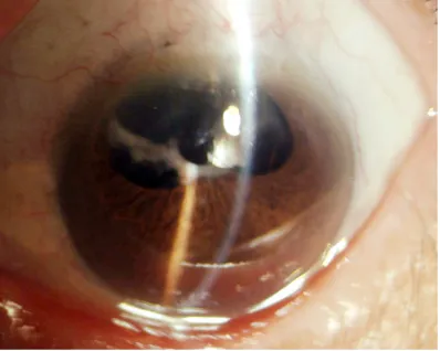
Figure 1. Anterior segment photography of eye under
mydriasis preoperatively. The eye occurred CCS after cataract phacoemulsification following trabeculecto
-my, hyperplasia and thickening of 270 degree fibrous
ring in the area of anterior capsule opening led to
follow-up patients deceased after phaco-emulsification surgery. In this study, 3 eyes in 4-5 weeks, 3 eyes in 7-9 weeks, 5 eyes in 11-14 weeks, 6 eyes in 18-22 weeks, and 2 eyes in 24-27 weeks (mean: 19±2 weeks) occurred with CCS follow-ing phacoemulsification surgery. By the slit-lamp examination, 4 eyes of 4 cases were with white anterior capsule though the rear edge of the pupil. Dilated eye examination showed the hyperplastic and white fibrous membrane-like tissues for anterior subcapsular, the hyperplastic and incrassate annular fibers for anterior capsule opening, and dislocation for anterior capsular opening. We also found that the eyes had decreased and deformed transparent areas with diame-ters of 1.5-2.5 mm, which caused con-traction and shrinkage for surrounding capsule or posterior capsular (Figure 1). Most of the patients were with difficulty visualizing the fundus. The study enrolled 19 eyes of 19 consecutive patients, and the clinical characteristics of the patients are demonstrated in Table 1.
Postoperative visual acuity
[image:3.612.89.330.85.468.2]After the capsular bag releasing surgery, the visual acuity of all the patients improved with varying degrees of suc-cess. At the end of follow-up, the median incorrected visual acuity significantly increased to 4.7±4.0 compared to preop-erative UCVA (u=5.435, P<0.01). The Visual clarity was also significantly im- proved compared to preoperative level. Moreover, the glare and monocular diplo-pia disappeared. The Table 2 lists the comparison between the preoperative UCVA and last follow-up UCVA in the patients.
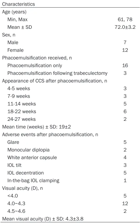
Table 1. Clinical characteristics of patients Characteristics
Age (years)
Min, Max 61, 78
Mean ± SD 72.0±3.2
Sex, n
Male 7
Female 12
Phacoemulsification received, n
Phacoemulsification only 16
Phacoemulsification following trabeculectomy 3
Appearance of CCS after phacoemulsification, n
4-5 weeks 3
7-9 weeks 3
11-14 weeks 5
18-22 weeks 6
24-27 weeks 2
Mean time (weeks) ± SD: 19±2
Adverse events after phacoemulsification, n
Glare 5
Monocular diplopia 2
White anterior capsule 4
IOL tilt 3
IOL decentration 5
In-the-bag IOL clamping 1
Visual acuity (D), n
<4.0 5
4.0~4.3 12
4.5~4.6 2
Mean visual acuity (D) ± SD: 4.3±3.8
Statistics analysis
Wilcoxon Rank-Sum test and the Wilcoxon Signed-Rank test were performed for visual acuity comparison between preoperative and postoperative examination. In this study, the significance (P<0.05) was considered.
Results
Patient characteristics
The time for the patients occurring CCS was
Postoperative follow-up
The follow-up was conducted at postoperative months 1, 3, 6, and the slit lamp examination was performed with normal pupil examination or dilated pupil every time. With the normal pupil, pupillary area was clear without white fibrous membrane. Under mydriasis condition, the fibrous ring of anterior capsule opening was releasing and capsular contraction disap-peared, and the area of the anterior capsule opening increased with the diameter of 5.0-6.0 mm (Figures 2, 3). IOL was located in the cap-Table 2. Comparison of preoperative and last follow-up
UCVA in 19 patients (P<0.01)
UCVA n<4.0 4.0~4.3 4.5~4.6 4.7~4.8 >4.8 Total
Preoperative 5 12 2 0 0 19
[image:3.612.89.330.513.554.2]optic portion was unfolded without the bag IOL clamping. Evaluation demonstrated that there was no cracks/breaks of the IOL’ optics and unexpected radial tears of anterior and poste-rior capsular. Posteposte-rior capsular was found firmly attached to the IOL surface. After radial incision releasing, cutting edge of the anterior capsular opening was serrated, however, with the second CCC tears, the cutting edge of the anterior capsular opening edge was smooth. At the end of follow-up, there was no CCS recur-rence. In all the cases, only one case with sec-ond CCC tears was found with a littering-like fibrous hyperplasia but no reduction in the area of capsular opening.
Discussion
CCS is characterized by shrinkage and whiten-ing of the anterior capsular openwhiten-ing after cata-ract surgery, which is associated with deteriora-tion in postoperative visual funcdeteriora-tion [3, 8]. It has been suggested that CCS is associated with IOL material stimulation, postoperative inflammation, damage of blood-retinal barrier and blood-aqueous barrier, small diameter of CCC or CCC decentration. These factors lead to proliferation of residual lens epithelial cells (LEC) in the anterior subcapsular resulting in formation of subcapsular fibrous plaque. CCS patients may have impaired visual acuity sec-ondary to opacity of the anterior capsular
mem-brane in the pupillary area and IOL decentra-tion within the capsular bag [4, 8]. The onset of CCS in the previously reported cases ranges from weeks to years after uneventful cataract surgery, with adverse events including blurry vision, glare, refractive changes, an eccentric IOL, and full flexion of the haptics [4, 9, 10]. Therefore, CCS is a unique clinical manifesta-tion, and LEC plays a leading role in capsular contraction.
[image:4.612.322.522.72.232.2]Currently, the major strategy for CCS treatment is Nd: YAG laser or surgery [11, 12]. Nd: YAG laser is an effective, safe, convenient and eco-nomical method for CCS treatment, which can enlarge the anterior capsular opening area of transparency and recover visual acuity [13]. However, some patients cannot receive Nd: YAG laser or achieve optimal therapeutic effect after Nd: YAG laser treatment even with increased dose of laser that may cause severe reaction and complications, poor releasing effect, difficult with reposition of displaced or clamped IOL, or even IOL damage [14]. Therefore, surgery treatment, especially CBRS, should be a better choice in these cases [15]. In this study, the patients who were not suitable for Nd: YAG laser capsulotomy, like thickness and hyperplasia of anterior capsule opening and anterior capsular, or annulus width more than 1.5 mm, or anterior capsular opening diameter less than 1.5 mm, were subjected to CBRS treatment.
Figure 2. Anterior segment photography of eye
un-dermydriasis postoperatively. The eye occurred CCS after cataract phacoemulsification following trabecu -lectomy, the combined surgeries with CBRS and sec-ond CCC tear were performed, resulting thatcapsular contraction disappeared, and the area of the ante-rior capsule opening increased.
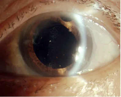
Figure 3. Anterior segment photography of eye under
the normal pupil condition. The eye occurred CCS after cataract phacoemulsification CCS following
[image:4.612.91.289.73.232.2]Treatment of capsule contraction syndrome depends on the degree of visual disability, the capsule and IOL stability, and the presence of other complications. Radial anterior capsuloto-my (surgically or with the Nd: YAG laser) and anterior capsule peeling have been reported to reverse the capsule contraction and its compli-cations [16]. To better control capsulorhexis parameters, such as size and shape, we per-formed capsular peeling, as described by Koizumi and coauthors in aphakic eyes [17]. The disadvantage of the surgery is that patient with suspensory ligament dysfunction may ca- use suspensory ligament injuries or IOL decen-tration. Especially, the surgery cannot be used for the patient with can-opener type of capsu-lotomy. The surgical complications include anterior vitreous prolapse, pupillary block, increased intraocular pressure, capsular bag rupture, suspensory ligament disruption, cor-neal edema, and macular edema. However, the operated patients in our study did not appear the complications mentioned above. The gold-en rule to avoid it is to carefully separate the adhesions and not to drag tough and thick fibrous membrane of anterior capsule with inci-sion or second CCC. For serious adheinci-sions between fibrous membrane and either anterior capsule or IOL, it is difficult to separate but necessary to use capsulotomy needle for care-fully dicing in advance and expose the anterior capsule opening edge for separating by capsu-lotomy scissors.
The proliferation and metaplasia ability of LEC could decrease at three months after cataract phacoemulsification resulting in low recurrence rate of CCS [11, 18, 19]. The amount of residu-al LEC depends on the capsulorhexis size, which should be between 5.5 and 6.0 mm and can be reduced by polishing the anterior cap-sule’s posterior surface [20]. To avoid CCS occurrence, the surgery should focus on pre-vention with perfect surgical procedure to clearly remove cortex and polish capsule bag, and implant IOL into the middle. Moreover, reg-ular follow-up should be applied after cataract phacoemulsification with IOL implantation. In our study, there was no appearance of CCS in all the 19 eyes.
Taken together, we reported favorable out-comes of CBRS for CCS after phacoemulsifica-tion surgery. It offers a safe, effective, and reversible option for CCS treatment.
Disclosure of conflict of interest
None.
Address correspondence to: Haijian Wu, Department
of Ophthalmology at Taizhou Municipal Hospital of Taizhou University, No. 38, Jiaoji Lane, Jiaojiang
Dis-trict, Taizhou 318000, Zhejiang Province, China. E-mail: [email protected]
References
[1] Alio J, Rodriguez-Prats JL, Galal A and Ramzy M. Outcomes of microincision cataract surgery
versus coaxial phacoemulsification. Ophthal -mology 2005; 112: 1997-2003.
[2] Hwang HS, Ahn YJ, Lee HJ, Kim MS and Kim EC. Comparison of macular thickness and
in-flammatory cytokine levels after microincision
versus small incision coaxial cataract surgery. Acta Ophthalmol 2016; 94: e189-94.
[3] Davison JA. Capsule contraction syndrome. J Cataract Refract Surg 1993; 19: 582-589. [4] Michael K, O’Colmain U, Vallance JH and
Cor-mack TG. Capsule contraction syndrome with haptic deformation and flexion. J Cataract Re -fract Surg 2010; 36: 686-689.
[5] Morris D, Fraser SG and Gray C. Cataract sur -gery and quality of life implications. Clin Interv Aging 2007; 2: 105-108.
[6] Altintas AG, Dal D and Simsek S. Significant in -traocular lens folding due to severe capsular contraction. Jpn J Ophthalmol 2008; 52: 134-136.
[7] Karahan E, Er D and Kaynak S. An Overview of Nd:YAG Laser Capsulotomy. Med Hypothesis Discov Innov Ophthalmol 2014; 3: 45-50. [8] Hayashi K and Hayashi H. Effect of anterior
capsule contraction on visual function after cataract surgery. J Cataract Refract Surg 2007; 33: 1936-1940.
[9] Nichamin LD. Reduction in the area of the an-terior capsule opening after polymethylmeth-acrylate, silicone, and soft acrylic intraocular lens implantation. Am J Ophthalmol 1997; 124: 710-711.
[10] Hayashi K, Hayashi H, Nakao F and Hayashi F.
Reduction in the area of the anterior capsule opening after polymethylmethacrylate, sili-cone, and soft acrylic intraocular lens implan-tation. Am J Ophthalmol 1997; 123: 441-447. [11] Pandey SK, Apple DJ, Werner L, Maloof AJ and
Milverton EJ. Posterior capsule opacification: a
review of the aetiopathogenesis, experimental and clinical studies and factors for prevention. Indian J Ophthalmol 2004; 52: 99-112. [12] Ram J, Sukhija J, Thapa BR and Arya VK. Com
[13] Balestrazzi A, Malandrini A, Martone G,
Mari-gliani D, Caporossi T and Tosi GM. Capsule
contraction syndrome with a microincision foldable hydrophilic acrylic intraocular lens: two case reports and review of the literature. Case Rep Ophthalmol 2014; 5: 329-335. [14] Keates RH, Steinert RF, Puliafito CA and Max
-well SK. Long-term follow-up of Nd:YAG laser posterior capsulotomy. J Am Intraocul Implant Soc 1984; 10: 164-168.
[15] Hayashi K, Yoshida M, Hirata A and Hayashi H. Anterior capsule relaxing incisions with neo-dymium: YAG laser for patients at high-risk for anterior capsule contraction. J Cataract Re-fract Surg 2011; 37: 97-103.
[16] Reyntjens B, Tassignon MJ and Van Marck E.
Capsular peeling in anterior capsule contrac-tion syndrome: surgical approach and histo-pathological aspects. J Cataract Refract Surg 2004; 30: 908-912.
[17] Koizumi K, Watanabe A, Koizumi N and
Kinoshita S. Peeling the fibrous membrane
from the anterior capsule for capsulorhexis
contraction after phacoemulsification in apha -kic patients. J Cataract Refract Surg 2002; 28: 1728-1732.
[18] Raj SM, Vasavada AR, Johar SR, Vasavada VA and Vasavada VA. Post-operative capsular
opacification: a review. Int J Biomed Sci 2007;
3: 237-250.
[19] Javadi MA and Zarei-Ghanavati S. Cataracts in diabetic patients: a review article. J Ophthal-mic Vis Res 2008; 3: 52-65.