Original Article
Comparison of open reduction and internal fixation in
treatment of delayed and early acetabular fractures
Bin Wu, Haibin Wang, Chunyang Meng, Cunling Jia, Yifeng Zhao
Department of Orthopedic Surgery, Affiliated Hospital of Jining Medical University, Jining 272029, Shandong,
China
Received June 29, 2015; Accepted September 10, 2015; Epub October 15, 2016; Published October 30, 2016
Abstract: To retrospectively compare the clinical efficacy of open reduction and internal fixation (ORIF) in treating delayed and early acetabular fractures. Ninety cases with delayed (n = 35) and early acetabular fractures (n = 55) undergoing ORIF between September 2009 and March 2013 were retrospectively analyzed. Patients in the delayed acetabular fracture group underwent ORIF at 22-65 d (mean 36 d) after injury and those in the early acetabular fracture group received ORIF at 3-20 d (mean: 8.1 d) after trauma. Ilioinguinal, Kocher-Langenbenk or combined approaches were adopted according to the types of fracture. Fracture reduction was evaluated using the Matta standard. Clinical efficacy was assessed by Matta modified D’Aubigne and Postel grading system. Postoperative follow-up endured for 18-36 months, 25 months on average. In the delayed fracture group, 15 (43%) cases had ana-tomical reduction, 17 (49%) were satisfied with the reduction whereas 3 (9%) were unsatisfied. In the early fracture group, 35 (64%) cases had anatomical reduction, 19 (35%) cases were satisfied with the reduction and only 1 (3%) patient was unsatisfied with no statistical significance between two groups. In the delayed fracture group, 16 cases obtained excellent outcomes, 15 good, 2 average and 2 had poor results, and 35 excellent, 18 good, 1 average and 1 poor in the early fracture group. Excellent rate did not significantly differ between two groups. Satisfactory and excellent rates of ORIF did not significantly differ between two groups. ORIF is an effective and feasible treatment of delayed acetabular fracture.
Keywords: Delayed acetabular fracture, early acetabular fracture, open reduction, internal fixation
Introduction
The incidence of acetabular injury complicated with severe damage in other organs has been dramatically increased along with the high-energy trauma occurring in the transportation and mining industries. Recent advances in sur-gical techniques have increased the use of
open reduction and internal fixation (ORIF) of acetabular fractures. Early internal fixation of acetabular fractures allows rapid mobilization
of severely injured patients and provides trau-ma patients optitrau-mal, long-term, functional results [1, 2]. However, the high-energy acetab-ular injury complicated with other organ trauma
is likely to delay the recovery of acetabular frac
-ture and negatively affect the surgical efficacy
[3-5]. Compared with early acetabular fracture,
it is more difficult and higher risk to treat
delayed acetabular fracture. In this retros- pective analysis, 35 patients diagnosed with
delayed and 55 early acetabular fractures
underwent ORIF at our institution between
September 2008 and March 2012. The effect of reduction and clinical outcomes were statis-tically compared between two pools of patients. Materials and methods
General data
In this study, 35 patients with delayed
acetabu-lar fracture were treated with ORIF, 23 males
and 12 females, aged 23-67 years, 40 years on average. Twenty seven were injured by road
traffic accidents and 8 by falling. According to Letournel and Judet classification, 25 cases
col-Table 1. General data of patients with acetabular fractures
Delayed acetabular fracture Early acetabular fracture
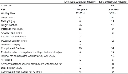
Cases (n) 35 55
Age 23-67 years 17-86 years
Waiting time 22-65 d 3-20 d
Traffic injury 27 36
Falling injury 8 19
Single fracture 25 16
Posterior wall injury 15 9
Anterior wall injury 4 3
Anterior column injury 3 1
Posterior column injury 1 2
Transverse injury 2 1
Complicated fracture 10 39
Posterior column complicated with posterior wall injury 6 14
Transverse complicated with posterior wall injury 1 12
“T” shape 1 5
Anterior/posterior column complicated with transverse 1 6
Dual column injury 1 6
Complicated with ischial nerve injury 6 9
Inclusion criteria: 6the subjects with acetabular fractures shift > 5 mm, which severely affected the movement of hip joint;
those with stable vital signs; those with heart and lung diseases or distal femur fracture were excluded; no alternative surgical
[image:2.612.89.522.86.337.2]contraindications could be noted; those who were able to independently work prior to the trauma and the patients whose fam -ily relatives required performing the surgery.
Table 2. Clinical data of ORIF
[image:2.612.91.524.415.466.2]Group n approachAnterior approachPosterior Combined approach Operation time Intraoperative blood loss Delayed acetabular fracture 35 25 (71.4%) 7 (20%) 3 (8.6%)* (180±67) min* (1500±700) mL* Early acetabular fracture 55 32 (58.2%) 10 (18.2%) 13 (23.6%) (120±45) min (500±200) mL Note: *in the delayed acetabular fracture group, operation time was significantly longer and intraoperative blood loss was larger compared with those in the early acetabular fracture group. Operation time and blood loss com-pared with delayed acetabular fractures, *P < 0.05 denotes a statistical significance. Postoperative processing.
Table 3. Clinical results of articular surface reduction
Group n Anatomic reduction Satisfactory reduction Unsatisfactory reduction Overall satisfactory rate Delayed acetabular fracture 35 15 (42.9%)* 17 (48.6%) 3 (8.5%)* 91.4% Early acetabular fracture 55 35 (63.7%) 19 (34.5%) 1 (1.8%) 98.2% Note: *denotes statistical significance. Overall satisfactory rate did not significantly differ between two groups (χ2 = 0.98, P > 0.05), whereas statistical significance was observed regarding the rates of anatomic reduction and unsatisfactory reduction
(all P < 0.05) between two groups.
Table 4. Clinical outcomes of ORIF
Group n Excellent rate Good rate Average rate Poor rate Overall satisfactory rate Delayed acetabular fracture 35 16 (45.7%)* 15 (42.9%) 2 (5.7%) 2 (5.7%) 31 (88.6%) Early acetabular fracture 55 35 (63.6%) 18 (32.8%) 1 (1.8%) 1 (1.8%) 53 (96.4%) Note: *No statistical significance was noted in overall satisfactory rate (excellent and good outcomes) between two groups (χ2
[image:2.612.90.522.537.588.2] [image:2.612.90.525.656.697.2]umn complicated with posterior wall fracture, 1
[image:3.612.89.524.72.248.2]transverse complicated with posterior wall frac- ture, 1 “T” shape fracture, 1 anterior and poste-rior column complicated with transverse frac-Figure 1.Preoperative CT scan. A. Preoperative 3D CT (frontal view). B. Preoperative 3D CT (posterior view).
[image:3.612.90.525.287.640.2]ture and 1 dual column fracture. Two cases were complicated with posterior dislocation of the femoral head, 5 central dislocation of vary-ing degree, 1 fracture of the femoral head and
6 ischial nerve injury. Five cases were compli -cated with brain trauma, 3 chest trauma and abdominal injury and 2 urinary injury. The injury of other positions was treated in other depart-ments or hospitals. The acetabular fractures were merely handled with traction treatment. They had to wait for 22 to 65 d before
undergo-ing the operation, 36 d on average. Fifty five
patients with early acetabular fracture under-went surgery, 39 males and 16 females, aged 17-86 years, 46 years on average. Among them,
36 were injured from road traffic and 16 from accidental falling. According to Letournel and Judet classification 16 cases had simple frac -ture, 9 posterior wall frac-ture, 3 anterior wall fracture, 2 posterior column fracture, 1 anterior column fracture and 1 transverse fracture; 39 patients were diagnosed with complicated frac-ture including 14 posterior column complicated with posterior wall fracture, vernolicacid trans-verse complicated with posterior wall fracture, 5 “T” shape fracture, 6 anterior and posterior column complicated with transverse fracture and 2 dual column fracture. Nine patients
suf-fered from ischial nerve injury. Fifteen cases
were complicated with cerebral trauma, 5 chest trauma, 3 abdominal trauma and 2 urinary
inju-ry. They had to wait for 3-20 d to undergo the surgery, 8.1 d on average (Table 1).
Surgical approach
Preoperative examinations: Conventional χ-ray
radiograph was conducted under AP pelvic, obturator oblique and iliac bone oblique views. Subsequently, 64-slice spiral CT scan and then three dimensional reconstruction was per-formed. Systemic conditions of the patients
were assessed to verify their tolerance to ORIF and identify the condition of the skin and soft
tissues in the surgical site. All contraindications should be excluded.
Surgical approach: The patients with
trans-verse facture and complicated acetabular frac-tures involving with two columns underwent
ORIF via anterior and posterior combined
approach. Those presenting with single column or/and anterior wall, anterior column, posterior wall or/and posterior column injuries were
sub-jected to ORIF via single surgical approach.
Intraoperatively, the fracture ends of the bone
were fully isolated and exposed. ORIF was first
delivered to treat the single fracture. Then, the
reduction and fixation of the acetabular frac -tures at the linear extension location were per-formed (from linear extension of the fracture to abarticular iliac bone, ischial bone, pubis and sacroiliac joint). The anatomic relationship of dislocated or disordered femoral head mortar was restored, bone fracture plate was
recon-structed and fixed with proper tensile force
simultaneously. Patients with posterior wall fracture defects received bone repairing and maintenance by using autologous iliac bone. Bone implantation was performed at the poste-rior wall to elevate the height of posteposte-rior wall. The posterior wall soft tissues repair was con-ducted to replace the function of joint capsule, aiming to prevent the posterior dislocation of hip joint and enhance the structural stability. The surgery was performed under general
anesthesia. The patients were kept in semi -prone position on the healthy side when single posterior approach was selected, in a lying pos-ture when the anterior approach was adopted and in a “drifting” posture when combined approach was chosen. The operating table
should be fully penetrable by the χ-ray. All
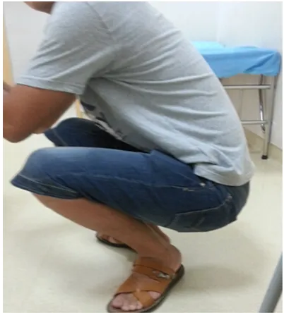
patients were subjected to re-infusion of autol-ogous blood during the surgery. Thus, the intra-Figure 3.The patient restored normal hip joint
[image:4.612.88.289.71.293.2]operative volume of blood transfusion only accounted for 1/3 to 1/2 of blood loss, which
significantly reduced surgical risk and trauma.
After the surgery, negative pressure drainage was placed, as shown in Table 2.
After the surgery, conventional application of broad-spectrum antibiotics was conducted for
48 h. During 48-72 h following the operation,
the catheter was withdrawn when 24
h-drain-age amount < 30 ml. Skin traction was per -formed to constrain the movement of
acetabu-lum according to the situation of fixation. On
the next day after surgery, lower extremity an-
kle pump exercises were taken by the patients.
At 72 h after removal of drainage catheter,
patients performed passive hip and ankle flex -ion exercises. Approximately after 1-month bed
rest, the patients were re-examined by χ-ray
and gradually started weight bearing exercise and recovery.
Follow-up and efficacy evaluation
The patients were subjected to outpatient and
telephone follow-up after the surgery. During each outpatient follow-up, χ-ray radiograph was
performed under AP pelvic, obturator oblique and iliac bone oblique views. The reduction of articular surface fracture was assessed by Matta standards [7]. Clinical outcomes were
evaluated by modified D’Aubigne and Postel
grading system.
Statistical analysis
Data analysis was conducted by using SPSS 20.0 statistical software (SPSS, Chicago, IL,
USA). Quantitative data were statistically com-pared between two groups by t-test. Comparison of qualitative data between two groups was performed by chi-square test. P < 0.05 was
considered as statistical significance.
Results
Articular surface fracture reduction by Matta
standards: horizontal and vertical shift < 1 mm was defined as anatomic reduction; horizontal
and vertical shift between 1 and 3 mm as
sat-isfactory reduction and horizontal and vertical
shift > 3 mm as unsatisfactory reduction, as illustrated in Table 3. Overall satisfactory rate
did not significantly differ between two groups (χ2 = 0.98, P > 0.05), whereas statistical signifi
-cance was observed regarding the rates of ana-tomic reduction and unsatisfactory reduction between two groups (both P < 0.05).
Clinical outcomes assessed by modified D’Aubigne and Postel grading system [7]
include pain, movement and range of joint motion, etc. Clinical results of acetabular
frac-tures were classified into four grades: excellent
(18 points), good (15-17 points), average (13-15 points) and poor (< 13 points), as illustrated in Table 4. Overall satisfactory rate (excellent
and good outcomes) did not significantly differ between two groups (χ2 = 1.02, P > 0.05),
whereas statistical significance was noted in
the excellent rate between two groups (P < 0.05).
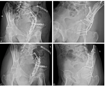
Typical case: a male patient aged 25 years were diagnosed with complex delayed acetabu-lar fractures complicated with infection and rupture of small intestine (Figure 1). He
under-went ORIF via anterior approach at 59 d after
injury in combination with postoperative
recov-ery exercises. During 3-month follow-up, bone
fracture was well healed with good hip joint function (Figures 2 and 3). During 2-year
follow-up, good hip joint function was obtained with no pain.
Discussion
A majority of acetabular fracture cases are caused by high-energy trauma and thus
con-servative therapy yields mild clinical efficacy [8,
9]. A small proportion of patients who are
com-plicated with severe chest, abdominal and skull trauma are likely to miss the optimal timing and postpone the surgery by 3 weeks even longer after bone fracture. De Bellis et al. [10] sum
-marized previous findings and concluded that
immediate total hip replacement (THR) can be successful in appropriately selected elderly patients or patients with extensive
osteoporo-sis, combined acetabular and femoral neck
fractures or pathological fractures. There is cur-rently a limited evidence base for immediate THR in patients with acetabular fractures.
Therefore, orthopedic surgeons’ practice and
expertise are the most useful tools in clinical practice.
In our institution, ORIF has been successfully
attempting to conduct ORIF in those with
delayed acetabular fracture. The reduction
quality and clinical efficacy were statistically
compared between these two populations.
Favorable therapeutic effects have been
obtained in both groups with no statistical
significance.
Selection of timing and approach of ORIF
Previous studies have demonstrated that
post-operative clinical efficacy of ORIF in treating acetabular fractures is significantly correlated
with the waiting time for surgery. The earlier the surgery is conducted, the higher rate of ana-tomic reduction will be achieved. Mears et al. [11] reported a rate of anatomic reduction up to 76% at 2 d after injury, 68% at 3-10 d and 54% at 11-12 d. In this investigation, the total rate of
anatomic reduction was 63.6% following ORIF, which is consistent with previous findings.
However, the total rate of anatomic reduction in the delayed acetabular fracture group was merely 42.8%, suggesting that it is not feasible
to perform ORIF immediately after injury
be-cause the acetabular fracture is be-caused by
high-energy shock frequently accompanied by
severe damages at multiple sites and unstable
vital signs. Hence, simple pelvic external fixa -tion is the only choice available. Peng et al. [12]
regarded the 1st week after injury as the opti
-mal timing for performing ORIF and insisted that it was difficult to conduct reduction suc -cessfully after 10 days after fracture, especially more challenging to achieve reduction
exceed-ing 3 weeks, which could be categorized into
delayed fracture. Zhou et al. [13] have
demon-strated that it is reasonable to perform ORIF within 1-2 weeks following the incidence of
complex acetabular fractures, whereas should
be highly alerted to conduct ORIF over 3 weeks after delayed acetabular fracture. For those
patients suffering from acetabular fractures for over 3-4 months due to poor systemic condi-tion or alternative causes, other clinical thera-pies, such as THR, should be considered rather
than ORIF. In this study, 55 patients diagnosed
with early acetabular fractures steadily under-went reduction and exposure of the fracture
ends, especially for those receiving ORIF within 1 week after fracture. Although significant frac -ture shifting may be observed, the operation
time and quality of reduction could be signifi -cantly improved compared with their
counter-parts undergoing ORIF during late period. Despite it was extremely challenging to suc
-cessfully perform ORIF for patients with delayed
acetabular fracture and those waited for 65 d prior to surgery, excellent therapeutic effect of anatomic reduction was obtained due to simple
fracture. Letournel and Letournel [14] catego
-rized the surgical procedures into three stages
according to the time interval between the inju-ry and surgeinju-ry: the 1st stage was 1-20 d after injury, the 2nd stage was 21-120 d and the 3rd stage over 120 d after fracture. It is convenient
and efficacious to perform ORIF for patients
with early fracture (belonging to the 1st stage).
For those left untreated for > 3 weeks after
bone fractures (acetabular fracture and dislo-cation, pubic branch fracture and dislodislo-cation, sacroiliac fracture and dislocation, as well as ala of ilium fracture, etc.), both the number and length of incision should be increased to guar-antee the high quality of reduction. In addition,
the delay of ORIF for acetabular fractures
threatens the survival of femoral head, espe-cially for those accompanied by persistent dis-location and semi-disdis-location, and leads to abrasion of articular cartilage, chondrolysis, osteonecrosis of femoral head and posterior wall necrosis, etc. Although it is still easy to identify the fracture line and obtain relatively high quality of reduction within 120 d after inju-ry, it becomes more challenging to identify the fracture line and achieve excellent anatomic reduction exceeding 120 days. Instead,
alter-native surgical approaches rather than ORIF
should be considered to treat hip joint cartilagi-nous injury and abrasion.
At present, joint arthroplasty and fusion are being adopted to treat delayed acetabular
frac-ture. During joint arthroplasty, the acetabular
structure was found to be severely disrupted, even requiring impaction bone grafting or cages to repair the acetabulum. Eventually, postoper-ative therapeutic effect was not satisfactory in terms of unstable hip joint structure and high revision rate. Joint fusion is a surgical proce-dure which completely destroys the function of joint. Therefore, it is rarely recommended ex-
cept specific conditions. Nowadays, osteotomy
has been attempted by most orthopedic sur-geons to perform three dimensional recon-struction of the acetabulum, whereas the
surgi-cal difficulty and risk are extremely high. ORIF is
anatomic position and creates favorable condi-tions for joint arthroplasty as a remedial sur-gery. The authors in this study suggested that
ORIF should be adopted to treat acetabular
fractures as possible by stripping callus and exposing the para-position of the fracture ends. This technique probably causes malreduction, whereas it offers solid foundation for hip joint arthroplasty. Based upon the experience from
this study, it is recommended to perform ORIF
to treat the acetabular fractures within 2 months after injury.
Surgical difficulty and approach selection ORIF
ORIF functions to attain fracture reduction,
restore the anatomic relationship of mortar,
stabilize the hip joint and recover the function
of hip joint. Whether anatomic reduction could
be obtained directly influences the functional recovery of the affected hip. We’ve accumulat -ed a substantial quantity of clinical experience in treating and curing patients diagnosed with early acetabular fractures. However, reduction of delayed acetabular fracture is even more challenging due to the following primary
fac-tors, such as formation of fibrous callus, sclero
-sis of fibrous scar tissue, contracture, serious tissue adhesion, blurry layer and margin, diffi -culty in stripping and eliminating callus, ana-tomical structural injury and massive
hemor-rhage, which collectively increase the difficulty
in exposure and reduction of acetabular
frac-ture [15, 16]. The clinical findings of 35 cases in
this study demonstrated that much attention should be paid to the followings. 1. Except ante-rior or posteante-rior column fracture, dual column fracture should be treated by combined
approach. ORIF via single approach is not rec -ommended to repair the anterior and posterior factures in spite of the simple fracture since the structure of delayed acetabular fracture is relatively stable. 2. The subperiosteum strip-ping should be performed more extensively. The soft tissue scars surrounding the acetabu-lum should be completely separated and
dislo-cated, and ORIF via an enlarged surgical
approach should be chosen when necessary to facilitate the reduction of bone fracture. 3. All the callus and scars adjacent to the site of
frac-ture should be explicitly identified and thor -oughly eliminated to fully expose the fracture ends. 4. Posterior wall defects should be repaired by elevating the height of posterior wall. The soft tissue repair in the posterior wall
should be conducted to replace the function of joint capsule, aiming to prevent the posterior dislocation of hip joint and enhance the struc-tural stability. A majority of patients with delayed acetabular fracture are complicated with serious multiple trauma and possess a relatively high tolerance to pain. They have a lower expectation of the surgical outcomes. Combined with active and effective recovery exercises, relatively good clinical outcomes
could be obtained. The excellent rates of ORIF efficacy did not significantly differ between the
delayed (88.6%) and early acetabular fracture groups (96.4%).
To sum up, compared with early acetabular
fracture, it is more difficult to treat delayed ace
-tabular fracture via ORIF with longer operation
time and more blood loss. A suitable surgical approach should be selected during acetabular fracture based upon systemic conditions and the type of fracture of the patients. After the surgery, favorable recovery training should be delivered to obtain satisfactory reduction and hip joint function. Although the reduction and excellent rates in the delayed acetabular frac-ture group were relatively low, they did not
sig-nificantly differ from those in the early aceta-bular fracture group. ORIF is an efficacious
approach in the treatment of delayed acetabu-lar fracture.
Study limitations
The sample size in this clinical trial is relatively
small (n = 35). The number of cases with com-plicated fracture is too small to integrate all types of delayed acetabular fractures. Next, a more comprehensive investigation with a larger
sample size is urgently required.
Acknowledgements
This work was supported by grants from the
Jining science and technology development
plan (Funding NO. [2011] 57#).
Disclosure of conflict of interest
None.
References
[1] Giannoudis PV, Nikolaou VS, Kheir E, Mehta S, Stengel D, Roberts CS. Factors determining quality of life and level of sporting activity after internal fixation of an isolated acetabular frac -ture. J Bone joint Surg Br 2009; 91: 1354-1359.
[2] Culemann U, Holstein JH, Kvhler D, Tzioupis CC, Pizanis A, Tosounidis G, Burkhardt M, Pohlemann T. Different stabilization tech -niques for typical acetabulur fractures in the elderly. A biomechanical assessment. Injury 2009; 25: 469-473.
[3] Ortega R, Loder RT, Louis DS. Open reduction and internal fixation of forearm fractures in children. J Pediatr Orthop 1996; 16: 651-654. [4] Koval KJ. Open reduction and internal Fixation.
J Orthop Trauma 2005; 19: 61-62.
[5] Grewal R, Perey B, Wilmink M, Stothers K. A randomized prospective study on the treat -ment of intra-articular distal radius fractures: open reduction and internal fixation with dor -sal plating versus mini open reduction, percu-taneous fixation, and external fixation. J Hand Surg Am 2005; 30: 764-772.
[6] Wang MY, Wu XB, Zhu SW, Cao QY, Wu HH, Rong GW. Operative treatment of delayed ace-tabular fractures. Chinese J Surg 2003; 41: 130-133.
[7] Metta JM. Fractures of the acetabulum: Reduction accuracy and clinical results of frac-tures operated within three weeks of injury. J Bone Joint Surg Am 1996; 78: 1632-1645.
[8] Bellabarba C, Berger RA, Bentley CD, Quigley LR, Jacobs JJ, Rosenberg AG, Sheinkop MB, Galante JO. Cementless acetabular recon-struction after acetabular fracture. J Bone Joint Surg Am 2001; 83-A: 868-76.
[9] Weber M, Berry DJ, Harmsen WS. Total hip ar -throplasty after operative treatment of an ace-tabular fracture. J Bone Joint Surg Am 1998; 80: 1295-1305.
[10] De Bellis UG, Legnani C, Calori GM. Acute total hip replacement for acetabular fractures: a systematic review of the literature. Injury 2014; 45: 356-261.
[11] Mears DC. Surgical treatment of acetabular fracture in elder patients with osteoporotic bone. J Am Acad Ortho Surg 1999; 7: 128-141. [12] Peng AQ, Pan JS. Surgical complications and
prevention of acetabular fracture. J Bone Joint Injury 2003; 18: 63-64.
[13] Zhou DS, Wang XQ, Wang BM, Wang LB. Treatment of complex acetabular fractures through combined approaches (46 cases re-ports). Orthoped J China 2004; 12: 170-172. [14] Letournel E, Judet R. Fractures of the acetabu
-lum, 2nd ed., New York: Springer-Verlag 2006; 5: 38-39.
[15] Liu YS, Yang SH. The surgical approach choic -es for acetabular fracture. Chin-ese J Joint Surg 2010; 4: 408-411.