Original Article
The effect of post-contrast washıng on post-endoscopıc
retrograde cholangıopancreatography pancreatıtıs
Hasan Ökmen1, Bünyamin Gürbulak1, Yiğit Düzköylü1, Esin Kabul Gürbulak2, Esra Paşaoğlu3, Hasan Bektaş1, Kenan Büyükaşık1, Acar Aren1
1Department of General Surgery, Istanbul Training and Research Hospital, Fatih 34098, Istanbul, Turkey;
2Department of General Surgery, ŞişliHamidiye Etfal Training and Research Hospital, Şişli 34371, Istanbul, Turkey; 3Department of Pathology, Istanbul Training and Research Hospital, Fatih 34098, Istanbul, Turkey
Received March 22, 2015; Accepted June 12, 2016; Epub August 15, 2016; Published August 30, 2016
Abstract: Objective: We aimed to evaluate the efficacy of dilutional washing with saline solution following the admin-istration of contrast agent for ERCP in decreasing the rate of pancreatitis, in our experimental rat model. Methods: Fourty Wistar-Albino® male rats of 250-300 g were divided into 4 equal groups. Group 1: Cannulation, Group 2: Cannulation+saline, Group 3: Cannulation+contrast agent, Group 4: Cannulation+contrast agent+saline. At the 24th hour following the procedures, the rats were sacrified and pancreatic tissues were examined histopathologi-cally, with the evaluation of blood levels of leukocyte, glucose, (SGOT), (LDH), amylase, C-reactive protein (CRP) and acid-base status. Histopathological grading of acute pancreatitis was performed using haematoxylin and eosin staining. Results: Mean levels of amylase, leukocyte, AST and LDH were found to be significantly higher in groups 2, 3, 4 when compared to group 1 (P<0.05). CRP level was found to be highest in group 3 (P>0.05). Histopathological grade of pancreatitis was found to be significantly higher in groups 2, 3, 4 when compared to group 1 (P<0.05). Scores of edema, acinar necrosis and amylase level were found to be higher in group 3 than group 4. Scores were similar in groups 2 and 4. Conclusion: We found that dilution with saline solution during ERCP procedure may be beneficial in decreasing the rate of post ERCP pancreatitis, which is shown with histopathological and laboratory findings. We suggest dilutional washing out of the contrast agent at low pressures following the ERCP procedure.
Keywords: Post ERCP pancreatitis, experimental pancreatitis, contrast induced pancreatitis
Introduction
The incidence of post-endoscopic retrograde cholangiopancreatography (ERCP) pancreatitis is reported to be between 2-10% (2-4% in low risk group and 0-5% in high risk group) [1]. Post-ERCP pancreatitis is a serious complication of the procedure and etiology is multifactorial [2]. The clinical progress is mild in 85-90% of the patients, and necrotising pancreatitis with multi-organ failure is encountered in 10-15% of the patients.
Although the underlying mechanisms are not clear yet, it is thought to be single or combined effects of mechanical, chemical, hydrostatic, enzymatic, microbiological, allergic and ther-mal mechanisms [3-5].
In ERCP procedure, contrast agent is adminis-tered directly into common bile duct ( CBD) and
Wirsung. Due to the suggested theory, contrast agent causes damage on the acinar cells with direct contact [5]. Although various medications and technical variations are suggested in means of minimizing the rate of post ERCP pan-creatitis, studies evaluating the effect of the dilution of the contrast agent are very limited [6, 7].
In our study, we aimed to evaluate the efficiency of the dilutional effect of saline solution in decreasing the toxic damage of contrast agent on pancreatic tissue, leading to pancreatitis, in an experimental rat model.
Material and methods
Faculty of Medicine, Istanbul, Turkey. Fourty Wistar-Albino® male rats of 250-300 g were seperated into 4 randomized groups, with 10 rats in each of them. All the experimental proto-cols were carried out in accordance with the Guide for the Care and Use of Laboratory Animals prepared by the National Academy of Sciences.
All rats were housed under standard laboratory conditions at room temperature with 12 h light/12 h dark cycle and allowed to have ad libitum food and water before and after surgery. During the experimental procedure, the ani-mals were individually placed in cages and kept at room temperature (22°C). All surgical proce-dures were performed under sterile condi- tions.
Experimental protocol
Before the experimental procedure, all animals were weighed and the results were recorded. Rats were anaesthetized with intramuscular injections of ketamine hydrochloride (50 mg/ kg, Ketalar; Parke-Davis, Morris Plains, NJ) and Xylazine (10 mg/kg, Rompun; Bayer, Istanbul). In our experimental model, bile duct was can-nulated transduodenally, and following the clamping of the hepatic duct with a bulldog clamp, saline solution and contrast agent were administered at 30 mmHg pressure. With this procedure, an ERCP procedure in obstructive cases that lead to retrograde flow of bile to the pancreatic duct, such as stasis due to chole-dochal stones, edema and tumors of pancreat-ic head was simulated (Figure 1).
A steady pressure of 30 mmHg was chosen as the most appropriate pressure in the light of the recent literature (5.lit.6.kaynak ve tufan
tezi). A sphygmomanometer cuff of a pediatric blood pressure device was prepared to deliver 100 cc of isotonic NaCl mediflex or 50% diluted contrast agent under 30 mmHg pressure. The contrast agent used was Ultravist® 300 (Schering, Germany).
Surgical procedure
Fourty Wistar-Albino® rats were assigned in four groups, as follows: The rats were shaved and then prepared with povidone-iodine. A mid-line (5 cm) laparotomy was performed. Then, the abdominal organs were explored. In all groups, common biliopancreatic duct was can-nulated transduodenally via median laparoto-my using 24 G catheters. For group 2, 3 and 4, solutions (contrast agent and isotonic NaCl) were infused following theclosure of both ends of the duct at 30 mmHg pressure.
Group 1 (Cannulation group, n=10): Common biliopancreatic ductwascannulated via median laparotomy by a 24 G cannula and the abdo-men was closed without performing any other procedure.
Group 2 (Isotonic group, n=10): Common bilio-pancreatic duct was cannulated transduode-nally via median laparotomy by a 24 G cannula. After applying a small bulldog clamp to the hepatic duct, 0.5 ml isotonic NaCl (saline) wasinjected at 30 mmHg pressure.
Group 3 (Contrast group, n=10): Common bilio-pancreatic duct wascannulated transduodenal-ly via median laparotomy by a 24 G cannula and after applying a small bulldog clamp to the hepatic duct, 0.5 ml 50% diluted contrast agent was injected at 30 mmHg pressure.
Group 4 (Contrast plusisotonic group, n=10): Common biliopancreatic duct was cannulated transduodenally via median laparotomy by a 24 G cannula after applying a small bulldog clamp to the hepatic duct and 0.5 ml 50% diluted con- trastagent was injected at 30 mmHg pressure. Later, 0.5 ml isotonic NaCl (saline) was injected at 30 mmHg pressure. The abdomen was closed with 3-0 silk suture in all of the groups. After 24 hours, all rats were re-anesthetized. Following laparotomy, blood samples were col-lected via intra-cardiac route for biochemical and blood gas analysis, then the rats were sac-rificed by cervical dislocation. The duodenal Figure 1. Transduodenal cannulation of common
[image:2.612.90.289.69.184.2]loop with whole pancreas was harvested as a sample, for histopathological confirmation of acute pancreatitis.
Biochemical analysis
Blood samples were taken for counting the blood levels of leukocyte, amylase, glucose, C-reactive protein (CRP), lactate dehydroge-nase (LDH), serum glutamic oxaloacetic trans-aminase (SGOT) and evaluation of acid-base status. Blood gases analysing was performed immediately with RAPIDLab248 blood gases analyser system (Siemens Healthcare, Erlingen,
Germany). For further research, samples were transferred toIstanbul Training and Research Hospital, Clinical Biochemistry laboratory under appropriate conditions. Levels of serum amy-lase, serum SGOT, LDH, and CRP levels were measured for estimating the frequency and severity of pancreatitis by standard laboratory methods.
Histopathological examination
Pancreatic tissue samples were placed in 10% neutral formalin solution for pathological asssesment. Specimens were routinely
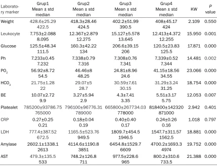
pro-Table 1. Mean and madian laboratory blood values of the groups
Laborato-ry marker
Grup1 Mean ± std
median
Grup2 Mean ± std
median
Grup3 Mean ± std
median
Grup4 Mean ± std
median KW
P
value Weight 428.6±25.29
422.5 418.3±28.46424.5 402.2±51.99390.5 408±45.17424 2.109 0.550
Leukocyte 7.753±2.088
8.095 12.367±2.87912.275 15.127±5.57813.645 12.413±4.37212.255 15.950 0.001 Glucose 125.5±48.34
111.5 160.3±42.22134 206.6±39.15206 120.5±23.83125.5 17.871 0.000 Ph 7.233±0.45
7.232 7.338±0.797.316 7.308±0.767.341 7.339±0.527.344 14.481 0.002 PO2 56.92±8.72
54.5 46.46±848.25 24.81±8.9624.6 41.15±18.5634.55 23.066 0.000
HCO3 21.75±1.28
22 29.07±528.7 30.59±7.6130.15 31.29±3.2431.25 18.754 0.000
BE 10.07±2.72
9.9 3.27±5.942.9 4.3±7.413.35 5.51±3.175.75 12.053 0.007 Platealet 785200±93786.75
765000 796100±96776.31789000 665800±267734.03778000 818400±142320871000 2.942 0.401
CRP 0.27±0.25
0.21 0.18±0.040.19 0.40±0.400.17 0.24±0.260.16 1.018 0.797
LDH 777.4±387.52
672.5 1165.5±523.76949.5 1809.7±454.51946.5 1547.7±311.571562.5 18.881 0.000 Amylase 2602.1±1338.1
2613 4114.6±1190.83851 6454.8±1529.76609 4700.2±1693.34974 19.752 0.000
AST 479.3±135.5
533 748.2±126.8711 977.5±228.6965 800.2±310.6733.5 21.388 0.000
[image:3.612.91.520.85.412.2]KW: Kruskal-Wallis. AST: Aspartate Aminotransferase. LDH: Lactate dehydrogenase. CRP: C-reactive protein.
Table 2. Intergroup comparison of blood analysis
Groups Weight Leukocyte Glucose Ph PO2 HCO3 BE Platelet CRP LDH Amylase AST
[image:3.612.91.524.458.543.2]cessed and embedded in paraffin wax. Pancreas tissue sections of 5 μm thickness were stained with hematoxylin and eosin. The specimens were examined under light micro-scope as a pathologist-blinded study. Presence and grade of acute pancreatitis were evaluated and documented in each of the tissue sections according to Schmidt’s method with regard to edema, acinar necrosis, hemorrhage, fat necro-sis, inflammation and perivascular infiltration scores which determines the severity of acute pancreatitis described previously [8].
Statistical analysis
Statistical analyses were carried out by using the Statistical Package for Social Sciences ver-sion 15.0 (SPSS for Windows 15.0, Inc, Chicago, IL, USA).
The data were evaluated by descriptive statisti-cal methods (Mean and standart deviation, Median), while intergroup comparisons were compared by Kruskal-Wallis test, and subgroup comparisons were performed with Mann Whitney U test. A ‘P’ value <0.05 was accepted as statistically significant.
Results
No complication was encountered and none of the animals died during the experimental pro-cedure. Histopathological examination of sp- ecimens revealed acute pancreatitis in all of the groups.
A homogeneous distribution was determined between the groups in means of body weight (P=0.55).
Leukocyte, glucose, SGOT, LDH, amylase, CRP, pH, PO2, HCO3 and base excess (BE) parame-ters were evaluated with Kruskal-Wallis test which showed significant difference be- tween the groups with regard to other parame-ters (P<0.05), while demonstrating no signifi- cant difference between the groups in terms of platealet counts and CRP. There was a slight increase in CRP levels especially in group 3, however the difference was not statistically sig-nificant (P>0.05) (Table 1).
Intergroup comparision of laboratory findings
Serum levels of amylase, leukocyte, pH, HCO3, SGOT and LDH were significantly lower in group 1 when compared to other groups (P<0.05). BE and PO2 levels were higher in group 1 (Table 2). While levels of glucose, amylase, AST, LDH were higher in group 3 than group 2, PO2 was found to be higher in group 2.
In group 3, glucose and amylase levels were higher, PO2 was found to be lower than group 4.
Histopathological assessment
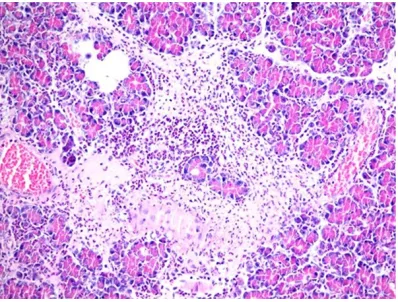
Histopathological assessment of the speci-mens of the pancreatic gland were consistent with acute pancreatitis leading to interstitial edema, inflammation and perivascular infiltra-tion, acinar necrosis, hemorrhage and fat ne- crosis in all experimental groups (Figures 2, 3). The difference was significant between gropus in means of acinar necrosis, inflammation and perivascular infiltration (p<0.05). Conversely, Figure 2. Hematoxylin-eosin (H-E) staining (200×).
Microscopic appearance. Significant leukocyte infil-tration, edema, focal fat necrosis, hemorrhage and congestion is apparent (H&E ×200).
[image:4.612.90.289.70.221.2] [image:4.612.89.289.288.439.2]Table 3. Mean pathological injury scores of the groups
Injuries Mean ± stdGrup1 median
Grup2 Mean ± std
median
Grup3 Mean ± std
median
Grup4 Mean ± std
median KW
P
value
Edema 1.95±0.59
2 2.35±0.412.5 3.1±0.393 2.6±0.212.5 21.156 0.000 Acinar necrosis 1.1±0.73
1 2.45±0.762.5 3.2±0.883.25 2.2±0.942 18.312 0.000 Hemorrhage and fat necrosis 0.95±0.59
1 0.95±0.651 2.05±1.032.25 1.15±0.621 7.671 0.053 Inflammation and perivascular infiltration 1±0.91
0.75 2.75±1.033.25 3.4±0.803.75 2.95±0.552.75 18.598 0.000
KW: Kruskal-Wallis.
there were not any significant differences in means of hemorrhage or fat necrosis (Table 3). In group 2, acinar necrosis, inflammation and perivacular necrosis scores were significantly higher than group 1 (P<0.05).
All of the damage scores ( edema, acinar necro-sis, inflammation, perivascular infiltration) were significantly higher in group 3, when compared to group 1 (P<0.05) (Figure 4).
se syndrome (SIRS) or multipl organ dysfunc-tion syndrome (MODS), caused by a cascade of reactions. It has been also reported that there may be seen life threatening attacks following ERCP, with a rate of 0-5% [9-13].
[image:5.612.90.374.224.409.2]In acute pancreatitis, following acinar cell dam-age, amylase enters into the systemic circula-tion. The increase in serum level starts in 2-12 hours and reaches its peak value between 12-72 hours. Renal excretion of amylase Figure 4. Comparison of the mean pathological injury scores of the groups.
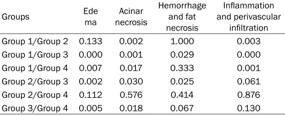
Table 4. Intergroup comparison of pathological injury scores
Groups Ede ma necrosisAcinar Hemorrhage and fat necrosis
Inflammation and perivascular
infiltration Group 1/Group 2 0.133 0.002 1.000 0.003 Group 1/Group 3 0.000 0.001 0.029 0.000 Group 1/Group 4 0.007 0.017 0.333 0.001 Group 2/Group 3 0.002 0.030 0.025 0.061 Group 2/Group 4 0.112 0.576 0.414 0.876 Group 3/Group 4 0.005 0.018 0.067 0.130
Mann Whitney U test.
In group 3, scores of edema, acinar necrosis, hemorrhage and fat necrosis were signifi-cantly higher than group 2 (P<0.05). Similarly, edema and acinar necrosis were found to be significantly higher than group 4 (P<0.05).
In group 4, edema, necrosis, inflammation and perivascular infiltration scores were found to be significantly higher than group 1 (P<0.05). Hemorrhage and fat necrosis were found to be similar (Table 4).
Discussion
[image:5.612.91.370.462.576.2]increases at the same time, leading to an increase in urinary concentration. In uncompli-cated cases, the levels come back to normal range in a few days duration [14, 15].
Hemorrhage, cholangitis, cholecystitis, perfora-tion and pancreatitis are among the complica-tions of ERCP. Post-ERCP pancreatitis is sus-pected and diagnosed with typical pain, amy-lase levels 3-times higher than normal, and symptoms starting in the following 24 hours, and can be graded clinically as low, mild or seri-ous [16].
Patient-related etiologic factors that lead to post-ERCP pancreatitis involve young females, SOD suspicion, normal bilirubin levels, previous post-ERCP or recurrent pancreatitis history, anatomic variations such as fusion anomalies of the pancreatobiliary duct, presence of ane-mia and history of cholecystectomy. Etiologic factors that are related with ERCP procedure are difficult cannulation, multipl attempts of cannulation, cannulation and injection of the pancreatic duct, pre-cut sphincterotomy and unexperienced endoscopist [17].
The mechanism that underlie post-ERCP pan-creatitis include traumatic effect of the instru-mentation of papilla and/or pancreatic sphinc-ter, hydrostatic damage due to injection of con-trast agent or saline solution, chemical and/or allergic damage caused by the injection of the contrast agent, obstruction of the pancreatic secretion due to edema or perforation casued by the thermal effect of electrosurgical unit on bile duct or ampulla [9, 18].
In previous studies, advantages of various sug-gestions have been reported, such as; appropr-iate selection of patients, preferring magnetic resonance cholangiopancreatography (MRCP) or endoscopic ultrasonography instead of ERCP, identifying of high-risk patients before the procedure, performing guide wire cannula-tion instead of contrast assisted cannulacannula-tion, injection of the contrast agent or saline solu-tion at low pressures, stenting of high-risk patients and/or cases that need multipl pan-creatic duct cannulations, and administration of somatostatin analogues, protease inhibitors or single dose of rectal indomethacin [11, 19]. The efficacy of stents in high-risk patients in preventing post-ERCP pancreatitis, is the deliv-erance of enzyme-rich pancreatic fluids away
from pancreas, without causing autodigestion. Sofuni et al. showed in their study that the rate of post-ERCP pancreatitis decreased to 3.2% from 13.6%, with stenting of the pancreatic duct [20].
In this experimental study, we evaluated the effects of direct trauma, high intraductal pres-sure caused by saline, chemical hazard of con-trast agent on the etiology of post-ERCP pan-creatitis, and compared the efficacy of washing with saline at low pressure, after the adminis-tration of contrast agent.
It is a known fact that contrast agents casue damage on the pancreas and microvascular bed in the other organs, when they are adminis-tered systemically. Conversely, in ERCP proce-dure, contrast agent is given directly into com-mon biliary duct and Wirsung. Although it has been reported in the literature that contrast agents have direct hazardous effects on acinar cells [5], the occurrence of post-ERCP pancre-atitis has been shown to be independent from the osmolarity of the contrast agent [21]. In our research, we performed dilutional washing with saline solution, following the direct administra-tion of saline and contrast agent at 30 mmHg pressure into the biliopancreatic duct, to eval-ute this effect. In group 3, damage scores, bio-chemical markers and leukocyte count were found to be considerably high. The similar dam-age scores, biochemical findings and leukocyte count in groups 2 and 4, may be an indication of the advantageous effect of saline washing in decreasing pancreatic damage.
As an acute phase reactant, CRP level is fol-lowed in evaluating the seriousity of acute pan-creatitis, with scoring systems such as Ranson and Apache [22, 23], and has been shown to be a significant indicator of the disease in various studies [24]. In our study, CRP levels were found to be significantly higher in group 3, when com-pared to other groups. Similarly, amylase levels were significantly lower in groups 2 and 4, than group 3. Other prognostic factor such as leuko-cyte, AST and LDH were similar in groups 2 and 4, lower than group 3.
we had aimed in our hypothesis, we have found that dilution of the contrast agent with saline solution, may be effective in decreasing the destructive results of the chemical effect. Similarly, the higher levels of damage scores in group 3 and 4 may be a predictor of the dam-age caused by the hydrostatic pressure of the contrast agent and saline solution, which are administered during the procedure.
In previous experimental studies concerning the effect of pressure on post-ERCP pancreati-tis, it has been shown that ductal injection at high pressures cause pancreatic damage, resulting in post-ERCP pancreatitis [25].
Although post-ERCP pancreatitis is a multi-fac-torial, iatrogenic disease, direct traumatic dam-age and high pressure caused by saline and contrast agent, are important etiologic factors. In our research on experimental pancreatitis model, we have shown that dilutional effect of saline has an advantageous effect in decreas-ing pancreatic damage. But further prospective randomized studies with large patient groups are needed for clinical confirmation.
Conclusion
We state that further studies are necessary to confirm our findings; and carrying out laborato-ry and clinical studies on large patient popula-tionsmay help defining the exact iatrogenic mechanisms resulting in pancreatitis.
Acknowledgements
This study has supported financially by Istanbul Training and Research Hospital.
Disclosure of conflict of interest
None.
Address correspondence to: Dr. Bünyamin Gürbulak, Department of General Surgery, Istanbul Training and Research Hospital, Fatih 34098, Istanbul, Turkey. E-mail: [email protected]
References
[1] Rabenstein T, Schneider HT, Bulling D, Nicklas M, Katalinic A, Hahn EG, Martus P, Ell C. Analysis of the risk factors associated with en-doscopic sphincterotomy techniques: prelimi-nary results of a prospective study, with
em-phasis on the reduced risk of acute pancreati-tis with lowdose anticoagulation treatment. Endoscopy 2000; 32: 10-19.
[2] Cheon YK, Cho KB, Watkins JL, McHenry L, Fogel EL, Sherman S, Lehman GA. Frequency and severity of post-ERCP pancreatitis corre-lated with extent of pancreatic ductal opacifi-cation. Gastrointest Endosc 2007; 65: 385-93.
[3] Osnes M, Skjennald A, Larsen S. A comparison of a new nonionic (metrizamide) and a disso-ciable (metrizoate) contrast medium in endo-scopic retrograde pancreatography (ERP). Scand J Gastroenterol 1977; 12: 821-5. [4] Borislow D. The etiology of post-ERCP
pancre-atitis. Gastrointest Endosc 1989; 35: 189-90. [5] Kivisaari L. Contrast absorption and
pancreat-ic inflammation following experimental ERCP. Invest Radiol 1979; 14: 493-497.
[6] Tarnasky PR. Mechanical prevention of post-ERCP pancreatitis by pancreatic stents: re-sults, techniques, and indications. JOP 2003; 4: 58-67.
[7] Meyerson SM, Geenen JE, Johnson GK, et al. Pancreatic duct stenting decreases the inci-dence of post-ERCP pancreatitis: a prospective randomized study. Gastroenterology 1998; 114: G1967.
[8] Schmidt J, Rattner DW, Lewandrowski K, Compton CC, Mandavilli U, Knoefel WT, War- shaw AL. Abettermodel of acute pancreatitis for evaluating therapy. Ann Surg 1992; 215: 44-56.
[9] Freeman ML and Guda NM. Prevention of post-ERCP pancreatitis: a comprehensive review. Gastrointest Endosc 2004; 59: 845-64. [10] Kochar B, Akshintala VS, Afghani E, Elmunzer
BJ, Kim KJ, Lennon AM, Khashab MA, Kalloo AN, Singh VK. Incidence, severity, and mortali-ty of post-ERCP pancreatitis: a systematic re-view by using randomized, controlled trials. Gastroint Endosc 2015; 81: 143-9.e9. [11] Dumonceau JM, Andriulli A, Elmunzer BJ,
Mariani A, Meister T, Deviere J, Marek T, Baron TH, Hassan C, Testoni PA, Kapral C; Europ- eanSociety of Gastrointestinal Endoscopy. Pro- phylaxis of post-ERCP pancreatitis: European Society of Gastrointestinal Endoscopy (ESGE) Guideline - Updated June 2014. Endoscopy 2014; 46: 799-815.
[12] Sherman S, Lehman GA. ERCP and en- doscopic sphincterotomy-induced pancreati-tis. Pancreas 1991; 6: 350-67.
[13] Brust R, Thomson AB, Wensel RH, Sherbaniuk RW, Costopoulos L. Pancreatic injury following ERCP. Failure of prophylactic benefit of Trasylol. Gastrointest Endosc 1977; 24: 77-79.
edition. Saunders, Philedelphia; 2001. pp. 1116-1125.
[15] Cevik Y, Kavalci C, Ozer M, Daş M, Kiyak G, Ozdoğan M. The role of urine trypsinogen-2 test in the differential diagnosis of acute pan-creatitis in the Emergency Department. Ulus Travma Acil Cerrahi Derg 2010; 2: 125-9. [16] Cotton PB, Lehman G, Vennes J, Geenen JE,
Russell RC, Meyers WC, Liguory C, Nickl N. Endoscopic sphincterotomy complications and their management: an attempt at consensus. Gastrointest Endosc 1991; 37: 383-93. [17] Rabenstein T, Schneider HT, Bulling D, Nicklas
M, Katalinic A, Hahn EG, Martus P, Ell C. Analysis of the risk factors associated with en-doscopic sphincterotomy techniques: prelimi-nary results of a prospective study, with em-phasis on the reduced risk of acute pancreati-tis with lowdose anticoagulation treatment. Endoscopy 2000; 32: 10-19.
[18] Cheng CL, Sherman S, Watkins JL, Barnett J, Freeman M, Geenen J, Ryan M, Parker H, Frakes JT, Fogel EL, Silverman WB, Dua KS, Aliperti G, Yakshe P, Uzer M, Jones W, Goff J, Lazzell-Pannell L, Rashdan A, Temkit M, Lehman GA. Risk factors for post-ERCP pan-creatitis: a prospective multicenter study. Am J Gastroenterol 2006; 101: 139-47.
[19] Adler DG, Baron TH, Davila RE, Egan J, Hirota WK, Leighton JA, Qureshi W, Rajan E, Zuckerman MJ, Fanelli R, Wheeler-Harbaugh J, Faigel DO; Standards of Practice Commit- tee of American Society for Gastrointestinal Endoscopy. ASGE guideline: the role of ERCP in diseases of the biliary tract and the pancreas. Gastrointest Endosc 2005; 62: 1-8.
[20] Sofuni A, Maguchi H, Itoi T, Katanuma A, Hisai H, Niido T, Toyota M, Fujii T, Harada Y, Takada T. Prophylaxis of post-endoscopic retrograde cho- langiopancreatography pancreatitis by an en-doscopic pancreatic spontaneous dislodge-ment stent. Clin Gastroenterol Hepatol 2007; 5: 1339-46.
[21] George S, Kulkarni AA, Stevens G, Forsmark CE, Draganov P. Role of osmolality of contrast media in the development of post-ERCP pan-creatitis: a metanalysis. Dig Dis Sci 2004; 49: 503-8.
[22] Gurleyik G, Cirpici OZ, Aktekin A, Saglam A. The value of Ranson and APACHE II scoring sys-tems, and serum levels of interleukin-6 and C-reactive protein in the early diagnosis of the severity of acute pancreatitis. Ulus Travma Derg 2004; 10: 83-8.
[23] Riché FC, Cholley BP, Laisné MJ, Vicaut E, Panis YH, Lajeunie EJ, Boudiaf M, Valleur PD. Inflammatory cytokines, C reactive protein, and procalcitonin as early predictors of necro-sis infection in acute necrotizing pancreatitis. Surgery 2003; 133: 257-62.
[24] Windsor JA, Hammodat H. Metabolic manage-ment of severe acute pancreatitis. World J Surg 2000; 24: 664-672.