organic papers
o916
N. Srinivasanet al. 2C9H11NO2H+NO3ÿ DOI: 10.1107/S1600536801014350 Acta Cryst.(2001). E57, o916±o918Acta Crystallographica Section E
Structure Reports
Online
ISSN 1600-5368
L
-Phenylalanine±nitric acid (2/1)
N. Srinivasan,aB. Sridharb and
R. K. Rajaramb*
aDepartment of Physics, Thiagarajar College,
Madurai 625 009, India, andbDepartment of
Physics, Madurai Kamaraj University, Madurai 625 021, India
Correspondence e-mail: [email protected]
Key indicators
Single-crystal X-ray study
T= 293 K
Mean(C±C) = 0.012 AÊ H-atom completeness 70%
Rfactor = 0.069
wRfactor = 0.220 Data-to-parameter ratio = 7.6
For details of how these key indicators were automatically derived from the article, see http://journals.iucr.org/e.
#2001 International Union of Crystallography Printed in Great Britain ± all rights reserved
In the title compound, 2 C9H11NO2H+NO3ÿ, both
phenyl-alanine residues have agaucheI conformation. The aggrega-tion of the hydrophilic zone is alongz= 0 and the hydrophobic zone is sandwiched between two such layers atz= 0 andz= 1.
Comment
The crystal structures of l-phenylalanine hydrochloride (Gurskaya & Vainshtein, 1963; Al-Karaghouli & Koetzle, 1975),l-phenylalaninel-phenylalaninium formate (Gorbitz & Etter, 1992), bis(l-phenylalanine) sulfate monohydrate (Nagashima et al., 1992) and l-phenylalanine l -phenyl-alaninium perchlorate (Srinivasan & Rajaram, 1997) have been reported. Even though the title compound, (I), has been studied (Srikrishnan et al., 1984), no structural data are available as the above publication does not contain the atomic coordinates and has a misprint in one of the cell dimensions. Therefore, in the present work, an independent X-ray diffraction study of the title compound was undertaken.
Both phenylalanine residues (1 and 2) have similar geometries (Fig. 1). In both residues, the bond distances CÐO [1.313 (6) and 1.311 (7) AÊ] and C O [1.202 (8) and 1.220 (7) AÊ] are nearly equal and the single-bonded O atoms of 1 and 2 (O12 O22) are at a distance of 2.426 (8) AÊ. This may refer to a short hydrogen bond between the singly bonded carboxyl O atoms (O12 O22), as found in l -phenylalanine l-phenylalaninium perchlorate (Srinivasan & Rajaram, 1997). However, this H atom could not be located unequivocally. Hence, taking into account the equality of the CÐO distances (see above), it can be surmised that the two residues may be connected by either a symmetric hydrogen bond or an asymmetric hydrogen bond with the H atom disordered over two positions.
The conformation angles 1 for residues 1 and 2 are ÿ9.9 (7) and 2.5 (8), respectively (see Table 1). This tendency
towards non-planarity is also found in various amino acids (Lakshminarayanan et al., 1967). The branched side confor-mation angle 1 shows a gauche I conformation for both
residues [68.2 (7) and 71.0 (7)], which agrees well with formate and perchlorate complexes. The 21 [89.0 (8) and
97.0 (8)] and 22 [ÿ92.3 (8) and ÿ84.3 (7)] torsion angles indicate that the residues have folded conformations.
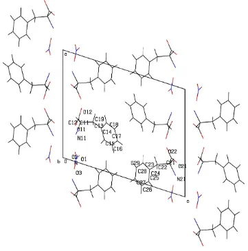
The aggregation of the hydrophilic zone is along thez= 0 plane. The hydrophobic zone atz =1
2consists of the phenyl
groups of both molecules and is sandwiched between two hydrophilic layers in the planes z= 0 andz= 1 (Fig. 2).
Experimental
The title compound was crystallized from an aqueous solution of a 2:1 stoichiometric ratio of l-phenylalanine and nitric acid by slow evaporation.
Crystal data 2C9H11NO2H+NO3ÿ
Mr= 393.39
Monoclinic,P21
a= 12.536 (16) AÊ b= 5.378 (4) AÊ c= 14.962 (9) AÊ = 107.83 (8)
V= 960.2 (15) AÊ3
Z= 2
Dx= 1.361 Mg mÿ3
Dm= 1.350 Mg mÿ3
Dmmeasured by ¯otation in a
mixture of carbon tetrachloride and xylene
MoKradiation Cell parameters from 25
re¯ections = 14.5±23.4 = 0.11 mmÿ1
T= 293 (2) K Needle, colorless 0.450.300.15 mm Data collection
Enraf±Nonius CAD-4 diffractometer !±2scans
Absorption correction: scan (Northet al., 1968) Tmin= 0.963,Tmax= 0.984
1895 measured re¯ections 1895 independent re¯ections
1356 re¯ections withI> 2(I) max= 25.0
h=ÿ14!14 k= 0!6 l= 0!17
3 standard re¯ections frequency: 60 min intensity decay: none Re®nement
Re®nement onF2
R[F2> 2(F2)] = 0.069
wR(F2) = 0.220
S= 1.09 1895 re¯ections 250 parameters
H-atom parameters constrained
w= 1/[2(F
o2) + (0.1548P)2]
whereP= (Fo2+ 2Fc2)/3
(/)max< 0.001
max= 0.30 e AÊÿ3
min=ÿ0.34 e AÊÿ3
Absolute structure: Flack (1983) Flack parameter = 0 (4)
Table 1
Selected geometric parameters (AÊ,).
O11ÐC11 1.202 (8)
O12ÐC11 1.313 (6) O21ÐC21O22ÐC21 1.220 (7)1.311 (7)
O11ÐC11ÐC12ÐN11 ÿ9.9 (7) N11ÐC12ÐC13ÐC14 68.2 (7) C12ÐC13ÐC14ÐC19 89.0 (8) C12ÐC13ÐC14ÐC15 ÿ92.3 (8)
O21ÐC21ÐC22ÐN21 2.5 (8) N21ÐC22ÐC23ÐC24 71.0 (7) C22ÐC23ÐC24ÐC29 97.0 (8) C22ÐC23ÐC24ÐC25 ÿ84.3 (7)
H atoms bonded to C atoms were placed in geometrically calcu-lated positions and allowed to ride on the attached atoms. Attempts to introduce H atoms attached to the amino N atom yielded a slight increase in all discrepancy values. Therefore, these H atoms were not included in the ®nal re®nement. The absolute con®guration is known from the synthesis and is indeterminate from the X-ray diffraction experiment.
Data collection: CAD-4 Software (Enraf±Nonius, 1989); cell re®nement: CAD-4 Software; data reduction: CAD-4 Software; program(s) used to solve structure: SHELXS97 (Sheldrick, 1997); program(s) used to re®ne structure:SHELXL97 (Sheldrick, 1997); molecular graphics:PLATON(Spek, 1999); software used to prepare material for publication:SHELXL97.
BS and RKR thank the Department of Science and Tech-nology (DST), Government of India, for ®nancial support.
References
Al-Karaghouli, A. R. & Koetzle, T. F. (1975).Acta Cryst.B31, 2461±2465. Enraf±Nonius (1989).CAD-4Software. Version 5.0. Enraf±Nonius, Delft, The
Netherlands.
Flack H. D. (1983).Acta Cryst.A39, 876±881.
Acta Cryst.(2001). E57, o916±o918 N. Srinivasanet al. 2C9H11NO2H+NO3ÿ
o917
organic papers
Figure 1
The structures of phenylalanine residues showing the atomic numbering scheme and 50% probability displacement ellipsoids (Johnson, 1976).
Figure 2
organic papers
o918
N. Srinivasanet al. 2C9H11NO2H+NO3ÿ Acta Cryst.(2001). E57, o916±o918Gorbitz, C. H. & Etter, M. C. (1992).Acta Cryst.C48, 1317±1320.
Gurskaya, G. V. & Vainshtein, B. K. (1963).Sov. Phys. Crystallogr.8, 288±291. Johnson, C. K. (1976).ORTEPII. Report ORNL-5138. Oak Ridge National
Laboratory, Tennessee, USA.
Lakshminarayanan, A. V., Sashisekaran, V. & Ramachandran, G. N. (1967). In Conformation of Biopolymers, edited by G. N. Ramachandran. London: Academic Press.
Nagashima, N., Sano, C., Kawakita, T. & Iitaka, Y. (1992).Anal. Sci.8, 723± 725.
North, A. C. T., Phillips, D. C. & Mathews, F. S. (1968).Acta Cryst.A24, 351± 359.
Sheldrick, G. M. (1997). SHELXL97 and SHELXS97. University of GoÈttingen, Germany.
Spek, A. L. (1999). PLATON for Windows. Utrecht University, The Netherlands.
Srikrishnan, T., Narasinga Rao, S. & Parthasarathy, R. (1984).Acta Cryst.A40, C-92.
supporting information
sup-1
Acta Cryst. (2001). E57, o916–o918
supporting information
Acta Cryst. (2001). E57, o916–o918 [doi:10.1107/S1600536801014350]
L
-Phenylalanine
–
nitric acid (2/1)
N. Srinivasan, B. Sridhar and R. K. Rajaram
S1. Comment
The crystal structures of L-phenylalanine hydrochloride (Gurskaya & Vainstein, 1963; Al-Karaghouli & Koetzle, 1975),
L-phenylalanine L-phenylalaninium formate (Gorbitz & Etter, 1992), bis(L-phenylalanine) sulfate monohydrate
(Nagashima et al., 1992) and L-phenylalanine L-phenylalaninium perchlorate (Srinivasan & Rajaram, 1997) have been
reported. Even though the title compound, (I), has been studied (Srikrishnan et al., 1984), no structural data are available
as the above publication does not contain the atomic coordinates and has a misprint in one of the cell dimensions.
Therefore, in the present work, an independent X-ray diffraction study of the title compound was undertaken.
Both phenylalanine residues (1 and 2) have similar geometries (Fig. 1). In both residues, the bond distances C—O
[1.313 (6) and 1.311 (7) Å] and C═O [1.202 (8) and 1.220 (7) Å] are nearly equal and the single-bonded O atoms of 1
and 2 (O12···O22) are at a distance of 2.426 (8) Å. This may refer to a short hydrogen bond between the singly bonded
carboxyl O atoms (O12···O22), as found in L-phenylalanine L-phenylalaninium perchlorate (Srinivasan & Rajaram,
1997). However, this H atom could not be located objectively. Hence, taking into account the equality of the C—O
distances (see above), it can be surmised that the two residues may be connected by either a symmetric hydrogen bond or
an asymmetric hydrogen bond with the H atom disordered over two positions.
The conformation angles ψ1 for residues 1 and 2 are -9.9 (7) and 2.5 (8)°, respectively (see Table 1). This tendency
towards non-planarity is also found in various amino acids (Lakshminarayanan et al., 1967). The branched side
conformation angle χ1 shows a gauche I conformation for both residues [68.2 (7) and 71.0 (7)°], which agrees well with
formate and perchlorate complexes. The χ21 [89.0 (8) and 97.0 (8)°] and χ22 [92.3 (8) and -84.3 (7)°] torsion angles
indicate that the residues have folded conformation.
The aggregation of hydrophilic zone is along z = 0 plane. The hydrophobic zone at z = 1/2 consists of the phenyl groups
of both molecules and is sandwiched between two hydrophilic layers at z = 0 and z = 1 planes (Fig. 2).
S2. Experimental
The title compound was crystallized from an aqueous solution of a 2:1 stoichiometric ratio of L-phenylalanine and nitric
acid by slow evaporation.
S3. Refinement
H atoms bonded to C atoms were placed in geometrically calculated positions and allowed to ride on the attached atom.
Attempts to introduce H atoms attached to the amino N atom yielded a slight increase in all discrepancy values.
supporting information
sup-2
[image:5.610.126.482.72.254.2]Acta Cryst. (2001). E57, o916–o918 Figure 1
The structures of phenylalanine residues showing the atomic numbering scheme and 50% probability displacement
ellipsoids (Johnson, 1976).
Figure 2
[image:5.610.125.486.307.672.2]supporting information
sup-3
Acta Cryst. (2001). E57, o916–o918
L-Phenylalanine–nitric acid (2/1)
Crystal data
2C9H11NO2·H+·NO3−
Mr = 393.39
Monoclinic, P21
a = 12.536 (16) Å
b = 5.378 (4) Å
c = 14.962 (9) Å
β = 107.83 (8)°
V = 960.2 (15) Å3
Z = 2
F(000) = 416
Dx = 1.361 Mg m−3
Dm = 1.350 Mg m−3
Dm measured by flotation in a mixture of carbon tetrachloride and xylene
Mo Kα radiation, λ = 0.71073 Å Cell parameters from 25 reflections
θ = 14.5–23.4°
µ = 0.11 mm−1
T = 293 K Needle, colorless 0.45 × 0.3 × 0.15 mm
Data collection
Enraf-Nonius sealed tube diffractometer
Radiation source: fine-focus sealed tube Graphite monochromator
ω–2θ scans
Absorption correction: ψ scan (North et al., 1968)
Tmin = 0.963, Tmax = 0.984 1895 measured reflections
1895 independent reflections 1356 reflections with I > 2σ(I)
Rint = 0.000
θmax = 25.0°, θmin = 2.5°
h = −14→14
k = 0→6
l = 0→17
3 standard reflections every 60 min intensity decay: none
Refinement
Refinement on F2 Least-squares matrix: full
R[F2 > 2σ(F2)] = 0.069
wR(F2) = 0.220
S = 1.09 1895 reflections 250 parameters 3 restraints
Primary atom site location: structure-invariant direct methods
Secondary atom site location: difference Fourier map
Hydrogen site location: inferred from neighbouring sites
H-atom parameters constrained
w = 1/[σ2(F
o2) + (0.1548P)2] where P = (Fo2 + 2Fc2)/3 (Δ/σ)max < 0.001
Δρmax = 0.30 e Å−3 Δρmin = −0.34 e Å−3
Absolute structure: Flack H D (1983) Absolute structure parameter: 0 (4)
Special details
Geometry. All e.s.d.'s (except the e.s.d. in the dihedral angle between two l.s. planes) are estimated using the full covariance matrix. The cell e.s.d.'s are taken into account individually in the estimation of e.s.d.'s in distances, angles and torsion angles; correlations between e.s.d.'s in cell parameters are only used when they are defined by crystal symmetry. An approximate (isotropic)
treatment of cell e.s.d.'s is used for estimating e.s.d.'s involving l.s. planes.
supporting information
sup-4
Acta Cryst. (2001). E57, o916–o918
Fractional atomic coordinates and isotropic or equivalent isotropic displacement parameters (Å2)
x y z Uiso*/Ueq
supporting information
sup-5
Acta Cryst. (2001). E57, o916–o918 Atomic displacement parameters (Å2)
U11 U22 U33 U12 U13 U23
N 0.041 (2) 0.050 (4) 0.059 (4) −0.010 (3) 0.012 (2) 0.006 (4) O1 0.080 (4) 0.064 (5) 0.177 (9) −0.014 (4) 0.036 (5) −0.011 (6) O2 0.093 (5) 0.070 (5) 0.223 (11) 0.037 (5) 0.072 (6) 0.003 (6) O3 0.042 (2) 0.066 (4) 0.094 (4) −0.004 (3) 0.013 (2) 0.004 (4) O11 0.048 (2) 0.031 (2) 0.042 (3) −0.009 (2) −0.0024 (18) −0.002 (2) O12 0.0278 (16) 0.050 (3) 0.098 (4) 0.002 (2) 0.020 (2) −0.019 (3) C11 0.029 (2) 0.039 (4) 0.029 (3) 0.016 (3) 0.004 (2) −0.005 (3) C12 0.040 (3) 0.034 (4) 0.037 (3) 0.001 (3) 0.017 (2) 0.003 (3) N11 0.040 (2) 0.033 (3) 0.054 (3) 0.002 (3) 0.025 (2) −0.004 (3) C13 0.055 (3) 0.028 (4) 0.052 (4) 0.003 (3) 0.020 (3) −0.011 (3) C14 0.053 (3) 0.042 (4) 0.043 (4) −0.011 (3) 0.021 (3) −0.002 (4) C15 0.051 (3) 0.058 (5) 0.060 (4) −0.002 (4) 0.022 (3) −0.005 (4) C16 0.090 (6) 0.130 (11) 0.060 (5) −0.029 (7) 0.043 (5) 0.006 (7) C17 0.123 (8) 0.087 (8) 0.052 (5) −0.033 (7) 0.033 (5) 0.003 (6) C18 0.117 (7) 0.052 (5) 0.057 (5) −0.027 (6) −0.008 (5) 0.024 (5) C19 0.076 (4) 0.046 (4) 0.022 (3) 0.005 (4) −0.005 (3) 0.000 (3) O21 0.048 (2) 0.035 (2) 0.041 (2) 0.006 (2) 0.0207 (19) 0.000 (2) O22 0.056 (2) 0.036 (3) 0.087 (4) 0.001 (2) 0.040 (3) 0.004 (3) C21 0.032 (2) 0.027 (3) 0.047 (4) −0.011 (3) 0.018 (2) −0.009 (3) C22 0.036 (2) 0.028 (3) 0.037 (3) 0.002 (3) 0.005 (2) −0.010 (3) N21 0.036 (2) 0.051 (3) 0.044 (3) 0.014 (3) 0.012 (2) −0.003 (3) C23 0.053 (3) 0.040 (4) 0.051 (4) 0.001 (3) 0.016 (3) 0.015 (4) C24 0.040 (3) 0.036 (4) 0.041 (4) 0.003 (3) −0.005 (3) 0.010 (3) C25 0.046 (3) 0.066 (5) 0.037 (4) −0.004 (4) 0.013 (3) −0.006 (4) C26 0.066 (5) 0.083 (8) 0.099 (8) −0.004 (6) −0.001 (5) −0.018 (7) C27 0.091 (5) 0.057 (5) 0.041 (4) −0.022 (5) 0.013 (4) −0.018 (4) C28 0.083 (5) 0.061 (6) 0.040 (4) 0.012 (5) 0.010 (4) −0.010 (4) C29 0.071 (4) 0.055 (5) 0.045 (4) 0.013 (4) 0.025 (4) −0.004 (4)
Geometric parameters (Å, º)
supporting information
sup-6
Acta Cryst. (2001). E57, o916–o918
C15—C16 1.382 (13) C26—C27 1.408 (10) C15—H15 0.9300 C26—H26 0.9300 C16—C17 1.307 (16) C27—C28 1.332 (11) C16—H16 0.9300 C27—H27 0.9300 C17—C18 1.421 (14) C28—C29 1.344 (12) C17—H17 0.9300 C28—H28 0.9300 C18—C19 1.400 (12) C29—H29 0.9300 C18—H18 0.9300
O2—N—O1 129.1 (6) C18—C19—H19 120.3 O2—N—O3 117.4 (7) O21—C21—O22 123.7 (6) O1—N—O3 113.5 (7) O21—C21—C22 123.6 (5) O11—C11—O12 127.0 (6) O22—C21—C22 112.6 (5) O11—C11—C12 122.9 (5) C21—C22—C23 114.7 (5) O12—C11—C12 109.8 (6) C21—C22—N21 106.4 (5) N11—C12—C13 111.2 (5) C23—C22—N21 111.5 (4) N11—C12—C11 106.8 (5) C21—C22—H22 108.0 C13—C12—C11 113.5 (5) C23—C22—H22 108.0 N11—C12—H12 108.4 N21—C22—H22 108.0 C13—C12—H12 108.4 C24—C23—C22 113.4 (6) C11—C12—H12 108.4 C24—C23—H23A 108.9 C14—C13—C12 116.3 (6) C22—C23—H23A 108.9 C14—C13—H13A 108.2 C24—C23—H23B 108.9 C12—C13—H13A 108.2 C22—C23—H23B 108.9 C14—C13—H13B 108.2 H23A—C23—H23B 107.7 C12—C13—H13B 108.2 C29—C24—C25 115.1 (7) H13A—C13—H13B 107.4 C29—C24—C23 123.0 (6) C19—C14—C15 118.8 (7) C25—C24—C23 122.0 (6) C19—C14—C13 122.4 (6) C26—C25—C24 120.2 (4) C15—C14—C13 118.9 (7) C26—C25—H25 119.9 C16—C15—C14 117.6 (9) C24—C25—H25 119.9 C16—C15—H15 121.2 C25—C26—C27 123.1 (4) C14—C15—H15 121.2 C25—C26—H26 118.4 C17—C16—C15 126.7 (9) C27—C26—H26 118.4 C17—C16—H16 116.7 C28—C27—C26 115.4 (7) C15—C16—H16 116.7 C28—C27—H27 122.3 C16—C17—C18 115.8 (9) C26—C27—H27 122.3 C16—C17—H17 122.1 C27—C28—C29 122.9 (8) C18—C17—H17 122.1 C27—C28—H28 118.6 C19—C18—C17 121.7 (10) C29—C28—H28 118.6 C19—C18—H18 119.1 C28—C29—C24 123.2 (7) C17—C18—H18 119.1 C28—C29—H29 118.4 C14—C19—C18 119.5 (8) C24—C29—H29 118.4 C14—C19—H19 120.3
supporting information
sup-7
Acta Cryst. (2001). E57, o916–o918