www.wjpr.net Vol 4, Issue 2, 2015. 1653
CEREBRAL ISCHEMIC-REPERFUSION INJURY AND
NEUROPROTECTIVE ACTIVITY OF HERBAL DRUGS
Bhagat Singh Jaiswal* and Mukul Tailang
S.O.S. in Pharmaceutical Sciences, Jiwaji University, Gwalior (M.P.) India.
ABSTRACT
Cerebral ischemia/reperfusion injury is one of the leading causes of human death and disability throughout the world and has attracted more and more attention in the field of drug discovery. Focal (stroke) and Global (cardiac arrest) cerebral ischemia represents diseases that are common in the human population. Stroke and cardiac arrest, which are major causes of death and disability, affect millions of individuals around the world and are responsible for the leading health care costs of all diseases. After cerebral ischemia/reperfusion, the reactive oxygen species production is dramatically increased and overwhelms endogenous antioxidant systems, leading to oxidative stress. The brain is particularly vulnerable to oxidative stress injury due to its high consumption of oxygen, abundant polyunsaturated fatty acids and low levels of endogenous antioxidants. Antioxidants are significant substances which can protect the body from oxidative damage. The extensive prescription of synthetic antioxidants is ruled out owing to their toxicity and unavoidable side effects. There is an increasing interest in the use of natural antioxidants for prophylaxis or treatment of oxidative stress-related diseases. According to the pharmacological mechanisms underlying neuroprotection, we evaluated some important natural drugs from traditional medicinal herb that posses the neuroprotective effects on cerebral ischemic-reperfusion injury.
KEYWORDS: Cerebral ischemia, Oxidative stress, Herbal drugs, Stroke.
INTRODUCTION
Stroke was the leading cause of death 30 years ago when hypertensive intracerebral hemorrhage was the most common cause of stroke deaths. At present, stroke is the second leading cause of death in most of the countries; it ranks behind only cancer deaths. The
Volume 4, Issue 2, 1653-1667. Research Article ISSN 2277– 7105
Article Received on 09 Dec 2014,
Revised on 03 Jan 2015, Accepted on 28 Jan 2015
*Correspondence for
Author
Bhagat Singh Jaiswal
S.O.S. in Pharmaceutical
Sciences, Jiwaji
University, Gwalior
www.wjpr.net Vol 4, Issue 2, 2015. 1654 decline in the death rate from stroke over the last 20 years has been attributed to improved control of hypertension and advances in the management of cerebral hemorrhage in the acute stage. However, the incidence of cerebral infarction resulting in disability in the survivors of ischemic stroke appears to be on the increase, at a time when the proportion of elderly individuals is rapidly increasing in India.[1] Of all types of stroke, the incidence of hemorrhagic cerebrovascular disease, including subarachnoid hemorrhage and intracerebral hemorrhage, is about 20-25%; the incidence of ischemic stroke is 75-80%. By 2020 heart disease and stroke will become the leading cause of death and disability worldwide with the number of fatalities projected to increases to over 20 million a year and by 2030 to over 24 million a year.[2] The increase in the number of individuals with mental and physical handicaps after stroke presents considerable problems in terms of quality of life and socioeconomic costs.
Permanent brain damage caused by ischemia and reperfusion accompanies disease processes such as stroke and resuscitation from cardiac. In ischemic stroke, only part of the brain is involved, and the ischemia is dense or complete only in the center of the affected area. This area of dense ischemia is surrounded by a penumbral zone in which the blood flow is diminished but not completely lost. In cardiac arrest and resuscitation, the entire brain is subjected to a transient period of complete ischemia followed by reperfusion.[3, 4]
During reperfusion after ischemia, while restoration of oxygen and glucose supply reinstates the oxidative phosphorylation that helps to normalize energy demanding physiologic processes, a parallel cascade of deleterious biochemical processes can be triggered that may paradoxically antagonize the beneficial effect of reperfusion.[5, 6]
This phenomenon has been demonstrated in various tissues, especially in the brain, and has been termed reperfusion injury (RI).[7] The understanding and treatment of RI is an important objective in the new era of reperfusion therapy for stroke. Recently it is demonstrated that 3 hours of focal ischemia followed by 3 hours of reperfusion in the rat produces more damage than 6 hours of continuous ischemia without reperfusion.[8]
Global and Focal cerebral ischemia
www.wjpr.net Vol 4, Issue 2, 2015. 1655 during extracorporeal circulation following cardiac arrest (CA) from ventricular fibrillation or asystole that lasts 5 to 10 minutes. Global ischemia from CA results in a predictable pattern of histologic injury in which specific neuronal populations are affected (selective ischemic necrosis) [9-11]. Although reperfusion restores CBF and it can lead to secondary brain injury from influx of neutrophils and to increases in reactive oxygen species (ROS), cerebral edema and hemorrhage. Elevated levels of ROS may lead to damage of intracellular proteins and DNA by way of oxidation and by activating a number of pathways that lead to cell death. Unlike global cerebral ischemia, focal cerebral ischemia entails reduction in regional CBF in a specific vascular territory and is usually encountered clinically as an ‘‘ischemic stroke’’ due
to thromboembolic or artherothrombotic vaso-occlusive disease. Normal CBF ranges from 50 to 75 ml/100 g of brain tissue per minute and can differ between the white and gray brain matter. Ischemic depolarization occurs when CBF decreases to levels of approximately 18 ml/100 g of brain tissue per minute, and neuronal cell death ensues if CBF is less than 10 ml/100 g of brain tissue per minute. In focal ischemia, the ischemic vascular bed comprises an area with severe CBF reduction that consists of an ‘‘ischemic core’’ and a more distal ‘‘ischemic penumbra’’ and includes regions that are marginally perfused and might be served
by collateral vascular channels. Histopathologic outcome following focal ischemia largely depends on ischemic severity and duration. Increasing durations of depolarizing ischemia are associated with a spectrum of histopathologic correlates from reversible injury to irreversible cerebral infarction.[9, 12]
In spite of the growing understanding of the mechanisms of neuronal damage and death accompanying brain ischemia and reperfusion, effective therapy has remained elusive. Preclinical and clinical trials of calcium channels blockers have shown some evidence of neuronal protection. Drugs that block glutamate receptors have been largely ineffective in neuronal preservation after complete ischemia, although they appear somewhat more promising in neuronal salvage in the penumbral zone surrounding the area of severe ischemia in stroke models.
www.wjpr.net Vol 4, Issue 2, 2015. 1656 recanalization caused by endogenous thrombolysis is a complex process that depends on the size, composition, and anatomic location of the thrombus, and frequently takes hours to days to occur.[13] Numerous preclinical and clinical studies using thrombolytic agents, especially tissue plasminogen activator, demonstrated pharmacological recanalization and restoration of cerebral circulation (reperfusion) within minutes of the onset of treatment. Tissue plasminogen activator recently was shown to be effective and approved for urgent treatment of ischemic stroke in humans. Because recanalization of occluded vessels in the clinic is becoming a daily practice, careful inspection of the possible negative influence of reperfusion (apart from the risk related to hemorrhage) must be taken into consideration.[14]
The lack of effective, widely applicable and safe pharmacological treatments for ischemic stroke patients may explain a growing interest towards the traditional medicines, for which extensive observation and vast experience has accumulated over the past years. It has been suggested that some herbal medicines or their products, may improve microcirculation in the brain, protect against ischemic reperfusion injury, possess neuroprotective properties and inhibit apoptosis, thus justifying their use in ischemic stroke patients.[15, 16]
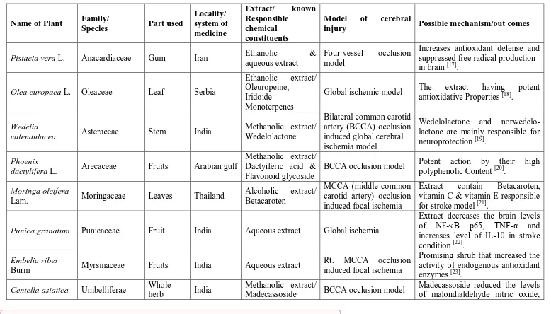
www.wjpr.net Vol 4, Issue 2, 2015. 1657 Table 1: Details of plants showing neuroprotection against cerebral ischemic-reperfusion injury
Name of Plant Family/
Species Part used
Locality/ system of medicine
Extract/ known Responsible
chemical constituents
Model of cerebral
injury Possible mechanism/out comes
Pistacia vera L. Anacardiaceae Gum Iran Ethanolic &
aqueous extract
Four-vessel occlusion model
Increases antioxidant defense and suppressed free radical production in brain [17].
Olea europaea L. Oleaceae Leaf Serbia
Ethanolic extract/ Oleuropeine,
Iridoide Monoterpenes
Global ischemic model The extract having potent antioxidative Properties [18].
Wedelia
calendulacea Asteraceae Stem India
Methanolic extract/ Wedelolactone
Bilateral common carotid artery (BCCA) occlusion induced global cerebral ischemia model
Wedelolactone and norwedelo-lactone are mainly responsible for neuroprotection [19].
Phoenix
dactylifera L. Arecaceae Fruits Arabian gulf
Methanolic extract/ Dactyiferic acid & Flavonoid glycoside
BCCA occlusion model Potent action by their high polyphenolic Content [20].
Moringa oleifera
Lam. Moringaceae Leaves Thailand
Alcoholic extract/ Betacaroten
MCCA (middle common carotid artery) occlusion induced focal ischemia
Extract contain Betacaroten, vitamin C & vitamin E responsible for stroke model [21].
Punica granatum Punicaceae Fruit India Aqueous extract Global ischemia
Extract decreases the brain levels of NF-κB p65, TNF-α and increases level of IL-10 in stroke condition [22].
Embelia ribes
Burm Myrsinaceae Fruits India Aqueous extract
Rt. MCCA occlusion induced focal ischemia
Promising shrub that increased the activity of endogenous antioxidant enzymes [23].
Centella asiatica Umbelliferae Whole
herb India
Methanolic extract/
Madecassoside BCCA occlusion model
www.wjpr.net Vol 4, Issue 2, 2015. 1658
pro-inflammatory cytokines and NF-κB p65 in ischemic condition
[24]
. Hypericum
perforatum Hypericaceae
Whole
Plant Ayurveda
Ethanolic extract/ Hyperforin
BCCA occlusion induced global cerebral ischemia
The presence of hyperforin in extract, increases in cyclic AMP levels that reversing the stroke [25].
Ocimum
basilicum L. Lamiaceae Leaf India Ethyl acetate extract Global cerebral ischemia
Plant showing the reduction of cerebral infarct size and lipid peroxidation, restoration of GSH content, and attenuation of motor dysfunctions [26].
Pongamia pinnata
(L) Pierre Leguminosae Root Ayurveda Ethanolic extract
BCCA occlusion induced global cerebral ischemia
Bioflavonoid contents of the root improved cerebral perfusion and microcirculation by inhibiting the lymphatic infiltration, astrocytosis & edema [27].
Eclipta alba
(Linn.) Hassk Asteraceae Aerial part India
Hydroalcoholic extract/
Wedelolactone
Global cerebral ischemia
The presence of Wedelolactone, that inhibit the pro-inflammatory cytokine IL-1β maturation and pathological neuronal apoptosis
[28]
.
Withania
somnifera Dunal Solanaceae Root Ayurveda
Aqueous extract/ Sitoindosides VII-X & Withaferin A
Acute
ischemia-reperfusion injury induced by BCCAO model
Plant shows beneficial in learning & memory symptoms of stroke [29].
Araucaria
bidwillii Hook Araucariaceae Leaves
Himalaya regions
Ethyl acetate
extract/ Biflavone Global ischemia
Extract Inhibit the free radical generation, reactive oxygen species scavenging and modulation of Intracellular antioxidant [30]. Artemisia
absinthium L. Asteraceae Aerial part
Traditional Chinese
Methanolic extract/ Flavonoid
MCCA occlusion
induced focal ischemia
www.wjpr.net Vol 4, Issue 2, 2015. 1659
medicine improved behavioral outcome
during cerebral I/R injury [31].
Momordica
charantia L. Cucurbitaceae Fruits
Asia, Africa and Latin America
Lyophilized juice/ Charantin, insulin-like peptide, Cucurbutanoids, Momordicin
BCCAO induced global ischemia
Potent action due to its high reactive oxygen species scavenging activity [32].
Cordyceps
militaris Cordyceps Fungus
Traditional Chinese medicine
Aqueous extract/
Cordycepine Global ischemia
The active cordycepine decreases the excitotoxicity of excitatory amino acids, inhibit the production of the matrix metalloproteinase -3 as well as block free radicals in stroke model [33].
Lithospermm
erythrorhizon Boraginaceae Root
Traditional Chinese medicine
Maize oil extract/
Shikonin Focal ischemia
Antioxidant ability appears to be a basic and important mechanism of the neuroprotective effect of shikonin [34].
Hemidesmus
indicus Asclepideaceae Root
Ayurveda, Unani & Siddha
Methanolic extract Four-vessel occlusion model
Drug decreases in the levels of Cholinesterase, Glutamate, Mono amino Oxidase-B, increases in levels of dopamine & serotonin, both ways get beneficial effect in stroke patients [35].
Ageratum
conyzoides L. Compositae
Whole
plant Kenya Pet. Ether extract
BCCAO induced Global ischemia
Plant shows the reduction in behavioral score, hyper locomotion and neuronal damage in brain injury [36].
Vanda tessellata Orchidaceae Leaves Ayurveda &
Unani Pet. Ether extract Global ischemia
Plant possesses significant activity against ischemic injury [37].
Allium sativum L. Alleaceae Fruits India Aqueous extract Focal ischemia Extract improves neurobehavior
www.wjpr.net Vol 4, Issue 2, 2015. 1660
aspartate levels, possibly by increasing the endogenous defensive capacity of the brain [38].
Nigella sativa L. Ranunculaceae Seeds Iran Aqueous and
ethanolic extracts
Four-vessel occlusion model
Both extract shows potent anti ischemic activity [39].
Allium cepa L. Alleaceae
Bulb (outer scales)
India
Methanolic extract & Flavonoid rich fraction
Global ischemia
Flavonoid rich fraction reduced the cerebral damage and oxidative stress [40].
Angelica Sinensis
Diels Umbelliferae
Whole plant
Traditional Chinese medicine
Ligustilide MCCAO induced focal ischemia
Ligustilide promoting EPO transcription via an ERK signaling pathway and inhibiting RTP801 expression in ischemic disorders
[41]
.
Litsea coreana
leve Lauraceae Aerial part
Traditional Chinese medicine
Total flavonoid
fraction Focal ischemia
Flavonoid suppressed the increase in the levels of nitrates, malondialdehyde and lactate dehydrogenase in cerebral injury
[42]
.
Vitis vinifera L. Vitaceae Seed
Ayurvedic medicine system
Ethanolic extracts Global ischemia
Promising effects of seed might be attributable partly to its antioxidant capacity and inhibitory effect on glutamate activities in the brain [43].
Cyperus rotundus Cyperaceae Rhizoms
Ayurvedic medicine system
Ethanolic extracts Global ischemia
Extract can reduce the ischemia-induced apoptosis in CA1 sector of hippocampus after transient, global cerebral Ischemia [44].
Luffa acutangula Cucurbitaceae Whole
plant
Tirupati, India
Petroleum ether
extract BCCAO ischemic model
Proves beneficial in hyper locomotion & brain damage [45].
Melissa officinalis Labiatae Leaves Iran Aqueous extract Global ischemia
www.wjpr.net Vol 4, Issue 2, 2015. 1661 Achyrocline
satureioides (Lam.) DC. (marcela)
Asteraceae Flower Uruguay Aqueous extract/
Quercetin Focal ischemia
Quercetin is responsible for the preventive benefits of extract due to the antioxidant and anti inflammatory Properties [47].
Dalbergia
latifoliais Fabeceae Bark
Bundelkhan d and Central India
Methanolic extract/
Flavonoid Global ischemia
Flavonoid fraction prevents the cerebral injury via potent antioxidant property [48].
Kaempferia
parviflora Zingiberaceae Rhizo-mes Thailand
Ethanolic extracts/ Dimethoxy flavone
MCCAO induced focal ischemia
Extract inhibit over production of cytotoxic nitric oxide, enhance free radical scavenger and antioxidant activity [49].
Ginkgo biloba Ginkgoceae Leaf
Traditional Chinese medicine
EGb761 extract MCAO model
EGb761 enhance the endogenous protective Gene, Heme oxygenase (HO-1) and reduced brain damage
[50]
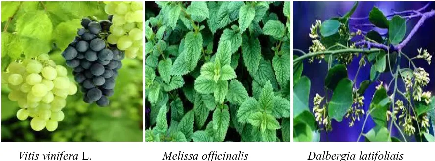
www.wjpr.net Vol 4, Issue 2, 2015. 1662 Pistacia vera L. Olea europaea L. Wedelia calendulacea
Phoenix dactylifera L. Punica granatum Embelia ribes
Pongamia pinnata Withania somnifera Momordica charantia L.
www.wjpr.net Vol 4, Issue 2, 2015. 1663 Vitis vinifera L. Melissa officinalis Dalbergia latifoliais
Figure 1: Parts of plants (selected) which are showing the Neuroprotective activity.
CONCLUSION
Herbs are staging a comeback and herbal ‘renaissance’ is happening all over the globe. The herbal products today symbolize safety in contrast to the synthetics that are regarded as unsafe to human and environment. Currently, there is an enormous increase in the use of herbs and other alternative medicines in the treatment of several disorders. As newer technologies developed, tremendous scope for further scientific exploration of therapeutic efficacy of herbal drugs generated. A review of the literature indicates that the medicinal plants are may be the most prolific source for treatment of stroke or related disorders. The Chinese and Indian system of medicine, especially Ayurveda, has several medicinal plants with proven beneficial claims towards these pathological conditions. The present paper directly reviewed and documented information on over medicinal plant species with links to cardiac arrest and stroke. Herbal extracts represent combinatorial chemistry of nature with vast complex effect on numerous cellular components and functions. There also difficulty in standardization, pharmacodynamics and pharmacokinetics of these herbal drugs. Therefore, the efficacy of herbals in the treatment of stroke needs to be further explored.
REFERENCES
1. Traystman R. Animal Models of Focal and Global Cerebral Ischemia. ILAR J, 2003; 44: 84-95.
2. Lloyd D, Adams RJ, Brown TM, Carnethon M, Dai S. Heart disease and stroke statistics: 2010 update. A report from American Heart Association. Circulation, 2009;
www.wjpr.net Vol 4, Issue 2, 2015. 1664 4. Krause S, Tiffany DR. Suppression of protein synthesis in the reperfused brain. Stroke,
1993; 24: 747-756.
5. Pulsinelli WA, Duffy TA. Regional Energy balance in rat brain after transient forebrain ischemia. J Neurochem, 1983; 40: 1500-1503.
6. Kinouchi H, Imaizumi S, Yoshimoto T, Yamamoto H, Motomiya M. Changes of polyphosphoinositides, Iysophospholipid, and free fatty acids in transient cerebral ischemia of rat brain. Mol Chem Neuropathol, 1990; 12: 215-228.
7. Karmazyn M. Ischemic and reperfusion injury in the heart: cellular mechanisms and pharmacological intervention. Can J Physiol Pharmacol, 1991; 69: 719-730.
8. Yang GY, Betz AL. Reperfusion-induced injury to the blood brain barrier after middle cerebral artery occlusion in rats. Stroke, 1994; 25: 1658-1665.
9. Pulsinelli WA. Selective neuronal vulnerability: morphological and molecular characteristics. Prog Brain Res, 1995; 63: 29-37.
10.Petito CK, Pulsinelli WA. Delayed neuronal recovery and neuronal death in rat hippocampus following severe cerebral ischemia: possible relationship to abnormalities in neuronal processes. J Cereb Blood Flow Metab, 1984; 4: 194-205.
11.Kaplan B, Brint S, Tanabe J. Temporal thresholds for neocortical infarction in rats subjected to irreversible focal cerebral ischemia. Stroke, 1991; 22: 1032-9.
12.Bhardwaj A, Alkayed NJ, Kirsch JR. Mechanisms of ischemic brain damage. Curr Cardiol Rep, 2003; 5: 160-7.
13.Aronowski J, Strong R, Grotta JC. Reperfusion injury: demonstration of brain damage produced by reperfusion after transient focal ischemia in rats. J Cereb Blood Flow Metab, 1997; 17: 1048-1056.
14.Wang NL, Liou YL, Lin MT, Lin CL, Chang CK. Chinese herbal medicine, Shengmai San, is effective for improving circulatory shock and oxidative damage in the brain during heatstroke. J Pharmacol Sci, 2005; 97: 253-65.
15.Kim H. Neuroprotective herbs for stroke therapy in traditional eastern medicine. Neurol Res, 2005; 27: 287-301.
16.Bei W, Peng W, Ma Y, Xu A. NaoXinQing, an anti-stroke herbal medicine, reduces hydrogen peroxide-induced injury in NG108-15 cells. Neurosci Lett, 2004; 33: 262-5. 17.Mansouri SM, Naghizadeh B, Hosseinzadeh H. The effect of Pistacia vera L. gum extract
www.wjpr.net Vol 4, Issue 2, 2015. 1665 18.Dekanskia D, Selakovi V, Piperskia V, Radulovi Z, Koreni A, Radenovi L. Protective effect of olive leaf extract on hippocampal injury induced by transient global cerebral ischemia and reperfusion in Mongolian gerbils. J Phymed, 2011; 18: 1137-1143.
19.Prakash T, Kotresha D, Nedendla R. Neuroprotective activity of Wedelia calendulacea on cerebral ischemia/reperfusion induced oxidative stress in rats. Indian J Pharmacol, 2011; 43(6): 676-682.
20.Pujari R, Vyawahare N, Kagathara V. Evaluation of antioxidant and neuroprotective effect of (Phonix dactylifera) date palm against bilateral common carotid artery occlusion in rat. Indian J Exp Biol, 2011; 49: 627-633.
21.Kirisattayakul W, Wattanathorn J, Tong-Un T, Muchimapura S, Wannanon P, Jittiwat J. Cerebroprotective effect of Moringa oleifera against focal ischemic stroke induced by middle cerebral artery occlusion. Oxid Med Cell Longev, 2013; 1-10.
22.Ahmed MA, Morsy EM, Ahmed AA. Pomegranate extract protects against cerebral ischemia/reperfusion injury and preserves brain DNA integrity in rats. Life Sci, 2014; 110(2): 61-69.
23.Bhandari U, Ansari MN. Protective effect of aqueous extract of Embelia ribes Burm fruits in middle cerebral artery occlusion-induced focal cerebral ischemia in rats. Indian J Pharmacol, 2008; 40(5): 215-220.
24.Luoa Y, Yanga YP, Liub J, Lia WH, Yanga J, Suia X. Neuroprotective effects of madecassoside against focal cerebral ischemia reperfusion injury in rats. Brain Res, 2014; 1565: 37-47.
25.Trigunayat A. Effect of ethanolic extract of H. perforatum on oxidative stress induced by cerebral ischemia-reperfusion in rats. Ann Neurosci, 2009; 16(1): 1-8.
26.Bora KS, Arora S, Shri R. Role of Ocimum basilicum L. in prevention of ischemia and reperfusion-induced cerebral damage and motor dysfunctions in mice brain. J Ethnopharmacol, 2011; 137: 1360-1365.
27.Raghvendra M, Trigunyat A, Mitra RK, S Mitra. Effect of ethanolic extract of root of Pongamia pinnata on oxidative stress, behavioral and histopathological alterations induces by cerebral-ischemic reperfusion and long term hypoperfusion in rat. I J Exp Biol, 2007; 45: 868-876.
www.wjpr.net Vol 4, Issue 2, 2015. 1666 29.Trigunayat A, Raghavendra M, Singh RK, Bhattacharya AK, Acharya SB. Neuroprotective effect of Withania somnifera (WS) in cerebral ischemia-reperfusion and long-term hypoperfusion induced alterations in rats. J Nat Remedies, 2007; 7(2): 234-246.
30.Mukherjee PK, Ahamed KF, Kumar V, Mukherjee K, Houghton PJ. Protective effect of biflavones from Araucaria bidwillii Hook in rat cerebral ischemia/reperfusion induced
oxidative stress. Behav Brain Res, 2007; 178: 221-228.
31.Bora KS, Sharma A. Neuroprotective effect of Artemisia absinthium L. on focal ischemia and reperfusion-induced cerebral injury. J Ethnopharmacol, 2010; 129: 403-409.
32.Malik ZA, Singh M, Sharma PL. Neuroprotective effect of Momordica charantia in global cerebral ischemia and reperfusion induced neuronal damage in diabetic mice. J Ethnopharmacol, 2011; 133: 729-734.
33.Cheng Z, He W, Zhou X, Lv Q, Xu X, Yang S, Zhao C, Guo L. Cordycepin protects against cerebral ischemia/reperfusion injury in vivo and in vitro. Eur J Pharmacol, 2011; 664: 20-28.
34.Wang Z, Liu T, Gan L, Wang T, Yuan X, Zhang B, Chen H, Zheng Q. Shikonin protects mouse brain against cerebral ischemia/reperfusion injury through its antioxidant activity. Eur J Pharmacol, 2010; 643: 211-217.
35.Sivaraman D, Shantha S, Muralidharan P, Rahman H. Effect of Hemidesmus indicus on cerebral infarct ischemia-reperfusion injured rats by four vessel occlusion method. Pharmacologia, 2012; 3(4): 91-102.
36.Angothu S. Protective effect of Ageratum conyzoides Linn. against bilateral carotid artery occlusion induced stroke in rats. IJCPCR, 2012; 2(1): 8-13.
37.Saravana A, Gandhimathi R. Protective effect of extract of Vanda tessellata against cerebral ischemia in rats. J Pharm Biol, 2012; 2(1): 1-6.
38.Saleem S, Ahmad M, Ahmad AS, Yousuf S, Ansari MA. Behavioral and histologic neuroprotection of aqueous garlic extract after reversible focal cerebral ischemia. J Med Food, 2006; 9(4): 537-544.
39.Hosseinzadeh H, Jaafaria MR, Khoeib AR, Rahmania M. Anti-ischemic effect of Nigella sativa L. seed in male rats. IJPR, 2006; 1: 53-58.
www.wjpr.net Vol 4, Issue 2, 2015. 1667 41.Wu X, Qian ZM, Zhu L, Du F, Yung WH, Gong Q, Ke Y. Neuroprotective effect of ligustilide against ischemia-reperfusion injury via up-regulation of erythropoietin and down-regulation of RTP801. BJP, 2011; 164: 332-343.
42.Dong S, Tong X, Li J, Huang C, Hu C, Jiao H, Gu Y. Total flavonoid of Litsea coreana leve exerts anti-oxidative effects and alleviates focal cerebral ischemia/reperfusion injury. Neural Regen Res, 2013; 8(34): 3193-3202.
43.Sarkaki A, Rafieirad M, Hossini SE, Farbood Y, Motamedi F, Mansouri SM, Naghizadeh B. Improvement in memory and brain long-term potentiation deficits due to permanent hypoperfusion/ischemia by grape seed extract in rats. Iran J Basic Med Sci, 2013; 16: 1004-1010.
44.Entezari M, Hashemi M, Sharifi ZN, Movassaghi S. Effect of Cyperus rotundus on ischemic brain injury in a rat model of transient global ischemia/ reperfusion. AFINIDAD, 2014; 80(566): 117-120.
45.Sathianarayanan S, Jose A, Rajasekaran A, George RM, Chittethu AB. Evaluation of protective effect of Luffa acutangula extract against bilateral carotid artery occlusion (BCCAO) induced stroke in rats. IJPSR, 2012; 2(1): 1-6.
46.Bayat M, Tameh AA, Ghahremani MH, Akbari M, Mehr SE, Khanavi M, Hassanzadeh G. Neuroprotective properties of Melissa officinalis after hypoxic-ischemic injury both in vitro and in vivo. DARUJPS, 2012; 20(42): 1-10.
47.Megret FR, Correa DT, Andrés J, Carriquiri A, Prunell G. Achyrocline satureioides (Lam.) DC. (marcela) reduces brain damage in permanent focal ischemia in rats. RCPM. 2013; 18(3): 445-460.
48.Kulkarni SC, Nagarathna PK, Sainadh NS, Vasanthakumar C. Evaluation of cerebroprotective effect of flavonoid of Dalbergia latifolia against cerebral ischemia re-perfusion induced cerebral infarction in rats by BCCAO method. RRJPTS, 2013; 1(1): 1-14.
49.Phachonpai W, Maharun S, Muchimapura S, Wattanathorn J, Tong-Un T. Effect of dietary Kaempferia parviflora on ischemic brain injury in the rat. OnLine J. Biol. Sci., 2012; 12(1): 27-33.