www.wjpr.net Vol 3, Issue 6, 2014. 1713
ANTIOXIDANT ACTIVITY OF ETHANOLIC EXTRACT OF
HERITIERA FOMES LEAVES
Bhupendra Nath Dwivedy1* , Prashant Dabral1, Rajeev Kumar1,2
1
Gyani Inder Singh Institute of Professional Studies, Dehradun, Uttarakhand, India.
2
Jodhpur National University, Jodhpur, Rajasthan, India.
ABSTRACT
Antioxidants have been widely used in the food industry to prolong shelf life. However, there is a widespread agreement that some synthetic antioxidants such as butyl hydroxyl anisole and butyl hydroxyl toluene (BHA and BHT, respectively) need to be replaced with natural antioxidants because of their potential health risks and toxicity. Thus, the search for antioxidants from natural resources has received much attention, and efforts have been made to identify new natural resources for active antioxidant compounds. Phenolic natural products such as flavonoids are of particular interest because of their antioxidant activity through scavenging oxygen radicals and inhibiting
peroxidation. Antioxidants that scavenge free radicals play an important role in prevention of cardiovascular disease, aging, cancer, and inflammatory disorders.
KEY WORDS: Antioxidant, Heritiera Fomes, DPPH, Ascorbic Acid.
1 INTRODUCTION
Antioxidants are any substance that delay or inhibits oxidative damage to a target molecule. At a time one antioxidant molecule can react with single free radicals and are capable to neutralize free radicals by donating one of their own electrons, ending the carbon-stealing reaction. Antioxidants prevent cell and tissue damage as they act as scavenger.[1] Cell produce defense against excessive free radicals by their preventative mechanisms, repair mechanisms, physical defenses and antioxidant defenses.A variety of components act against free radicals to neutralize them from both endogenous and exogenous in origin.[2] These include:
Volume 3, Issue 6, 1713-1723. Research Article ISSN 2277 – 7105
Article Received on 27 June 2014,
Revised on 22 July 2014, Accepted on 17 August 2014
*Correspondence for
Author
Bhupendra Nath Dwivedy
Gyani Inder Singh Institute of
Professional Studies,
www.wjpr.net Vol 3, Issue 6, 2014. 1714
1. Endogenous enzymatic antioxidants.
2. Non enzymatic, metabolic and nutrient antioxidants.
3. Metal binding proteins like ferritin, lactoferrin, albumin and ceruloplasmin.
Free radicals are produced under certain environmental conditions and during normal cellular function in the body. These molecules are missing an electron, giving them an electric charge. To neutralize this charge, free radicals try to steal an electron from, or donate an electron to, a neighboring molecule. This process, called oxidation, creates a new free radical from the neighboring molecule. The newly created free radical, in turn, searches out another molecule and steals or donates an electron, setting off a chain reaction that can damage hundreds of molecules. Antioxidants halt this chain reaction. Some antioxidants are themselves free radicals, donating electrons to stabilize and neutralize the dangerous free radicals. Other antioxidants work against the molecules that form free radicals, destroying
them before they can begin the domino effect that leads to oxidative damage.[3]
2 Qualitative assay 2.1 Principle
A Suitably diluted stock solutions were spotted on pre-coated Silica gel TLC plates and the plates were developed in solvent systems of different polarities (polar, medium polar and non-polar) to resolve polar and non-polar components of the extract. The plates were dried at room temperature and were sprayed with 0.02% 2, 2-diphenyl-1-picryl hydrazyl (DPPH) in ethanol. Bleaching of DPPH by the resolved bands was observed for 10 minutes and the color changes
(yellow on purple background) were noted.[4] DPPH forms deep pink color when it is dissolved
in ethanol. When it is sprayed on the chromatogram of the extract, it forms pale yellow or yellow color which indicates the presence of antioxidants.[5]
2.2 Thin Layer Chromatography (TLC) Technique
Thin-layer chromatography (TLC) is a chromatographic technique that is useful for
separating organic compounds. Because of the simplicity and rapidity of TLC, it is often used to monitor the progress of organic reactions and to check the purity of products. The main purpose of this technique is to detect polar, non-polar and medium polar groups present in the
www.wjpr.net Vol 3, Issue 6, 2014. 1715 2.3 Solvent system used
Groups Solvent system Ratio
Polar Chloroform: Ethanol : Water 40:10:1
Medium polar Chloroform: Ethanol 5:1
Nonpolar n- Hexane : Ethyl acetate 2:1
3 Methods
3.1 TLC plate preparation
Commercially available TLC plate is cut into appropriate size of 10 cm long and 8 cm wide.
3.2 Sample application
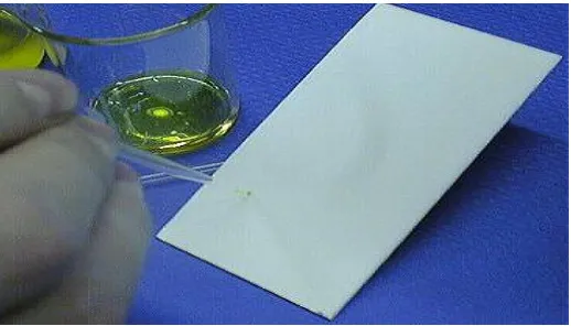
A fine capillary tube was used as spotter for sample application in the TLC plates.
A very little amount of plant extract was taken in a small vial and diluted suitably with ethanol.
The sample was spotted in uniform size (about 0.3 cm) on TLC plates.
The sample was applied several times in each spot to get better chromatogram. Each spot was dried before applying another volume of solution to the same spot.
[image:3.595.159.421.400.538.2][image:3.595.161.420.577.725.2]
Fig.1.1: Capillary tube as sample spotter.
www.wjpr.net Vol 3, Issue 6, 2014. 1716 3.3 Development of chromatogram
[image:4.595.163.438.102.261.2]The chromatogram was developed by ascending technique.
Fig. 1.3: A typical chromatogram by ascending technique.
1. Three solvent systems in the ratio mentioned before were kept in three jars due to the presence of different groups of compound in the plant extract.
2. The plates were placed in each jar in such a manner that the sample spots were just above
the solvent surface.
3. Filter papers were kept into each jar by wetting with respective solvent systems to keep
those jars saturated and the jars were closed tightly.
4. The solvents were allowed to move up the plates by capillary action until they have traveled a distance of about 7-8 cm from the point of application of the sample on a 10 cm plate.
5. The plates were then removed from the jars and dried with a current of air suitably.
6. After developing the chromatogram one of the plate was sprayed with 0.02 % DPPH
solution of ethanol on it by a spray gun. And the other plate was sprayed with 10% H2SO4.
Again two plates were spotted by the process mentioned above and chromatogram was run in solvent system chloroform and methanol (5:1). After developing the chromatogram one of the plate was sprayed with 0.02 % DPPH solution of ethanol on it by a spray gun. And the
other plate was sprayed with 10% H2SO4. Following the similar manner again two plates were
www.wjpr.net Vol 3, Issue 6, 2014. 1717 3.4 UV positive components detection
After drying the plates, three plates from three solvent systems were observed visually under UV light and various regions were marked.
After these plates ware sprayed with 10% H2SO4 and dried on hot plate at 110-120C for
some time.[7]
3.5 Antioxidant positive components detection
Remaining three plates from three solvent systems were observed visually under UV light and various regions were marked.
After these plates ware sprayed with 0.02% DPPH anddried on hot plate at 110-120C for
some time.[8]
3.6 Observation
The observed plates are given below which were viewed under UV detector both in short (254 nm) and long (360 nm) wavelength. When the plates were viewed under UV detector a lot of colored and fluorescent positive components were found in short and long wavelengths respectively. After applying DPPH on the TLC plate, yellow color on purple background was
observed which indicated the presence of antioxidant components in the extract of H. fomes.
www.wjpr.net Vol 3, Issue 6, 2014. 1718
Sample Standard (Ascorbic acid)
Fig.1.4: Comparison of TLC plate for H. fomes leaves with Standard (Ascorbic acid) after applying DPPH.
Sample¹ Sample² Sample³ Sample¹:
Sample²:
Sample³:
Fig.1.5: TLC plates of H. fomes leaves after applying 10% H2SO4.
4 Quantitative analysis 4.1 Principle
The anti-oxidant potential of the ethanolic extract was determined on the basis of their scavenging activity of the stable 2, 2-diphenyl-1-picryl hydrazyl (DPPH) free radical. DPPH
(Chloroform: Ethanol: = 5:1 as solvent)
(Chloroform: Ethanol: Water = 40:10:1 as solvent) (n- hexane : ethyl acetate = 2:1 as solvent)
www.wjpr.net Vol 3, Issue 6, 2014. 1719
is a stable free radical containing an odd electron in its structure and usually utilized for detection of the radical scavenging activity in chemical analysis. The aliquots of the different concentrations (1-500 µg/ml) of the extract were added to 3 ml of a 0.004% w/v solution of
DPPH. Absorbance at 517 nm was determined after 30 min, and IC50 (Inhibitory conc. 50%)
was determined. IC50 value denotes the concentration of sample required to scavenge 50% of
the DPPH free radicals.[9] The formula used for % inhibition ratio is-
% inhibition = (Blank absorbance - Sample absorbance/ Blank absorbance) × 100
4.2 Method
4.2.1 Preparation of solution
At first 10mg extract of H. fomes was measured by electronic balance and mixed with 20 ml
of ethanol (99-100%) to prepare 500 µg/ml solution of extract as stock solution.
Another six concentrations of solutions were prepared by proper dilution method. These concentrations were 200 µg/ml, 100 µg/ml, 50 µg/ml, 10 µg/ml, 5 µg/ml, 1 µg/ml. The following technique was followed to prepare different concentrations of solutions from stock solution:
= × Desired volume
In the same way, various concentrations of ascorbic acid solutions were prepared.
20 mg DPPH powder was measured by electronic balance and mixed with 500 ml of ethanol (99-100%) to prepare 0.004% DPPH solution. It should be kept in cool, dry and dark place.[10]
4.2.2 Working procedure
1. Seven test tubes were taken and labeled for 500 µg/ml, 200 µg/ml, 100 µg/ml, 50 µg/ml,
10 µg/ml, 5 µg/ml, 1 µg/ml.
2. Then 2 ml of solution of each concentration was taken into test tubes designed for each
concentration.
3. 6 ml of DPPH solution was then added to every test tube and kept for 30 minutes at dark
place.
4. In the same manner solutions of ascorbic acid were taken in seven test tubes and DPPH
solution was added and kept for 30 minutes at dark place.
6 DPPH was also applied on the blank test tubes at the same time where only ethanol was
taken as blank.
Desired concentration Supplied concentration Volume which we have
www.wjpr.net Vol 3, Issue 6, 2014. 1720
7 After 30 minutes, absorbance of each test tube was determined by UV spectrophotometer
at 517 nm. Noted that before taking absorbance UV spectrometer reading was nullified with blank solvent i.e. ethanol.
8 The readings were noted down carefully. Then % of inhibition was calculated as-
% inhibition = [(Blank absorbance - Sample absorbance) / Blank absorbance] X 100
9 IC50 was determined from % inhibition vs. conc. graph.
5 RESULT AND DISCUSSION
The DPPH antioxidant assay is based on the ability of 2,2-diphenyl-1 picrylhydrazyl (DPPH), a stable free radical, to decolorize in the presence of antioxidants. The DPPH radical contains an odd electron which is responsible for the absorbance at 517 nm, and also for visible purple yellow color. When DPPH accepts an electron donated ion by an antioxidant compound, the DPPH is decolorized which can be quantitatively measured from the change in the
absorbance. In the TLC-based qualitative antioxidant assay using DPPH assay, H. fomes
leaves showed the free radical scavenging properties indicated by the presence of strong yellow spot on a purple background on the TLC plate.
UV spectrophotometer reading of absorbance for Blank solution:
Table 5.1: DPPH Scavenging Assay of Ascorbic acid. Conc. (ascorbic acid)
(µg / ml) Log Conc.
Absorbance 1st reading
Absorbance 2nd reading
Absorbance
Average % inhibition
1 0 0.730 0.732 0.731 10.19
5 0.699 0.657 0.655 0.656 19.41
10 1 0.502 0.503 0.503 30.20
50 1.699 0.060 0.063 0.062 92.38
100 2 0.033 0.032 0.033 95.94
200 2.301 0.028 0.028 0.028 96.56
500 2.699 0.025 0.025 0.025 96.92
1st reading 2nd reading Average
www.wjpr.net Vol 3, Issue 6, 2014. 1721 Fig.1.5:Absorbance Vs Log Concentration of Ascorbic acid and H. fomes
Table 5.2: DPPH Scavenging Assay of H. fomes leaves Conc. (ascorbic
acid) (µg / ml)
Log Conc.
Absorbance 1st reading
Absorbance 2nd reading
Absorbance
Average % inhibition
1 0 0.720 0.719 0.7195 11.60
5 0.699 0.661 0.662 0.6615 18.73
10 1 0.596 0.597 0.5965 26.72
50 1.699 0.263 0.267 0.265 67.45
100 2 0.099 0.100 0.0995 87.77
200 2.301 0.095 0.096 0.0955 88.26
500 2.699 0.097 0.098 0.0975 88.022
Fig.1.6: Comparison of % inhibition Vs log concentration graph for standard (Ascorbic acid) Vs H. fomes leaves ( Sample).
Towards y-axis 1block = 0.01
Towards x-axis 1block = 0.05
Towards y-axis 1block = 2
IC50
(14.13) IC50
www.wjpr.net Vol 3, Issue 6, 2014. 1722 6 CONCLUSION
Antioxidants have been widely used in the food industry to prolong shelf life. However, there is a widespread agreement that some synthetic antioxidants such as butyl hydroxyl anisole and butyl hydroxyl toluene (BHA and BHT, respectively) need to be replaced with natural antioxidants because of their potential health risks and toxicity. Thus, the search for antioxidants from natural resources has received much attention, and efforts have been made to identify new natural resources for active antioxidant compounds. Phenolic natural products such as flavonoids are of particular interest because of their antioxidant activity through scavenging oxygen radicals and inhibiting peroxidation. Antioxidants that scavenge free radicals play an important role in prevention of cardiovascular disease, aging, cancer, and inflammatory disorders. Recent researches have shown that the antioxidants of plant origin
with free-radical scavenging properties could have great importance as therapeutic agents in several diseases caused due to oxidative stress. Plant extracts and phytoconstituents found
effective as radical scavengers and inhibitors of lipid peroxidation. Many synthetic
antioxidant compounds have shown toxic and/or mutagenic effects, which have stimulated the interest of many investigators to search natural antioxidant. In the TLC-based qualitative antioxidant assay using DPPH assay, H. fomes leaves showed the free radical scavenging properties indicated by the presence of strong yellow spot on a purple background on the TLC plate. In the quantitative assay, H. fomes leaves displayed a very free radical
scavenging activity in the DPPH assay (IC50 = 26.30 approx. µg/ml) which is comparable to
that of ascorbic acid (IC50 = 14.13 approx. µg/ml), a well-known standard antioxidant.
REFERENCES
1. Jacob R.A., 1995, The integrated antioxidant system, Nutrition Research, 15: 755-766.
2. Prakash A., 2001, Antioxidant Activity, Analytical progress report of Medallion
Laboratories, 19 (2): 1-2.
3. Sadhu S. K., Okuyama E., Fujimoto H. and Ishibashi M., 2003, Seperation of Leucas
aspera, a medicinal plant of Bangladesh, guided by prostaglandin inhibitory and
antioxidant activities. Chem Pharm Bull. 51(5): 595-598.
4. Badami S., Gupta M. K., Suresh B., 2003, Antioxidant activity of the ethanolic extract of
Striga orobanchioides. Journal of Ethnopharmacology Volume 85, Issues 2-3, Pages 227-230.
5. Dudonne S., Vitrac S., Coutiere P., Woillez M., Merillon J.M., 2009, Comparative study
www.wjpr.net Vol 3, Issue 6, 2014. 1723
interest using DPPH, ABTS, FRAP, SOD and ORAC assays. J. Agric. Food Chem. 57: 1768-1774.
6. Cioffi G., D’Auria M., Braca A., Mendez J., Castillo A., Morelli I., De Simone F., De Tommasi N., 2002, Antioxidant and Free Radical Scavenging Activity of Constituents of
the Leaves of Tachigalia paniculata, J. Nat. Prod. 65: 1526-1529.
7. Ramchoun M., Harnafi H., Alem C., Benlys M., Elrhaffari L., Amrani S., 2009, Study on
antioxidant and hypolipidemic effects of polyphenol rich extract from Thymus vulgaris
and Lavendula multifida, Pharmacognosy Research, 1: 106-112.
8. Dash D.K., Yeligar V.C., Nayak S.S., Ghosh T., Rajalingam D., Sengupta P., Maiti B.C.,
Maity T.K., 2007, Evaluation of hepatoprotective and antioxidant activity of Ichnocarpus
frutescens (Linn.) R.Br. on paracetamol-induced hepatotoxicity in rats, Tropical Journal
of Pharmaceutical Research, 6: 755-765.
9. Yildirim A., Oktay M., Bulaloulu V., 2001, The antioxidant activity of the leaves of
Cydonia vulgaris, Turkish Journal of Medical Sciences, 31: 23-27.
10. Nagulendran K., Velavan S., Mahesh R., Begum V.H., 2007, Invitro antioxidant activity
and total polyphenolic content of Cyperus rotundus rhizomes, E-Journal of Chemistry, 4;