Susceptibility of
Mycoplasma bovis
field isolates
to antimicrobial agents
J. Siugzdaite, A. Gabinaitiene, S. Kerziene
Lithuanian University of Health Science, Veterinary Academy, Kaunas, Lithuania
ABSTRACT: The purpose of this study was to determine the antibacterial susceptibility of field isolates of Myco-plasma bovis originating from the upper respiratory tract of cattle of different ages. Bacteriological examination of 90 nasal swabs collected from calves at three months of age identified M. bovis in 31 (34.44%) samples. Seventeen (18.88%) of these animals still housed M. bovis in their nasal cavity at nine months and five animals (5.55%) still at seventeen months of age. M. bovis were confirmed by biochemical and antigenic methods. To confirm that these belonged to the M. bovis species isolated mycoplasmas were tested using the PCR method. Fifteen field strains of
M. bovis isolated from the same cattle at three, nine and seventeen months (five strains from each age group) were selected for antibacterial susceptibility testing against six groups of antimicrobial agents using an agar dilution method. The MIC90 ranges established for tylosin, tulathromycin, enrofloxacin, florfenicol and lincomycin were 0.39–0.78 µg/ml, 0.50–1.00 µg/ml, 0.78–1.56 µg/ml, 3.12 µg/ml and 0.39–0.78 µg/ml, respectively. The range of MIC90 for oxytetracycline was from 50 to 100 µg/ml. Preliminary examination of the antimicrobial susceptibility of field strains of M. bovis did not reveal significant differences between different age groups of cattle. After evalu-ation of the MIC90 data with the SPSS 13.0 statistical package it was found that M. bovis isolates from animals at three, nine and seventeen months were similarly susceptible to tylosin and tulathromycin. Statistically significant differences in susceptibility of M. bovis isolated from cattle of different ages were found to florfenicol compared with tulathromycin (P < 0.01), lincomycin (P < 0.01) and enrofloxacin (P < 0.05). The susceptibility of all M. bovis
isolates to oxytetracycline and penicillin G significantly differed from the sensitivity to all other antimicrobial agents used in the present study (P < 0.05). The in vitro susceptibility test showed that field isolates of M. bovis
isolated from cattle of different ages were similarly sensitive to tylosin, tulathromycin, lincomycin and enrofloxacin. It was also determined that the field strains are resistant to oxytetracycline.
Keywords: Mycoplasma bovis; cattle; minimal inhibitory concentration
The most commonly encountered pathogenic my-coplasma in cattle that causes significant commercial losses in the cattle industry is Mycoplasmabovis
(M. bovis) (Francoz et al. 2005). Several studies have shown that M. bovis is the most common bacterium identified in feedlot cattle affected by chronic pneu-monia and in veal calves with fatal bronchopneumo-nia (Gerchman et al. 2009). M. bovis is also present in the respiratory tract of healthy cattle where it causes a subclinical upper respiratory infection and may provoke mastitis and metritis in caws and arthritis or tenosinovitis in fattening cattle (Fox et al. 2005; Gagea et al. 2006). From 2007 through to 2009, stud-ies in Lithuania revealed that M. bovis was present in the upper respiratory tract of 52.5 percent (%) of healthy cattle (Gabinaitiene et al. 2011).
Although appropriate vaccines are available to reduce the consequences of infection, antimicro-bial cure it is still a common practice for treat-ing and controlltreat-ing cattle infection. Infection with Mycoplasma is very difficult to cure because many commonly used antibiotics are not effective against the bacterium, as Mycoplasma do not have a nor-mal cell wall. However, like other mycoplasmas,
antibiotics that are commonly used for treatment of infection, particularly erythromycin, spectino-mycin and tilmicosin. Intermediate occurrence of resistance to oxytetracycline and chlortetracycline, and infrequent or no resistance to enrofloxacin, florfenicol was reported (Hirose et al. 2003; Francoz et al. 2005; Rosenbusch et al. 2005; Gerchman et al. 2009). Antimicrobial susceptibility of M. bovis
field isolates determined by Ter Laak et al. (1993), Ayling et al. (2000), Hirose et al. (2003), Francoz et al. (2005) and Gerchman et al. (2009) showed that the use of antimicrobials results in the selection of resistant and susceptible bacteria.
The in vivo activity of antimicrobial agents for the treatment of M. bovis-induced pneumonia has been reported frequently, some of these values are com-parable with in vitro susceptibility data (Stipkovits et al. 2005). The efficacy of tulathromycin and tilmico-sin for naturally occurring acute bovine respiratory disease treatment was also confirmed (Godinho et al. 2005; Caswell and Archambault 2008).
The purpose of this study was to determine the antibacterial susceptibility of field isolates of M. bo-vis originating from the upper respiratory tract of cattle of different ages.
MATERIAL AND METHODS
Mycoplasma bovis isolation
The study was carried out from 2008–2009. Nasal swabs for the isolation of Mycoplasma were collect-ed three times at a cattle brecollect-eding farm in Lithuania from 90 healthy cattle: when the cattle were aged three, nine and seventeen months, respectively. Fifteen field strains of M. bovis isolated from the same cattle were selected for antibacterial suscepti-bility testing. Mycoplasma isolation procedures and the testing of susceptibility to antimicrobial agents using an agar dilution method were carried out in the microbiology laboratory of the Lithuanian University of Health Science, Veterinary Academy, Department of Infection Diseases.
Mycoplasma cultivation procedures were per-formed according to the method described by Friis (1975). Isolation of Mycoplasma bovis was performed in NHS20 broth media by carrying out 10-fold dilutions from 10–1 to 10–4 and
inoculat-ing the last dilution onto NHS20 solid media agar. Inoculated broth media were cultivated under aero-bic conditions. Solid media were incubated under
microaerophillic conditions for 7 to 14 days. All media were incubated at 37 °C.
To identify microorganisms ofthe Mollicutes
class, a polymerase chain reaction (PCR) meth-od was applied. DNA from isolated microorgan-isms was extracted with a 5% Chelex solution (Sigma, USA). Isolated microorganisms were analyzed by PCR using the forward primer, MW28 (5'-CCAGACTCCTACGGGAGGCA-3') and reverse oligonucleotide primer MW29 (5'-T GCGAGCATACTACTCAGGC-3') (Grida Lab, Lithuania) that are specific for the Mollicutes class (Bashiruddin et al. 2005; Gabinaitiene et al. 2011). This primer pair generates a 560 bp product. The purity of Mycoplasma cultures was determined ac-cording to the recommendations of Goll (1994). PCR-positive Mycoplasma cultures were inocu-lated onto NHS-20 agar plates. Five Mycoplasma colonies were picked from each plate after three to seven days of cultivation and subcultured in NHS-20 broth. This procedure was repeated three times. In order to separate Mycoplasma bovis strains from others species of Mycoplasma, isolates were tested for their biochemical properties (glucose fer-mentation, arginine hydrolysis, phosphatase activ-ity, tetrazolium reduction and production of spots and films) while the disc growth inhibition (DGI) test was also used (Clyde 1964; Aluotto et al. 1970). Paper discs with antiserum against the following refer-ence strains were used: Mycoplasma arginini G230,
Mycoplasma bovigenitalium PG11, Mycoplasma bovirhinis PG43, Mycoplasma bovis Donetta and
Mycoplasma dispar 462/2. All strains biochemi-cally and antigenibiochemi-cally classified as Mycoplasma bovis were confirmed by the amplification of a 734 bp amplicon. Two oligonucleotide primers targeting the 16S rRNA gene, that are specific for
M. bovis species were used: forward primer MboF2 (5'-GAAGAAAAAGTAGCATAGGAAATGAT-3') and reverse primer MboR2 (5'-CGTCGTCCC- CACCTTCCTCCCG-3') (Timenetsky et al. 2006).
For further analysis pure M. bovis culture colo-nies were then picked from plates and transferred to NHS-20 broth medium. Isolates were stored at –70 °C until used.
the growth-induced pH shift exchange in the broth medium (from red to orange due to fermentation of glucose) (Francoz et al. 2005). After incubation 1 ml of fresh Mycoplasma cultures were added to 9 ml of selective medium and vortexed. Then, 0.1 ml of vor-texed suspensions were diluted ten-fold from 10–2 to
10–9 in 0.9 ml of selective medium. For Mycoplasma
colony counting 2 μl of each Mycoplasma dilution were inoculated onto the surface of freshly prepared Mycoplasma agar plates (in triplicate) and incu-bated at 37 °C under microaerophillic conditions. Inoculated plates were incubated until the plates with the lower dilutions of the Mycoplasma suspen-sions had well-developed Mycoplasma colonies. The number of colonies was counted using a microscope. For antimicrobial susceptibility testing a 105 CFU/ml
Mycoplasma inoculum was prepared (Hannan 2000).
Antimicrobial agents
The minimal inhibitory concentration of each antimicrobial agent was determined by applying an agar dilution method with some modifications and in accordance with the recommendations of Hannan (2000) and Kobayashi et al. (1996). Seven antimicrobial agents were included in the test-ing: tulathromycin (Pfizer, United States), tylosin (Chemifarma, Italia), lincomycin (Pfizer, United States), enrofloxacin (Vetoquinol, Austria), florfen-icol (Krka, Slovenia), oxytetracycline (Chemifarma, Italia),) and penicillin G (Actavis Baltics, Lithuania). The absence of a cell wall protects Mycoplasma from the influence of beta lactam antibiotics. Consequently, penicillin G was included in this study as a negative control. Antimicrobial agent concentrations ranged from 0.39 to 100 μg/ml for lincomycin, enrofloxacin, tylosin, florfenicol, oxy-tetracycline, penicillin G, and for tulathromycin concentrations from 0.0625 to 64 μg/ml were test-ed. The procedures were performed as described by Hannan (2000). All antimicrobial agents were dis-solved in the optimum diluent at 100 µg/ml, except tulathromycin (at 64 µg/ml) that was subjected to a two-fold serial dilution in test tubes containing NHS-20 broth without any selective supplement.
Agar dilution test
One millilitre of each antimicrobial dilution was mixed with 9 ml of NHS-20 agar medium without
selective supplement. NHS-20 agar plates without antimicrobial agents served as controls. Prepared agar plates with different antimicrobial concentra-tions were inoculated with 5 µl 105 CFU/ml of the
Mycoplasma culture. When the inoculum droplets had been absorbed the plates were incubated at 37 °C under microaerophilic condition. Inoculated plates were observed daily for a period of three to five days, by which time Mycoplasma species formed clearly visible colonies. The MIC for each isolate was defined as the lowest concentration of antibiotics to completely inhibit visible growth on agar. The MIC50 and MIC90 values were defined as the lowest concentrations capable of inhibiting the growth of 50% and 90% of isolates, respectively (Hannan 2000; Hirose et al. 2003; Gerchman et al. 2009). The tests were performed three times.To ensure the validity of the agar dilution method for determining antimicrobial susceptibility the M. bo-vis reference type strain Donetta was included in the tests.
Interpretation of MIC results
Clinical and Laboratory Standards Institute (CLSI, formerly known as NCCLS) criteria for veterinary pathogenic bacteria in cattle were used to interpret the results (NCCLS 2002). The MIC breakpoints of each antimicrobial agent group, tet-racyclines, macrolides, quinolones, phenicols were 4–16, 8–32, 0.25–2 and 2–8 µg/ml, respectively. The MIC breakpoints for penicillin (0.25–8 µg/ml) were based on guidelines for testing the suscepti-bility of bacteria that affect humans (Ter Laak et al. 1993). The MIC breakpoints of lincosamides (1–2 µg/ml) were evaluated as recommended by Hirose et al. (2003). If the MIC of tetracycline was 4 μg/ml or lower, the isolate was considered sus-ceptible; if the MIC was 16 μg/ml or higher, the isolate was considered resistant.
Statistical analysis
The antibacterial susceptibility of the M. bovis
field isolates was evaluated using the SPSS 13.0 Windows statistical package (2004). The Wilcoxon two-sample-test and the Kruskal-Wallis test were used to examine the equality among the isolate me-dians for each antibacterial material. Data with a
RESULTS
Mycoplasma was isolated from 41 (45.56%) nasal swabs collected from 90 calves at three months of age. Twenty four (26.67%) of these animals still housed Mycoplasma in their nasal cavity at nine and seventeen months of age. For each isolate, its identity as a member of the Mollicutes class was confirmed by PCR. Examination of the biochemical and antigenic characteristics of these isolates re-vealed that M. bovis was isolated from 31 (34.44%) nasal cavities of calves at three months of age. Seventeen (18.88%) of these animals still housed
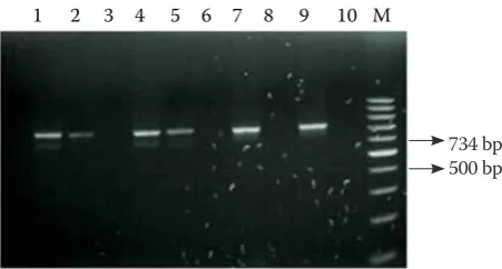
M. bovis in their nasal cavity at nine months and five animals (5.55%) still at seventeen months of age. All 53 (100%) isolates were confirmed as M. bo-vis by PCR, i.e., based on the amplification of a product of 734 bp (Figure 1).
The susceptibility to 15 field isolates of M. bovis
from the same cattle at three, nine and seventeen months of the age, respectively, to seven different antimicrobial agents is shown in Table 1.
All (100%) isolatesof M. bovis were observed to be susceptible to tylosin. The MIC90 for tylosin of field strains of M. bovis isolated from cattle aged three and seventeen months was identical (0.39 µg/ ml), while for nine months the MIC90 was 0.78 µg/ ml. One hundred percent of isolates were found to be sensitive to tulathromycin. The MIC90 range determined for tulathromycin was 0.50–1.00 µg/ ml. According to the breakpoints of the MICs for quinolone, 100% of isolates could be considered sensitive to enrofloxacin, too. The MIC90 range
de-termined for enrofloxacin was 0.78–1.56 µg/ml. All (100%) field strains of M. bovis isolated from cattle of different ages were found to be sensitive to florfenicol and lincomycin. The range of MICs for florfenicol and lincomycin was 0.78–3.12 µg/ ml and 0.39–1.56 µg/ml.
All (100%) field strains of M. bovis isolated from cattle of different ages were determined to be re-sistant to oxytetracycline. The range of MICs for oxytetracycline was 6.25–100 µg/ml and the MIC90 was from 50 to 100 µg/ml. This was higher than the breakpoints for the tetracyclines (4–16 µg/ml). All (100%) field strains of M. bovis were found to be resistant to penicillin G and no growth inhibition with penicillin G was observed. In all cases the MIC90 was >100 µg/ml.
To ensure the credibility of our antimicrobial sus-ceptibility test, procedures were performed initially with the Donetta M. bovis reference type strain. MIC90 values obtained by agar dilution method of ty-losin (0.78 µg/ml) and enrofloxacin (0.39 µg/ml), lin-comycin (0.39 µg/ml), oxytetracycline (1.56 µg/ml) and penicillin G (> 100 µg/ml) were comparable to previously reported MIC values for the same antimicrobials against this same reference type strain by Ter Laak et al. (1993) using a serial broth dilution test.
[image:4.595.63.534.551.727.2]Preliminary examination of the antimicrobial sus-ceptibility of field strains of M. bovis did not reveal any significant differences between different age groups of cattle. Upon evaluation of the MIC90 data with the SPSS 13.0 statistical package it was found that M. bovis isolates from animals aged three and
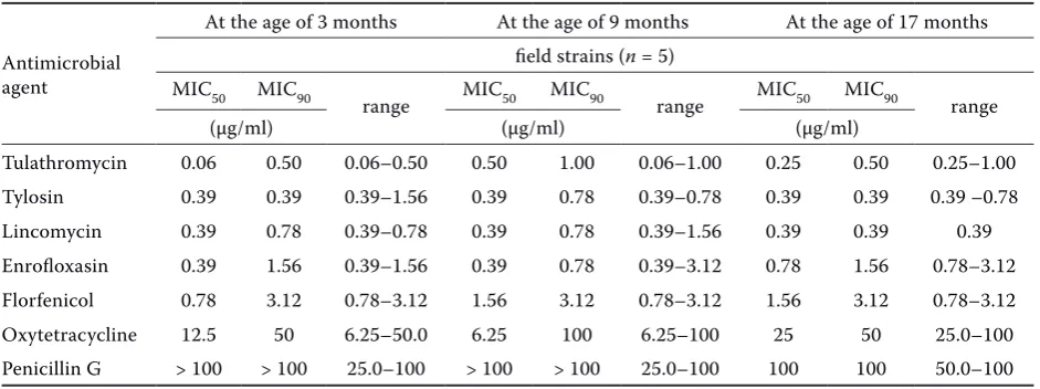
Table 1. The MICs of antimicrobial agents used against field isolates of M. bovis from cattle at 3, 9 and 17 months of age, respectively. The MIC values were estimated for five different isolates from each age group
Antimicrobial agent
At the age of 3 months At the age of 9 months At the age of 17 months
field strains (n = 5) MIC50 MIC90
range MIC50 MIC90 range MIC50 MIC90 range
(µg/ml) (µg/ml) (µg/ml)
Tulathromycin 0.06 0.50 0.06–0.50 0.50 1.00 0.06–1.00 0.25 0.50 0.25–1.00
Tylosin 0.39 0.39 0.39–1.56 0.39 0.78 0.39–0.78 0.39 0.39 0.39 –0.78
Lincomycin 0.39 0.78 0.39–0.78 0.39 0.78 0.39–1.56 0.39 0.39 0.39
Enrofloxasin 0.39 1.56 0.39–1.56 0.39 0.78 0.39–3.12 0.78 1.56 0.78–3.12
Florfenicol 0.78 3.12 0.78–3.12 1.56 3.12 0.78–3.12 1.56 3.12 0.78–3.12
Oxytetracycline 12.5 50 6.25–50.0 6.25 100 6.25–100 25 50 25.0–100
Penicillin G > 100 > 100 25.0–100 > 100 > 100 25.0–100 100 100 50.0–100
nine months were similarly susceptible to tylosin, tulathromycin and enrofloxacin. The strains of M. bovis isolated from seventeen month old cattle were found to be the most susceptible to tulathromycin and tylosin. Statistically significant differences in susceptibility of M. bovis isolated from cattle of different ages were found for florfenicol compared to tulathromycin (P < 0.01), lincomycin (P < 0.01) and enrofloxacin (P < 0.05). The susceptibility of
M. bovis isolates from seventeen month old cat-tle was also significantly different for enrofloxacin compared to tulathromycin (P < 0.05), lincomycin (P < 0.01) and tylosin (P < 0.05). The susceptibility of all M.bovis isolates to oxytetracycline and peni-cillin G significantly differed from the sensitivity to all other antimicrobial agents used in the present study (P < 0.05).
DISCUSSION
Mycoplasma bovis may be isolated from the upper respiratory tract, trachea, and lower respiratory tract of cattle without clinical disease or gross lesions, although its presence in the lower respiratory tract may cause subclinical inflammation. In this study the largest percentage (26.67%) of field strains of M. bovis was isolated from the upper respiratory tract of cattle at three months of age. Further investigation of the upper respiratory tract of these cattle showed that M. bovis is spread among animals which were the subject of this study. These observations are simi-lar to those reported from several other countries where Mycoplasma have been isolated from 3 to 79% of the respiratory tracts of clinically healthy cattle (Ter Laak et al., 1992; Siugzdaite 2002; Thomas et al. 2002; Arcangioli et al. 2008).
In this study, the susceptibility of M. bovis field isolates was determined to seven antimicrobial agents in vitro. The field strains of M. bovis (100%) isolated from the cattle of different ages revealed comparable susceptibility to tylosin, tulathromycin and enrofloxacin.
Varying sensitivity ranges were observed for field strains of M. bovis to tylosin and tulathromycin for cattle at different ages. M. bovis isolates from three and seventeen month old cattle had lower MIC90 values for tylosin (0.39 µg/ml) than the strains iso-lated from the same cattle aged nine months (MIC90 0.78 µg/ml). Similar results were obtained by valu-ation of Mycoplasma sensitivity to tulathromycin. However, in both cases the MIC90 value of M. bovis
to tylosin and tulathromycin differs by one dilution and was lower than the breakpoint of the MIC value for macrolides (8–32 µg/ml). This confirms that M. bovis strains isolated from all age groups are sensi-tive to tylosin and tulathromycin. Gerchman et al. (2009) tested M. bovis strains isolated from cattle grown in Lithuania and examined respiratory tract sensitivity to antibacterials using a micro-broth di-lution procedure for MIC detection. It was shown that, similarly to our investigated strains, 66.7% of
M. bovis strains were sensitive to tylosin (MIC90 was 0.5 µg/ml).
The present study revealed that tylosin, the oldest antibiotic in veterinary medicine, can still be used for the treatment Mycoplasmal infections of cattle affecting the respiratory tract. Tulathromycin, a semisynthetic, tribasic, macrolide antimicrobial, was effective against all M. bovis isolates and its MIC90 for each isolate was low (0.5–1 µg/ml). The results of our study were comparable with those reported by Kilgore et al. (2005) and the MIC90 of
M. bovis was established as 1 µg/ml. In in vivo in-vestigations tulathromycin has also demonstrated a broad spectrum of activity in the therapy of bovine respiratory disease (Kilgore et al. 2005; Skogerboe et al. 2005).
We isolated some field strains of M. bovis sensitive to enrofloxacin. The MIC90 range of M. boviswas fractionally (by one dilution) different between the different age groups, but in all age groups the MIC value was lower than the breakpoints of quinolone (0.25–2 µg/ml). In a previous study (Gerchman et al. 2009), field strains of M. bovis isolated from cattle raised in Lithuania were also sensitive to quinolone. In our study, the MIC values of M. bovis to enrofloxa-cin were higher than those reported by Francoz et al. (2005) (0.19 µg/ml to 0.25 µg/ml), Rosenbusch et al.
[image:5.595.64.290.81.202.2]734 bp 500 bp
Figure 1. Mycoplasma bovis identification by PCR. Lane M = GeneRuler TM 100 bp DNA Ladder (MBI, Fermen-tas) marker; lanes 1, 2, 4, 5, 7 = Mycoplasma bovis iso-lates; lane 9 = positive control; lane 10 = negative control
(2005) (0.25 µg/ml), Hirose et al. (2003) (0.2 µg/ml) and Ayling et al. (2000) (0.5 µg/ml to 1 µg/ml). In Mycoplasma species acquired resistance is usually due to alterations of the target enzymes or the in-duction of active efflux systems (Reinhardt et al. 2002; Hirose et al. 2004). Thomas et al. (2003) iso-lated enrofloxacin-resistant strains from bovines. It has also been shown that, in the presence of enro-floxacin, in vitro passaging of Mycoplasmas results in thedevelopment of fluoroquinolone resistance (Gautier-Bouchardon et al. 2002).
The MIC90 value of M. bovis field isolates from catlle of different ages to lincomycin either did not differ or differed by only one dilution. It was de-termined that the MIC values of lincomycin for all
M. bovis field strains were lower than the break-points of lincosamides (1–2 μg/ml) and lincomycin had a good effect against all field isolates. This is in agreement with the results of Hirose et al. (2003) and Ter Laak et al. (1993).
All field isolates of M. bovis were sensitive to florfenicol, as the MIC90 value (3.12 µg/ml) was lower than the breakpoints of phenicol (2–8 µg/ml). The MIC90 of florfenicol for all M. bovis field isolates in the present study was lower than the MICs to phenicol reported by Hirose et al. (2003). Florfenicol is exclusively used in veterinary medi-cine. The compound is analogous to chlorampheni-col but does not cause irreversible depression of the bone marrow and can therefore be used in food-producing animals. Florfenicol is a strong inhibitor of microbial protein synthesis through irreversible binding with the 50S subunit of ribosomes, abol-ishing the activity of peptidyl transferase (Liu et al. 2003). No resistance against florfenicol associated with Mycoplasma infection has been described.
Tetracycline has been reported to be effective against Mycoplasmas (Hirose et al. 2003). Our study did not confirm that this antimicrobial agent is ef-fective against field strains of M. bovis. In our study, the MICs for oxytetracycline of all isolates of M. bo-vis were higher than the breakpoint (4–16 µg/ml). In veterinary medicine, increases in tetracycline resistance have been described for M. bovis,
M. hyopneumoniae, M. bovirhinis and M. alkale-scens (Inamoto et al. 1994; Hirose et al. 2003; Thomas et al.2003). Oxytetracycline-sensitive spe-cies of M. bovis isolated from 2005 to 2007 from Lithuanian cattle were reported by Gerchman et al. (2009). It is suggested that oxytetracycline-resistant Mycoplasmas are produced by the administration of oxytetracycline to cattle affected by respiratory
disease. The results of the present study with re-gard to the resistance of M. bovis strains against tetracycline are in accordance with several previous studies, such as Hirose et al. (2003), Rosenbusch et al. (2005), Thomas et al. (2003) and Ayling et al. (2000). Resistance to antimicrobials is achieved by bacteria due to mutation. These abilities are typi-cal for the mycoplasma (Kaluina 1998). Resistance is transferred by R-plasmids and mutant strains, especially with sub-therapeutic or sub-inhibitory concentrations. Resistant Mycoplasmas exhibit a reduced uptake of tetracycline into cells, lowering the initial concentration, and have acquired the ability to excrete the drug out of the cell, (Boothe 1998). A possible explanation for the high preva-lence of oxytetracycline resistance in the present study may be the frequent use of oxytetracycline for the treatment of respiratory disease infection, gastroenteritis, metritis and mastitis in cattle.
In the present study, all M. bovis isolates were resistant to penicillin G. This confirms that the lack of a cell wall makes the Mycoplasma resistant to beta-lactam antimicrobials, and underlines the fact that penicillin G can be used in antimicrobial susceptibility testing only as a negative control.
CONCLUSION
In this study, the antibacterial susceptibility of field isolates of M. bovis originating from the up-per respiratory tract of cattle of different ages was determined. In vitro susceptibility tests showed that field isolates derived from cattle of different ages were sensitive to tylosin, tulathromycin, lincomycin and enrofloxacin. It was also determined that the field strains are resistant to oxytetracycline.
REfERENCES
Aluotto BB, Wittler RG, Williams CO, Faber JE (1970): Standardized bacteriologic techniques for the charac-terization of Mycoplasma species. International Jour-nal of Systematic Bacteriology 20, 35–58.
Arcangioli MA, Duet A, Meyer G, Dernburg A, Bezille P, Poumarat F, Le Grand D (2008): The role of Myco-plasma bovis in bovine respiratory disease outbreaks in veal calf feedlots. Veterinary Journal 177, 89–93. Aylingas RD, Baker SE, Nicholas RA, Peek ML, Simon
and tilmicosin against Mycoplasma mycoides subspe-cies mycoides small colony type. Veterinary Record 146, 243–246.
Bashiruddin JB, Frey J, Konigsson MH, Johansson KE, Hotzel H, Diller R, Santis P, Botelho A, Ayling RD, Nicholas RAJ, Thiaucourt F, Sachse K (2005): Evaluation of PCR systems for the identification and differentiation of Mycoplasma agalactiae and Mycoplasma bovis: a col-laborative trial. Veterinary Journal 169, 268–275. Boothe D (1998): The Marck Veterinary Manual. In:
Asiello S (eds.): Antibacterial Agents. Merck and Co, Inc, Philadelphia. 1745–1788.
Caswell JL, Archambault M (2008): Mycoplasma bovis pneumonia in cattle. Animal Health Research Reviews 8, 161–186.
Clyde WA (1964): Mycoplasma species identification based upon growth inhibition by specific antisera. Journal of Immunological Methods 92, 958–965. Fox LK, Kirk JH, Britten A (2005): Mycoplasma mastitis:
a review of transmission and control. Journal of Vet-erinary Medicine Series B 52, 153–160.
Francoz D, Fortin M, Fecteau G, Messier S (2005): De-termination of Mycoplasma bovis susceptibilities against six antimicrobial agents using the E test method. Veterinary Microbiology 105, 57–64. Friis NF (1975): Some recommendations concerning
primary isolations of Mycoplasma suipneumoniae and Mycoplasma flocculare. Nord Veterinary Medicine 27, 337–339.
Gabinaitiene A, Siugzdaite J, Zilinskas H (2011): Labora-tory diagnosis of Mycoplasma infection in young cat-tle. Polish Journal of Veterinary Sciences 14, 87–93. Gagea MI, Bateman KG, Van Dreumel T, McEwen BJ,
Carman S, Archambault M, Shanahan RA, Caswell JL (2006): Diseases and pathogens associated with mor-tality in Ontario beef feedlots. Journal of Veterinary Diagnostic Investigation 18, 18–28.
Gautier-Bouchardon AV, Reinhardt AK, Kobisch M, Kempf I (2002): In vitro development of resistance to enrofloxacin, erythromycin, tylosin, tiamulin and oxytetracycline in Mycoplasma gallisepticum, Myco-plasma iowae and MycoMyco-plasma synoviae. Veterinary Microbiology 88, 47–58.
Gerchman I, Levisohn S, Mikula I, Lysnyansky I (2009): In vitro antimicrobial susceptibility of Mycoplasma bovis isolated in Israel from local and imported cattle. Veterinary Microbiology 137, 268–275.
Godinho KS, Wolf LG, Sherington J, Rowan TG, Sun-derland SJ, Evans NA (2005): Efficacy of tulathromycin in the treatment and prevention of natural outbreaks of bovine respiratory disease in European cattle. Vet-erinary Therapeutics 6, 122–135.
Goll FJR (1994): Identification of Mycoplasmas isolated from domestic animals. In: Whitford HW, Rosenbusch RF, Lauerman LH (eds.): Mycoplasmosis in Animals: Laboratory Diagnosis. Iowa State University Press, Iowa, 15–30.
Hannan PC (2000): Guidelines and recommendations for antimicrobial minimum inhibitory concentration (MIC) testing against veterinary Mycoplasma species. International Research Programme on Comparative Mycoplasmology. Veterinary Research 31, 373–395. Hirose K, Kobayashi H, Ito N, Kawasaki Y, Zako M,
Ko-tani K, Ogawa H, Sato H (2003): Isolation of Myco-plasmas from nasal swabs of calves affected with respiratory diseases and antimicrobial susceptibility of their isolates. Journal of Veterinary Medicine B 50, 347–351.
Hirose K, Kawasaki Y, Kotani K (2004): Characterization of a point mutation in the parC gene of Mycoplasma bovirhinis associated with fluoroquinolone resistance. Journal of Veterinary Medicine B 51, 169–175. Inamoto T, Takahashi K, Yamamoto K, Nakai Y, Ogimoto
K (1994): Antibiotic susceptibility of Mycoplasma hyopneumoniae isolated from swine. Journal of Vet-erinary Medical Science 56, 393–394.
Kaluina V (1998): Avian mycoplasmosis (in Lithuanian). Margi Rastai, Vilnius. 215 pp.
Kilgore WR, Spensley MS, Sun F, Nutsch RG, Rooney KA, Skogerboe TL (2005): Therapeutic efficacy of tu-lathromycin, a novel triamilide antimicrobial, against bovine respiratory disease in feeder calves. Veterinary Therapeutics 6, 143–153.
Kobayashi H, Morozumi T, Munthali G, Mitani K, Ito N, Yamamoto K (1996): Macrolide susceptibility of Mycoplasma hyorhinis isolated from piglets. Antimi-crobial Agents and Chemotherapy 40, 1030–1032. Liu J, Fung KF, Chen Z, Zeng Z, Zhang J (2003):
Phar-macokinetics of florfenicol in healthy pigs and in pigs experimentally infected with Actinobacillus pleuro-pneumoniae. Antimicrobial Agents and Chemother-apy 47, 820–823.
Maunsell FP, Donovan GA, Risco C, Brown MB (2009): Field evaluation of a Mycoplasma bovis bacterin in young dairy calves. Vaccine 27, 2781–2788.
Rosenbusch RF, Kinyon JM, Apley M, Funk ND, Smith S, Hoffman LJ (2005): In vitro antimicrobial inhibition profiles of Mycoplasma bovis isolates recovered from various regions of the United States from 2002 to 2003. The Journal of Veterinary Diagnostic Investigation 17, 436–441.
Siugzdaite J (2002): Detection of Mycoplasma bovis from Hayflick-agar media by polymerase chain reaction. Zemes Ukio Mokslai 1, 67–70.
Skogerboe LT, Rooney AK, Nutsch RG, Weigel DJ, Ga-jewski K, Kilgore WR (2006): Comparative efficacy of tulathromycin versus florfenicol and tilmicosin against undifferentiated bovine respiratory disease in feedlot cattle. Veterinary Therapeutics 6, 180–196.
Stipkovits L, Ripley PH, Tenk M, Glavits R, Molnar T, Fodor L (2005): The efficacy of valnemulin (Econor) in the control of disease caused by experimental infec-tion of calves with Mycoplasma bovis. Research in Veterinary Science 78, 207–215.
Tenk M (2005): Examination of Mycoplasma bovis infec-tion in cattle. [Doctoral Thesis.] Budapest, 70 pp. Ter Laak EA, Noordergraaf JH, Dieltjes RP (1992):
Prevalance of Mycoplasmas in the respiratory tract of
pneumonic calves. Zentralblatt fur Veterinarmedizin Reihe B 39, 553–562.
Ter Laak EA, Noordergraaf JH, Verschure MH (1993): Susceptibilities of Mycoplasma bovis, Mycoplasma dispar, and Ureaplasma diversum strains to antimi-crobial agents in vitro. Antimiantimi-crobial Agents and Chemotherapy 37, 317–321.
Thomas A, Ball H, Dizier I, Trolin A, Bell C, Mainil J, Linden A (2002): Isolation of Mycoplasma species from the lower respiratory tract of healthy cattle and cattle with respiratory disease in Belgium. Veterinary Record 151, 472–476.
Thomas A, Nicolas C, Dizier I, Mainil J, Linden A (2003): Antibiotic susceptibilities of recent isolates of Myco-plasma bovis in Belgium. Veterinary Record 153, 428–431.
Timenetsky J, Santos LM, Buzinhani M, Mettifogo E (2006): Detection of multiple Mycoplasma infection in cell cultures by PCR. Brazilian Journal of Medical and Biological Research 39, 907–914.
Received: 2012–05–03 Accepted after corrections: 2012–10–30
Corresponding Author:
Ausra Gabinaitiene, Lithuanian University of Health Science Veterinary Academy, Department of Infectious Diseases, Tilzes str. 18, LT-47181 Kaunas, Lithuania