Ovarian dysgerminoma with retroperitoneal metastases
in a bitch: a case report
R. Novotny, R. Vitasek, A. Bartoskova
University of Veterinary and Pharmaceutical Sciences, Brno, Czech Republic
ABSTRACT: A four-year old, 26.5 kg, Boxer bitch was presented to the Department of Reproduction in the Clinic of Dogs and Cats with a six month history of vulvar swelling and vaginal discharge. General gynaecological examination showed an extremely swollen, oedematous and tough-elastic highly irritable vulva. A vaginal smear revealed the presence of superficial cells and red blood cells. After repeated unsuccesful administrations of HCG and GnRH the owner of the bitch agreed to surgical treatment. An ovariohysterectomy was performed and on the left ovary a grapefruit-size structure was found. Three nodular structures were found retroperitoneally and were also dissected. Histological examination showed a disgerminoma in metastasises, mitotically active with a bad prognosis. However, at a check up twenty months subsequently the patient was still alive.
Keywords: dog; ovary; tumour; metastase; vulvar discharge
Ovarian dysgerminoma is a rare tumour in dogs as well as in other species. It belongs to the group of germ cell origin tumours and is a counterpart of testicular seminoma in male dogs. Dysgerminomas represent 6–12% of ovarian tumours in dogs (Dehner et al., 1970), and range in size from 2 to 30 cm, with a mean of 9.5 cm. Grossly, dysgermi-nomas are nodular masses with slightly bulging, tan-coloured cut surfaces. Variable amounts of haemorrhage and necrosis have commonly been observed (Dehner et al., 1970). Histopathologically, dysgerminomas are composed of a uniform popula-tion of polyhedral cells that are arranged in sheets, cords, or alveoli, highly vascularized scant fibrous stroma, while mitoses are numerous and sometimes have an aberrant appearance. Additionally, there may be regions of necrosis and hemorrhage within the tumour. Despite the presence of a high mitotic rate, necrosis, and hemorrhage (all suggestive of a more malignant tumour), the majority of dysger-minomas are clinically benign. The histological ap-pearance of benign and malignant dysgerminomas is similar; in canines 10–20% of dysgerminomas are malignant (McEntee, 1990). Metastases (three out of 14 cases in one report) have been identi-fied in the regional, mesenteric, and mediastinal lymph nodes, abdominal organs (liver, kidney,
ad-renal), omentum, serosal surface of the intestinal tract, and lung (McEntee, 2002). Clinical signs in patients with dysgerminomas may include a pal-pable abdominal mass, bloody vaginal discharge, polyuria, polydipsia, emesis, weight loss, diarrhoea and lethargy. Cystic endometrial hyperplasia, and signs of persistent proestrus/oestrus are seen in some patients, compatible with hormone produc-tion. In some dogs (five out of seven in one study) concurrent pyometra can be present, which may be the origin of some of the clinical signs (Greenlee and Patnaik 1985).
Case description
A four-year old, 26.5 kg, Boxer bitch was pre-sented to the Department of Reproduction in the Clinic of Dogs and Cats with a six-month history of vulvar swelling and discharge of incarnadine col-our. Polakisuria was also reported. Apart from the above symptoms the animal was vivid, with a good appetite and had no other problems.
a concentration of 25 UI per kg was administered on the two consequent days.
During the second visit ten days later, the vulva was found to be less swollen but still very irritable. The vaginal cytology image was similar to that of the first examination (90–95% of cells superficial, approx. 50% anuclear, sporadic intermedial cells and neutrofils). A repeat ultrasound examination showed no pathological findings.
Ten days subsequent to this no changes in the size of vulvar swelling were found. The Irritability of the vulva persisted and the vaginal cytological image was unchanged. The pink vaginal discharge was also still present which was strongly attracting other dogs. The owner still refused an ovariohys-terectomy, thus gonadotropine releasing hormone (GnRH) (Supergestran, Nordic Pharma) at a dose of 100 µg pro toto was administered at eight hour intervals for two days.
At the next visit two weeks later the bitch was still in heat, with the vulva permanently swollen and irritable. The vaginal cytologic examination corre-sponded to previous findings (100% superficial cells, 40% anuclear). The owner was once again offered an ovariohysterectomy, but insisted on medicamentous therapy. HCG at 25 UI per kg was administered using the same scheme as after the first visit.
After another two weeks no changes in clinical findings were noted, whilst the owner persisted with her request for HCG therapy despite the in-efficacy. The possibility of ovariohysterectomy was again strongly recommended, but rejected.
[image:2.595.80.268.82.305.2]After almost two months the bitch was presented to our departement for unwanted mating. The clini-cal examination revealed that her health state was unchanged. She showed intensive oestrogenisa-tion with vulvar swelling, and the presence of pink discharge was noted. Vaginal cytology, ultrasono-graphic and X-ray examinations were performed.
[image:2.595.64.292.512.724.2]Figure 2. Right ovary with present disgerminoma, grape-fruit size
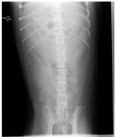
Figure 1. X-ray image of an abdominal mass in the right flank region
(Haemacolor, Merck) revealed the presence of su-perficial cells (100%, 40–50% anuclear) and red blood cells (RBC). Due to the length of the proc-ess and the possibility of pathological structures unviewable by ultrasound imaging, a ovariohys-terectomy was recommended to the owner as the most suitable procedure. Although she was in-formed about the possible risks, she chose medi-camentous treatment. Therefore, human chorionic gonadotropine (HCG) (Pregnyl 1500, Organon) at
[image:2.595.306.531.615.723.2]The presence of superficial, anuclear (approx. 50%) cells was found in the vaginal smear, X-ray exami-nation revealed an intraabdominal mass caudally from the right kidney (Figure 1). On the ultrasound image a structure with many of cavities 2–4 mm in diameter was visible. After discussion the owner agreed to a ovariohysterectomy.
A laparotomy was performed immediately. No abnormal findings were noted at the presurgi-cal examination. An anaesthesia premedicant of butorphanol (Butomidor, Richter) 0.1 mg/kg, di-azepam (Apaurin, Krka) 0.3 mg/kg and ketamine (Narketan, Vetoquinol) at 4 mg/kg were admin-istered. Anaesthesia was maintained with inhala-tional isoflurane (Isofluran, Piramal). An incision was made in the linea alba and an approximately 1–2 cm thick uterus was visualised. The left ovary was normal in size, while the right ovary was of a grapefruit size with a rough surface (Figure 2). After securing artheriae ovaricae and artheriae uterinae the ovariohysterectomy was performed. Three dark coloured nodular structures were found retroperitoneally and removed after blunt dissec-tion of the adjacent urether (Figure 3). After surgery amoxycillin clavulanate (Synulox, Pfizer) 15 mg/kg and meloxicam (Metacam, Boehringer Ingelheim) 0.2 mg/kg were administered.
[image:3.595.65.292.84.256.2]After transection of the right ovary numerous small cavities were found. Histological examination revealed a mitotically active tumour resembling aseminoma, neoplastic cells with clear cytoplasm creating pseudolobular structures devided by connective tissue septa, numerous haemorrhages with foci of extravasation and coagulation necro-sis. Also, clusters of lymphocytes were present
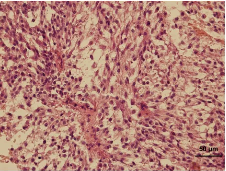
(Figure 4). Therefore, a dysgerminoma in metas-tasises (Figure 5) was diagnosed with a bad prog-nosis.
On the next day clinical examination after sur-gery showed a good health condition and the bitch urinated, ate and drank normally. Amoxycillin cla-vulanate at 15 mg/kg was administered by injection and amoxycillin clavulanate (Augmentin 625 mg tbl., GlaxoSmithKline) at 15 mg/kg twice daily was prescribed as homecare.
One week later the stitches were removed, and the suture line was observed to be healing well. The vulva was smaller, but still a bit swollen, with a small amount of whitish serous vaginal discharge.
The patient recovered very well and twenty months after surgery, despite the poor prognosis, was still alive.
DISCUSSION AND CONCLUSIONS
Dysgerminomas in dogs are reported rarely, but have a distinctive, recognizable, cytologic appear-ance and should be included in the differential diag-nosis of intra-abdominal masses in a reproductively intact female dog (Brazzell and Borjesson, 2006). Dysgerminomas are in most cases incidental find-ings when a laparotomy is performed due to other reasons. Hormone production by the tumour may also predispose the bitch to the development of a pyometra (Jackson et al., 1985).
[image:3.595.305.532.84.257.2]Figure 5. Metastasis in a regional lymph node – the growth is composed of uniform large cells with oval to spheroid-shaped nuclei and a rim of faintly staining cytoplasm. Mitotic figures are frequent. The cells, which stand out individually, are grouped into nests by delicate fibrous stroma
In our case, persistent oestrogenisation gave rise to hormonally active structure in the reproductive tract. When an ovarian neoplasm is suspected based on clinical findings, the determination of hormone levels would be useful in learning more about the biological nature of the tumour. All dysgerminomas should be treated as malignant tumours with the potential to become very large locally and/or to metastasize (Jackson et al., 1985).
The issue of ovarian malignancies is described in richer detail in human medicine. According to FIGO (International Federation of Gynecology and Obstetrics) ovarian neoplastic processes are classi-fied into four stages (Table 1, Heintz et al., 2006). For each stage an appropriate surgical therapy and possible adjuvant chemotherapy is prescibed. In ac-cordance with FIGO classification, our case equates to IIb stage, that should, in human medicine, be treated by surgical dissection of neoplastic struc-tures combined with adjuvant chemotherapy.
The treatment of ovarian tumours in dogs most commonly entails surgical resection either ova-riectomy or more commonly ovariohysterectomy.
Ovariohysterectomy is indicated particularly in dogs with pyometra secondary to altered hormonal function due to an ovarian tumour (McEntee, 1990). Dysgerminomas are typically treated by surgical re-section alone. It is thought that, because dysgermi-nomas are similar histologically to semidysgermi-nomas, they might be amenable to radiation therapy if alternate nonsurgical treatment is indicated (Buergelt, 1968).
REFERENCES
Brazzell JL, Borjesson DL (2006): Intra-abdominal mass aspirate from an alopecic dog. Veterinary Clinical Pa-thology 35, 259–262.
Buergelt C (1968): Dysgerminomas in two dogs. Journal of the American Veterinary Medical Association 153, 553–555.
Dehner LP, Norris HJ, Garner FM, Taylor HB (1970): Comparative pathology of ovarian neoplasms. III. Germ cell tumours of canine, bovine, feline, rodent and human species. Journal of Comparative Pathology 80, 299–310.
Table 1. Carcinoma of the ovary: FIGO nomenclature (Rio de Janeiro, 1988)
Stage I Growth limited to the ovaries
Ia Growth limited to one ovary: no ascites present containing malignant cells. No tumor on the external surface; capsule intact
Ib Growth limited to both ovaries: no ascites present containing malignant cells. No tumor on the external sur-faces; capsules intact
Ic* Tumor either Stage Ia or Ib, but with tumor on surface of one or both ovaries, or with capsule ruptured, or with ascites present containing malignant cells, or with positive peritoneal washings
Stage II Growth involving one or both ovaries with pelvic extension IIa Extension and/or metastases to the uterus and/or tubes IIb Extension to other pelvic tissues
IIc* Tumor either Stage IIa or IIb, but with tumor on surface of one or both ovaries, or with capsule(s) ruptured, or with ascites present containing malignant cells, or with positive peritoneal washings
Stage III Tumor involving one or both ovaries with histologically-confirmed peritoneal implants outside the pelvis and/or positive retroperitoneal or inguinal nodes. Superficial liver metastases equals Stage III. Tumor is limited to the true pelvis, but with histologically-proven malignant extension to small bowel or omentum
IIIa Tumor grossly limited to the true pelvis, with negative nodes, but with histologically-confirmed microscopic seeding of abdominal peritoneal surfaces, or histologic proven extension to small bowel or mesentery
IIIb Tumor of one or both ovaries with histologically confirmed implants, peritoneal metastasis of abdominal peritoneal surfaces, none exceeding 2 cm in diameter: nodes are negative
IIIc Peritoneal metastasis beyond the pelvis N2 cm in diameter and/or positive retroperitoneal or inguinal nodes
Stage IV Growth involving one or both ovaries with distant metastases. If pleural effusion is present, there must be positive cytology to allot a case to Stage IV. Parenchymal liver metastasis equals Stage IV
Greenlee PG, Patnaik AK (1985): Canine ovarian tumors of germ cell origin. Veterinary Pathology 22, 117–122. Heintz APM, Odicino F, Maisonneuve P, Quinn MA,
Benedet JL, Creasman WT, Ngan HYS, Pecorelli S, Beller U (2006): Carcinoma of the ovary. International Journal of Gynecology and Obstetrics 95 (Suppl. 1), 161–192.
Jackson ML, Mills JHL, Fowler JD (1985): Ovarian dys-germinoma in a bitch. Canadian Veterinary Journal 26, 285–287.
McEntee K (1990): Reproductive Pathology of Domestic Animals. 1st ed. Academic Press, New York. 31–93.
McEntee M (2002): Reproductive oncology. Clinical Techniques in Small Animal Practice 17, 133–149.
Received: 2010–05–11 Accepted after corrections: 2011–02–28
Corresponding Author:
MVDr. Robert Novotny, University of Veterinary and Pharmaceutical Sciences, Faculty of Veterinary Medicine, Palackeho 1/3, 612 42 Brno, Czech Republic