Methyl
3-[3-(ethoxycarbonyl)thioureido]-1
H
-pyrazole-5-carboxylate
Buwen Huang,aPei-Pei Kung,aArnold L. Rheingold,b Antonio DiPasqualeb and Alex Yanovskya*
aPfizer Global Research and Development, La Jolla Labs, 10770 Science Center Drive, San Diego, CA 92121, USA, andbDepartment of Chemistry and Biochemistry, University of California, San Diego, 9500 Gilman Drive, La Jolla, CA 92093, USA Correspondence e-mail: alex.yanovsky@pfizer.com
Received 1 May 2009; accepted 4 May 2009
Key indicators: single-crystal X-ray study;T= 208 K; mean(C–C) = 0.003 A˚; Rfactor = 0.040;wRfactor = 0.113; data-to-parameter ratio = 16.0.
The title compound, C9H12N4O4S, was proven to be the
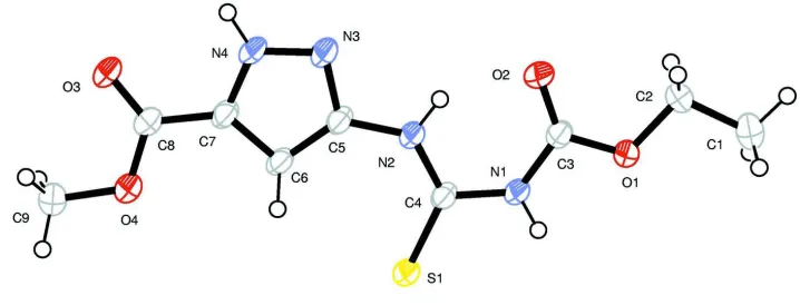
product of the reaction of methyl 5-amino-1H -pyrazole-3-carboxylate with ethyl isothiocyanatocarbonate. All non-H atoms of the molecule are planar, the mean deviation from the least squares plane being 0.048 A˚ . The intramolecular N— H O bond involving the NH-group, which links the thiourea and pyrazole fragments, closes a six-membered pseudo-heterocyclic ring, and two more hydrogen bonds (N—H O with the participation of the pyrazole NH group and N— H S involving the second thiourea NH group) link the molecules into infinite chains running along [120].
Related literature
For the structures of similar N-pyrazole-substituted thiourea derivatives, see: Pasket al.(2006); Wenet al.(2006).
Experimental
Crystal data
C9H12N4O4S Mr= 272.29
a= 8.0855 (8) A˚
b= 9.0035 (8) A˚
c= 9.5959 (9) A˚ = 64.510 (1) = 82.294 (1) = 78.716 (1)
Z= 2
MoKradiation = 0.28 mm1
T= 208 K
0.200.150.10 mm
Data collection
Siemens P4 diffractometer with APEX CCD detector Absorption correction: multi-scan
(SADABS; Bruker, 2001)
Tmin= 0.947,Tmax= 0.973
5852 measured reflections 2653 independent reflections 2255 reflections withI> 2(I)
Rint= 0.044
Refinement
R[F2> 2(F2)] = 0.040
wR(F2) = 0.113
S= 1.04 2653 reflections
166 parameters
H-atom parameters constrained
max= 0.39 e A˚3
min=0.28 e A˚3
Table 1
Hydrogen-bond geometry (A˚ ,).
D—H A D—H H A D A D—H A
N1—H1 S1i
0.87 2.51 3.347 (1) 161 N2—H2 O2 0.87 1.92 2.657 (2) 141 N4—H4 O3ii
0.87 2.03 2.876 (2) 164
Symmetry codes: (i)xþ1;yþ1;zþ1; (ii)xþ2;y1;zþ1.
Data collection:SMART(Bruker, 1997); cell refinement:SAINT
(Bruker, 1997); data reduction:SAINT; program(s) used to solve structure: SIR2004 (Burla et al., 2005); program(s) used to refine structure: SHELXL97 (Sheldrick, 2008); molecular graphics:
ORTEP-32(Farrugia, 1997); software used to prepare material for publication:WinGX(Farrugia, 1999).
Supplementary data and figures for this paper are available from the IUCr electronic archives (Reference: DN2451).
References
Bruker (1997).SMARTandSAINT. Bruker AXS Inc., Madison, Wisconsin, USA.
Bruker (2001).SADABS. Bruker AXS Inc., Madison, Wisconsin, USA. Burla, M. C., Caliandro, R., Camalli, M., Carrozzini, B., Cascarano, G. L., De
Caro, L., Giacovazzo, C., Polidori, G. & Spagna, R. (2005).J. Appl. Cryst.38, 381–388.
Farrugia, L. J. (1997).J. Appl. Cryst.30, 565. Farrugia, L. J. (1999).J. Appl. Cryst.32, 837–838.
Pask, C. M., Camm, K. D., Kilner, C. A. & Halcrow, M. A. (2006).Tetrahedron Lett.2531–2534.
Sheldrick, G. M. (2008).Acta Cryst.A64, 112–122.
Wen, L.-R., Li, M., Zhou, J.-X. & Liu, P. (2006).Acta Cryst.E62, o940–o941. Structure Reports
Online
supporting information
Acta Cryst. (2009). E65, o1249 [doi:10.1107/S1600536809016742]
Methyl 3-[3-(ethoxycarbonyl)thioureido]-1
H
-pyrazole-5-carboxylate
Buwen Huang, Pei-Pei Kung, Arnold L. Rheingold, Antonio DiPasquale and Alex Yanovsky
S1. Comment
The reaction of methyl 5-amino-1H-3-carboxylate with ethyl isothiocyanatocarbonate produces the
pyrazole-thiourea deivative; its structure was established by the present X-ray study (Fig.1).
All non-H atoms of the molecule are planar (mean deviation from its least squares plane is 0.048 Å), in contrast to
previously studied pyrazole-thiourea derivative (Wen et al., 2006), where the pyrazole fragment has a nitrile substituent
in position 4 and pyrazole/thiourea fragments form dihedral angle of 46.2°. Another similar compound, where pyrazole
has no substituents in position 4 (Pask et al., 2006), is also essentially planar, just like the title compound.
There are three NH-groups in the molecule which are responsible for the formation of three independent H-bonds in the
crystal (Table 2). The intramolecular N2—H2···O2 bond closes the 6-membered pseudo-cycle, whereas two
intermolecular H-bonds each produce typical centrosymmmetric pairing motive, and their combination thus gives rise to
infinite chains running along the [1,-2,0]. direction in the crystal (Fig. 2).
S2. Experimental
A suspension of methyl 5-amino-1H-pyrazole-3-carboxylate (2.0 g, 14.2 mmol) in 10 ml of ethyl acetate and 40 ml of
benzene was cooled to 0°C and stirred. To this solution, ethyl isothiocyanatocarbonate (2.04 g, 15.6 mmol) in 10 ml
benzene was added dropwise. The resulting reaction mixture was allowed to warm up to room temperature, and stirring
was continued for 5 h. The reaction mixture was filtered, and washed with plenty of ether to afford the desired product
(3.32 g, 12.2 mmol, 86.0% yield). 1H NMR (400 MHz, DMSO-d
6) δ p.p.m.: 13.99 (br. s., 1 H), 12.12 (br. s., 1 H), 11.48
(br. s., 1 H), 7.51 (s, 1 H), 4.22 (q, J=7.07 Hz, 2 H), 3.85 (s, 3 H), 1.26 (t, J=7.07 Hz, 3 H).
S3. Refinement
All H atoms were placed in geometrically calculated positions (N—H 0.87 Å, C—H 0.94 Å, 0.97 Å, 0.98 Å, for
aromatic, methyl and methylene H atoms respectively) and included in the refinement in riding motion approximation.
The Uiso(H) were set to 1.2Ueq of the carrying atom for aromatic, methylene, methyne and amine groups, and 1.5Ueq for
Figure 1
Molecular structure of the title compound showing 50% probability displacement ellipsoids and atom numbering scheme;
Figure 2
Packing diagram for the title compound viewed approximately along the a axis; H-bonds are shown as dashed lines.
Methyl 3-[3-(ethoxycarbonyl)thioureido]-1H-pyrazole-5-carboxylate
Crystal data
C9H12N4O4S
Mr = 272.29
Triclinic, P1 Hall symbol: -P 1 a = 8.0855 (8) Å b = 9.0035 (8) Å c = 9.5959 (9) Å α = 64.510 (1)° β = 82.294 (1)°
γ = 78.716 (1)° V = 617.39 (10) Å3
Z = 2 F(000) = 284 Dx = 1.465 Mg m−3
Mo Kα radiation, λ = 0.71073 Å Cell parameters from 3767 reflections θ = 2.5–27.8°
Block, colorless
Data collection
Siemens P4
diffractometer with APEX CCD Radiation source: fine-focus sealed tube Graphite monochromator
φ and ω scans
Absorption correction: multi-scan (SADABS; Bruker, 2001) Tmin = 0.947, Tmax = 0.973
5852 measured reflections 2653 independent reflections 2255 reflections with I > 2σ(I) Rint = 0.044
θmax = 28.2°, θmin = 2.4°
h = −5→10 k = −11→11 l = −11→12
Refinement
Refinement on F2
Least-squares matrix: full R[F2 > 2σ(F2)] = 0.040
wR(F2) = 0.113
S = 1.04 2653 reflections 166 parameters 0 restraints
Primary atom site location: structure-invariant direct methods
Secondary atom site location: difference Fourier map
Hydrogen site location: inferred from neighbouring sites
H-atom parameters constrained w = 1/[σ2(F
o2) + (0.0521P)2 + 0.1805P]
where P = (Fo2 + 2Fc2)/3
(Δ/σ)max = 0.001
Δρmax = 0.39 e Å−3
Δρmin = −0.28 e Å−3
Extinction correction: SHELXL (Sheldrick, 2008), Fc*=kFc[1+0.001xFc2λ3/sin(2θ)]-1/4
Extinction coefficient: 0.064 (8)
Special details
Geometry. All e.s.d.'s (except the e.s.d. in the dihedral angle between two l.s. planes) are estimated using the full covariance matrix. The cell e.s.d.'s are taken into account individually in the estimation of e.s.d.'s in distances, angles and torsion angles; correlations between e.s.d.'s in cell parameters are only used when they are defined by crystal symmetry. An approximate (isotropic) treatment of cell e.s.d.'s is used for estimating e.s.d.'s involving l.s. planes.
Refinement. Refinement of F2 against ALL reflections. The weighted R-factor wR and goodness of fit S are based on F2,
conventional R-factors R are based on F, with F set to zero for negative F2. The threshold expression of F2 > σ(F2) is used
only for calculating R-factors(gt) etc. and is not relevant to the choice of reflections for refinement. R-factors based on F2
are statistically about twice as large as those based on F, and R- factors based on ALL data will be even larger.
Fractional atomic coordinates and isotropic or equivalent isotropic displacement parameters (Å2)
x y z Uiso*/Ueq
C1 0.7861 (3) 0.9454 (2) −0.0198 (2) 0.0539 (6)
H1A 0.6653 0.9725 −0.0320 0.081*
H1B 0.8446 1.0106 −0.1156 0.081*
H1C 0.8125 0.9706 0.0626 0.081*
C2 0.8414 (3) 0.7653 (2) 0.0194 (2) 0.0451 (5)
H2A 0.8135 0.7375 −0.0619 0.054*
H2B 0.9638 0.7366 0.0301 0.054*
C3 0.7832 (2) 0.5090 (2) 0.22047 (19) 0.0319 (4)
C4 0.6784 (2) 0.27556 (19) 0.44802 (18) 0.0285 (4)
C5 0.7965 (2) −0.00499 (19) 0.46055 (19) 0.0300 (4)
C6 0.7316 (2) −0.1229 (2) 0.5968 (2) 0.0310 (4)
C7 0.8050 (2) −0.2725 (2) 0.59165 (19) 0.0312 (4)
C8 0.7909 (2) −0.4462 (2) 0.6958 (2) 0.0328 (4)
C9 0.6497 (3) −0.6292 (2) 0.9141 (2) 0.0435 (5)
H9A 0.6341 −0.6894 0.8550 0.065*
H9B 0.5509 −0.6267 0.9831 0.065*
H9C 0.7486 −0.6844 0.9741 0.065*
N1 0.69023 (19) 0.44206 (16) 0.35749 (16) 0.0322 (3)
H1 0.6316 0.5127 0.3917 0.039*
N2 0.77543 (19) 0.16936 (16) 0.39615 (16) 0.0327 (3)
H2 0.8340 0.2152 0.3099 0.039*
N3 0.9003 (2) −0.07384 (17) 0.37579 (17) 0.0360 (4)
N4 0.9033 (2) −0.23814 (17) 0.46052 (17) 0.0344 (3)
H4 0.9626 −0.3141 0.4333 0.041*
O1 0.75193 (17) 0.67399 (14) 0.16595 (14) 0.0374 (3)
O2 0.87813 (18) 0.43041 (15) 0.15826 (15) 0.0425 (3)
O3 0.87755 (18) −0.56425 (14) 0.67884 (15) 0.0407 (3)
O4 0.67259 (17) −0.46002 (15) 0.80911 (15) 0.0397 (3)
S1 0.54880 (6) 0.22717 (5) 0.60563 (5) 0.03395 (17)
Atomic displacement parameters (Å2)
U11 U22 U33 U12 U13 U23
C1 0.0681 (15) 0.0334 (10) 0.0449 (11) −0.0077 (10) 0.0106 (10) −0.0060 (8) C2 0.0512 (12) 0.0338 (9) 0.0352 (9) −0.0029 (8) 0.0133 (8) −0.0061 (8) C3 0.0347 (9) 0.0267 (8) 0.0311 (8) 0.0000 (7) 0.0010 (7) −0.0119 (6) C4 0.0312 (9) 0.0247 (7) 0.0300 (8) 0.0008 (6) −0.0025 (7) −0.0137 (6) C5 0.0340 (9) 0.0240 (8) 0.0333 (8) 0.0005 (6) −0.0013 (7) −0.0154 (7) C6 0.0343 (9) 0.0257 (8) 0.0346 (8) 0.0005 (6) 0.0007 (7) −0.0171 (7) C7 0.0346 (9) 0.0259 (8) 0.0359 (9) −0.0010 (7) −0.0003 (7) −0.0174 (7) C8 0.0356 (9) 0.0295 (8) 0.0373 (9) −0.0036 (7) 0.0003 (7) −0.0189 (7) C9 0.0499 (12) 0.0310 (9) 0.0460 (11) −0.0095 (8) 0.0089 (9) −0.0147 (8) N1 0.0393 (8) 0.0234 (7) 0.0306 (7) −0.0001 (6) 0.0074 (6) −0.0127 (6) N2 0.0405 (8) 0.0234 (7) 0.0313 (7) −0.0012 (6) 0.0061 (6) −0.0124 (6) N3 0.0436 (9) 0.0251 (7) 0.0383 (8) −0.0008 (6) 0.0038 (7) −0.0160 (6) N4 0.0403 (9) 0.0256 (7) 0.0396 (8) −0.0003 (6) 0.0042 (7) −0.0195 (6) O1 0.0432 (7) 0.0254 (6) 0.0341 (6) −0.0013 (5) 0.0113 (5) −0.0091 (5) O2 0.0514 (8) 0.0321 (7) 0.0377 (7) 0.0001 (6) 0.0138 (6) −0.0160 (6) O3 0.0490 (8) 0.0263 (6) 0.0472 (7) −0.0015 (6) 0.0073 (6) −0.0204 (6) O4 0.0455 (8) 0.0274 (6) 0.0448 (7) −0.0050 (5) 0.0098 (6) −0.0174 (5) S1 0.0400 (3) 0.0257 (2) 0.0328 (2) −0.00173 (17) 0.00760 (18) −0.01324 (18)
Geometric parameters (Å, º)
C1—C2 1.486 (3) C5—N2 1.401 (2)
C1—H1A 0.9700 C6—C7 1.380 (2)
C1—H1B 0.9700 C6—H6 0.9400
C1—H1C 0.9700 C7—N4 1.343 (2)
C2—H2B 0.9800 C8—O4 1.329 (2)
C3—O2 1.214 (2) C9—O4 1.452 (2)
C3—O1 1.3278 (19) C9—H9A 0.9700
C3—N1 1.374 (2) C9—H9B 0.9700
C4—N2 1.338 (2) C9—H9C 0.9700
C4—N1 1.387 (2) N1—H1 0.8700
C4—S1 1.6617 (16) N2—H2 0.8700
C5—N3 1.340 (2) N3—N4 1.344 (2)
C5—C6 1.397 (2) N4—H4 0.8700
C2—C1—H1A 109.5 N4—C7—C6 107.60 (14)
C2—C1—H1B 109.5 N4—C7—C8 119.72 (14)
H1A—C1—H1B 109.5 C6—C7—C8 132.67 (16)
C2—C1—H1C 109.5 O3—C8—O4 123.84 (16)
H1A—C1—H1C 109.5 O3—C8—C7 123.32 (16)
H1B—C1—H1C 109.5 O4—C8—C7 112.84 (14)
O1—C2—C1 106.83 (15) O4—C9—H9A 109.5
O1—C2—H2A 110.4 O4—C9—H9B 109.5
C1—C2—H2A 110.4 H9A—C9—H9B 109.5
O1—C2—H2B 110.4 O4—C9—H9C 109.5
C1—C2—H2B 110.4 H9A—C9—H9C 109.5
H2A—C2—H2B 108.6 H9B—C9—H9C 109.5
O2—C3—O1 125.25 (16) C3—N1—C4 127.95 (13)
O2—C3—N1 125.62 (15) C3—N1—H1 116.0
O1—C3—N1 109.13 (13) C4—N1—H1 116.0
N2—C4—N1 114.69 (14) C4—N2—C5 129.41 (14)
N2—C4—S1 126.73 (12) C4—N2—H2 115.3
N1—C4—S1 118.59 (11) C5—N2—H2 115.3
N3—C5—C6 112.89 (14) C5—N3—N4 103.49 (14)
N3—C5—N2 114.22 (15) C7—N4—N3 112.76 (13)
C6—C5—N2 132.89 (15) C7—N4—H4 123.6
C7—C6—C5 103.26 (14) N3—N4—H4 123.6
C7—C6—H6 128.4 C3—O1—C2 116.14 (14)
C5—C6—H6 128.4 C8—O4—C9 115.46 (13)
Hydrogen-bond geometry (Å, º)
D—H···A D—H H···A D···A D—H···A
N1—H1···S1i 0.87 2.51 3.347 (1) 161
N2—H2···O2 0.87 1.92 2.657 (2) 141
N4—H4···O3ii 0.87 2.03 2.876 (2) 164