1-Phenyl-2-(1
H
-1,2,4-triazol-1-yl)ethanol
O¨ zden O¨ zel Gu¨ven,aHakan Tahtacı,aSimon J. Colesband Tuncer Ho¨kelekc*
a
Zonguldak Karaelmas University, Department of Chemistry, 67100 Zonguldak, Turkey,bDepartment of Chemistry, Southampton University, Southampton SO17 1BJ, England, andcHacettepe University, Department of Physics, 06800 Beytepe, Ankara, Turkey
Correspondence e-mail: [email protected]
Received 15 May 2008; accepted 9 June 2008
Key indicators: single-crystal X-ray study;T= 294 K; mean(C–C) = 0.002 A˚; Rfactor = 0.042;wRfactor = 0.115; data-to-parameter ratio = 12.9.
In the title compound, C10H11N3O, the planar five- and
six-membered rings are nearly parallel to each other, making a dihedral angle of 2.52 (5). Weak intermolecular C—H O
hydrogen bonds link the molecules into centrosymmetric dimers and strong intermolecular O—H N hydrogen bonds link the dimers into infinite chains along thebaxis.
Related literature
For general backgroud, see: Hollaet al.(1996); Senguptaet al.
(1978); Paulvannan et al. (2001); Sui et al. (1998); Bodey (1992). For related literature, see: Peeters et al. (1979a,b); Cairaet al.(2004); Freeret al.(1986); Peeterset al.(1996).
Experimental
Crystal data
C10H11N3O
Mr= 189.22 Monoclinic,P21=c
a= 11.5356 (2) A˚
b= 10.1173 (2) A˚
c= 8.7127 (2) A˚
= 108.581 (1)
V= 963.85 (3) A˚3
Z= 4
MoKradiation
= 0.09 mm1
T= 294 (2) K 0.550.250.10 mm
Data collection
Bruker–Nonius KappaCCD diffractometer
Absorption correction: multi-scan (SADABS; Sheldrick, 2007)
Tmin= 0.972,Tmax= 0.989
13352 measured reflections 2208 independent reflections 1647 reflections withI> 2(I)
Rint= 0.040
Refinement
R[F2> 2(F2)] = 0.042
wR(F2) = 0.115
S= 1.03 2208 reflections
171 parameters
All H-atom parameters refined
max= 0.16 e A˚
3
min=0.21 e A˚
3
Table 1
Hydrogen-bond geometry (A˚ ,).
D—H A D—H H A D A D—H A
O—H N2i
0.88 (2) 2.00 (2) 2.8645 (17) 166 (2) C10—H10 Oii
0.959 (16) 2.566 (16) 3.3198 (17) 135.6 (13)
Symmetry codes: (i)x;yþ1 2;z
1
2; (ii)xþ2;y;z.
Data collection: COLLECT (Hooft, 1998); cell refinement:
DENZO(Otwinowski & Minor, 1997) andCOLLECT; data reduc-tion:DENZO andCOLLECT; program(s) used to solve structure:
SHELXL97(Sheldrick, 2008); program(s) used to refine structure:
SHELXL97 (Sheldrick, 2008); molecular graphics: ORTEP-3 for Windows (Farrugia, 1997); software used to prepare material for publication:WinGX(Farrugia, 1999).
The authors acknowledge Zonguldak Karaelmas University Research Fund for support.
Supplementary data and figures for this paper are available from the IUCr electronic archives (Reference: FL2201).
References
Bodey, G. P. (1992).Clin. Infect. Dis.14, S161–S169.
Caira, M. R., Alkhamis, K. A. & Obaidat, R. M. (2004).J. Pharm. Sci.93, 601– 611.
Farrugia, L. J. (1997).J. Appl. Cryst.30, 565. Farrugia, L. J. (1999).J. Appl. Cryst.32, 837–838.
Freer, A. A., Pearson, A. & Salole, E. G. (1986).Acta Cryst.C42, 1350–1352. Holla, B. S., Poojary, K. N., Kalluraya, B. & Gowda, P. V. (1996).Farmaco,51,
793–799.
Hooft, R. W. W. (1998).COLLECT. Nonius BV, Delft, The Netherlands. Otwinowski, Z. & Minor, W. (1997). Methods in Enzymology, Vol. 276,
Macromolecular Crystallography, Part A, edited by C. W. Carter Jr & R. M. Sweet, pp. 307–326. New York: Academic Press.
Paulvannan, K., Hale, R., Sedehi, D. & Chen, T. (2001).Tetrahedron,57, 9677– 9682.
Peeters, O. M., Blaton, N. M. & De Ranter, C. J. (1996).Acta Cryst.C52, 2225– 2229.
Peeters, O. M., Blaton, N. M. & De Ranter, C. J. (1979a).Bull. Soc. Chim. Belg. 88, 265–272.
Peeters, O. M., Blaton, N. M. & De Ranter, C. J. (1979b).Acta Cryst.B35, 2461–2464.
Sengupta, A. K., Bajaj, O. P. & Chandra, U. (1978).J. Indian Chem. Soc.55, 962–964.
Sheldrick, G. M. (2007).SADABS. Bruker AXS Inc., Madison, Wisconsin, USA.
Sheldrick, G. M. (2008).Acta Cryst.A64, 112–122.
Sui, Z. H., Guan, J. H., Hlasta, D. J., Macielag, M. J., Foleno, B. D., Goldschmidt, R. M., Loeloff, M. J., Webb, G. C. & Barrett, J. F. (1998).
Bioorg. Med. Chem. Lett.8, 1929–1934. Acta Crystallographica Section E
Structure Reports
Online
supporting information
Acta Cryst. (2008). E64, o1254 [doi:10.1107/S1600536808017303]
1-Phenyl-2-(1
H
-1,2,4-triazol-1-yl)ethanol
Ö
zden
Ö
zel G
ü
ven, Hakan Tahtac
ı
, Simon J. Coles and Tuncer H
ö
kelek
S1. Comment
Azole derivatives continue to occupy an important place among systemic antifungal drugs. 1,2,4-triazoles are biologically
interesting and their chemistry is receiving considerable attention due to their antihypertensive, antifungal and
antibacterial properties (Holla et al., 1996; Sengupta et al., 1978; Paulvannan et al., 2001; Sui et al., 1998). The azole
antifungals possessing an imidazole or triazole ring (such as miconazole, ketoconazole, fluconazole, econazole and
itraconazole) inhibit the synthesis of sterols in fungi by inhibiting cytochrome P-450-dependent 14α-lanosterol
demethyl-ase (P-45014DM) and prevent cytochrome P-450 activity (Bodey, 1992). The crystal structures of miconazole (Peeters et
al., 1997a), ketoconazole (Peeters et al., 1979b), fluconazole (Caira et al., 2004), econazole (Freer et al., 1986) and
itraconazole (Peeters et al., 1996) have already been reported. This paper describes the crystal structure of a 1,2,4-triazole
derivative, (I).
In (I) the bond lengths and angles are generally within normal ranges (Fig. 1). The 1,2,4-triazole and benzene rings, A
(N1—N3/C1/C2) and B (C5—C10), are planar and nearly parallel to each other with a dihedral angle of A/B = 2.52 (5)°.
Atoms C3 and C4 are 0.040 (1) Å and -0.046 (1) Å away from the ring planes of A and B, respectively indicating that
they are coplanar with the adjacent rings. The N1—C3—C4 [111.53 (10)°] and C3—C4—C5 [109.94 (10)°] bond angles
are a little different from each other, while O—C4—C3 [109.53 (11)°] and O—C4—C5 [110.01 (10)°] bond angles are
nearly equal. In ring A, the equivalent N1—N2—C1 [102.24 (12)°] and C1—N3—C2 [102.29 (13)°] bond angles are
narrowed and approximately equal to one another, while the N3—C2—N1 [111.04 (15)°] and N3—C1—N2
[115.33 (15)°] bond angles are quite different and larger than normal, probably due to the strong intermolecular O—H···N
hydrogen bonds (Table 1).
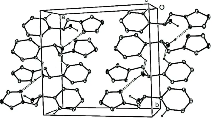
In the crystal packing weak intermolecular C—H···O hydrogen bonds (Table 1) link the molecules into centrosymmetric
dimers and strong intermolecular O—H···N hydrogen bonds (Table 1) link the dimers along the b axis (Fig. 2).
S2. Experimental
For the preparation of the title compound, a mixture of 1-phenyl-2-(1H-1,2,4 -triazol-1-yl)ethanone (800 mg, 4.27 mmol)
and sodiumborohydride (324 mg, 8.54 mmol) in ethanol (13 ml) was refluxed for 5 h. After evaporation of solvent, the
mixture was neutralized with dilute HCl and then refluxed for 30 min. After the mixture was cooled, the solution was
alkalinized with NaOH and the precipitate was collected and crystallized from benzene to obtain colorless crystals (yield;
577 mg, 71%).
S3. Refinement
H atoms were located in difference syntheses and refined isotropically [O—H = 0.88 (2) Å, Uiso(H) = 0.096 (7) Å2 and C
Figure 1
The molecular structure of the title molecule with the atom-numbering scheme. Displacement ellipsoids are drawn at the
50% probability level.
Figure 2
A packing diagram of (I). Hydrogen bonds are shown as dashed lines.
1-Phenyl-2-(1H-1,2,4-triazol-1-yl)ethanol
Crystal data
C10H11N3O
Mr = 189.22 Monoclinic, P21/c
Hall symbol: -P 2ybc
a = 11.5356 (2) Å
b = 10.1173 (2) Å
c = 8.7127 (2) Å
β = 108.581 (1)°
V = 963.85 (3) Å3
Z = 4
F(000) = 400
Dx = 1.304 Mg m−3
Mo Kα radiation, λ = 0.71073 Å Cell parameters from 12727 reflections
θ = 2.9–27.5°
µ = 0.09 mm−1
[image:3.610.129.484.288.488.2]Data collection
Bruker–Nonius Roper CCD camera on κ -goniostat
diffractometer
Radiation source: Bruker-Nonius FR591 rotating anode
Graphite monochromator
Detector resolution: 9.091 pixels mm-1
φ and ω scans
Absorption correction: multi-scan (SADABS; Sheldrick, 2007)
Tmin = 0.972, Tmax = 0.989
13352 measured reflections 2208 independent reflections 1647 reflections with I > 2σ(I)
Rint = 0.040
θmax = 27.5°, θmin = 3.2°
h = −14→14
k = −13→12
l = −11→10
Refinement
Refinement on F2
Least-squares matrix: full
R[F2 > 2σ(F2)] = 0.042
wR(F2) = 0.115
S = 1.03 2208 reflections 171 parameters 0 restraints
Primary atom site location: structure-invariant direct methods
Secondary atom site location: difference Fourier map
Hydrogen site location: inferred from neighbouring sites
All H-atom parameters refined
w = 1/[σ2(F
o2) + (0.0583P)2 + 0.1287P]
where P = (Fo2 + 2Fc2)/3
(Δ/σ)max < 0.001
Δρmax = 0.16 e Å−3
Δρmin = −0.21 e Å−3
Special details
Geometry. All e.s.d.'s (except the e.s.d. in the dihedral angle between two l.s. planes) are estimated using the full covariance matrix. The cell e.s.d.'s are taken into account individually in the estimation of e.s.d.'s in distances, angles and torsion angles; correlations between e.s.d.'s in cell parameters are only used when they are defined by crystal symmetry. An approximate (isotropic) treatment of cell e.s.d.'s is used for estimating e.s.d.'s involving l.s. planes.
Refinement. Refinement of F2 against ALL reflections. The weighted R-factor wR and goodness of fit S are based on F2,
conventional R-factors R are based on F, with F set to zero for negative F2. The threshold expression of F2 > σ(F2) is used
only for calculating R-factors(gt) etc. and is not relevant to the choice of reflections for refinement. R-factors based on F2
are statistically about twice as large as those based on F, and R- factors based on ALL data will be even larger.
Fractional atomic coordinates and isotropic or equivalent isotropic displacement parameters (Å2)
x y z Uiso*/Ueq
C6 1.12425 (12) 0.22987 (15) 0.42648 (16) 0.0480 (3) H6 1.0823 (15) 0.2996 (17) 0.467 (2) 0.070 (5)* C7 1.24947 (13) 0.21333 (16) 0.49370 (19) 0.0572 (4) H7 1.2961 (16) 0.2732 (19) 0.584 (2) 0.079 (5)* C8 1.30805 (13) 0.11523 (17) 0.4370 (2) 0.0566 (4) H8 1.3973 (17) 0.1018 (18) 0.484 (2) 0.078 (5)* C9 1.24185 (13) 0.03446 (15) 0.31286 (19) 0.0529 (4) H9 1.2826 (15) −0.0328 (17) 0.269 (2) 0.065 (5)* C10 1.11643 (12) 0.05072 (13) 0.24487 (16) 0.0426 (3) H10 1.0711 (14) −0.0032 (15) 0.1553 (19) 0.055 (4)*
Atomic displacement parameters (Å2)
U11 U22 U33 U12 U13 U23
O 0.0489 (6) 0.0914 (8) 0.0316 (5) 0.0183 (6) 0.0065 (4) 0.0033 (5) N1 0.0314 (5) 0.0530 (6) 0.0427 (6) −0.0020 (4) 0.0077 (5) 0.0030 (5) N2 0.0371 (6) 0.0646 (8) 0.0512 (7) −0.0016 (5) 0.0095 (5) −0.0048 (6) N3 0.0349 (6) 0.0949 (11) 0.0780 (10) −0.0047 (7) 0.0036 (6) −0.0166 (8) C1 0.0365 (7) 0.0801 (11) 0.0692 (10) 0.0026 (7) 0.0124 (7) −0.0067 (9) C2 0.0399 (8) 0.0711 (10) 0.0607 (9) −0.0100 (7) 0.0061 (7) −0.0121 (8) C3 0.0321 (6) 0.0542 (8) 0.0438 (7) 0.0021 (5) 0.0075 (5) 0.0098 (6) C4 0.0363 (6) 0.0399 (7) 0.0345 (6) 0.0042 (5) 0.0084 (5) 0.0021 (5) C5 0.0347 (6) 0.0380 (6) 0.0345 (6) −0.0004 (5) 0.0122 (5) 0.0041 (5) C6 0.0440 (7) 0.0515 (8) 0.0465 (7) −0.0005 (6) 0.0116 (6) −0.0084 (6) C7 0.0441 (8) 0.0671 (10) 0.0537 (8) −0.0100 (7) 0.0060 (6) −0.0084 (8) C8 0.0337 (7) 0.0728 (10) 0.0603 (9) 0.0009 (7) 0.0106 (6) 0.0085 (8) C9 0.0456 (8) 0.0579 (9) 0.0585 (8) 0.0132 (7) 0.0212 (7) 0.0042 (7) C10 0.0425 (7) 0.0424 (7) 0.0417 (7) 0.0038 (6) 0.0117 (6) 0.0006 (6)
Geometric parameters (Å, º)
O—C4 1.4144 (15) C4—H4 0.966 (13) O—H 0.88 (2) C5—C4 1.5128 (16) N1—N2 1.3602 (16) C5—C6 1.3869 (18) N1—C2 1.3257 (18) C5—C10 1.3866 (18) N1—C3 1.4565 (16) C6—C7 1.385 (2) N2—C1 1.3178 (18) C6—H6 0.982 (18) N3—C2 1.322 (2) C7—H7 1.01 (2) C1—N3 1.341 (2) C8—C7 1.378 (2) C1—H1 0.96 (2) C8—C9 1.376 (2) C2—H2 0.961 (19) C8—H8 0.989 (19) C3—H31 1.012 (17) C9—H9 0.974 (18) C3—H32 0.977 (17) C10—C9 1.3875 (19) C4—C3 1.5194 (18) C10—H10 0.959 (16)
N2—N1—C3 120.31 (11) C6—C5—C4 119.78 (11) C1—N2—N1 102.24 (12) C10—C5—C4 121.28 (11) C2—N3—C1 102.29 (13) C10—C5—C6 118.92 (12) N2—C1—N3 115.33 (15) C5—C6—H6 119.2 (10) N2—C1—H1 120.7 (11) C7—C6—C5 120.68 (13) N3—C1—H1 123.9 (11) C7—C6—H6 120.1 (10) N1—C2—H2 123.7 (11) C6—C7—H7 119.0 (10) N3—C2—N1 111.04 (15) C8—C7—C6 120.00 (14) N3—C2—H2 125.2 (11) C8—C7—H7 121.0 (10) N1—C3—C4 111.53 (10) C7—C8—H8 120.9 (11) N1—C3—H31 107.0 (9) C9—C8—C7 119.79 (13) N1—C3—H32 108.5 (9) C9—C8—H8 119.3 (11) C4—C3—H31 109.9 (9) C8—C9—C10 120.47 (14) C4—C3—H32 110.9 (9) C8—C9—H9 120.6 (9) H31—C3—H32 109.0 (13) C10—C9—H9 118.9 (9) O—C4—C3 109.53 (11) C5—C10—C9 120.14 (13) O—C4—C5 110.01 (10) C5—C10—H10 119.5 (9) O—C4—H4 109.6 (7) C9—C10—H10 120.3 (9)
C2—N1—N2—C1 0.35 (16) C6—C5—C4—C3 −92.44 (14) C3—N1—N2—C1 178.19 (12) C10—C5—C4—O −34.88 (15) N2—N1—C2—N3 −0.37 (19) C10—C5—C4—C3 85.83 (14) C3—N1—C2—N3 −177.91 (14) C4—C5—C6—C7 178.13 (12) C2—N1—C3—C4 94.22 (18) C10—C5—C6—C7 −0.2 (2) N2—N1—C3—C4 −83.09 (15) C6—C5—C10—C9 0.30 (19) N1—N2—C1—N3 −0.24 (19) C4—C5—C10—C9 −177.99 (12) C1—N3—C2—N1 0.2 (2) C5—C6—C7—C8 −0.2 (2) N2—C1—N3—C2 0.0 (2) C9—C8—C7—C6 0.4 (2) O—C4—C3—N1 −69.27 (14) C7—C8—C9—C10 −0.3 (2) C5—C4—C3—N1 169.73 (11) C5—C10—C9—C8 −0.1 (2) C6—C5—C4—O 146.85 (12)
Hydrogen-bond geometry (Å, º)
D—H···A D—H H···A D···A D—H···A
O—H···N2i 0.88 (2) 2.00 (2) 2.8645 (17) 166 (2)
C10—H10···Oii 0.959 (16) 2.566 (16) 3.3198 (17) 135.6 (13)