4-Hydroxy-3-(3-methoxybenzoyl)-2-[(3-methoxybenzoyl)methyl]-2
H
-1,2-benzothiazine 1,1-dioxide
Salman Gul,aHamid Latif Siddiqui,a* Matloob Ahmad,b Muhammad Nisarcand Masood Parvezd
aInstitute of Chemistry, University of the Punjab, Lahore 54590, Pakistan,bInstitute of Chemistry, University of the Punjab, Lahore-54590, Applied Chemistry Research Centre, PCSIR Laboratories Complex, Lahore 54600, Pakistan,cInstitute of Chemical Sciences, University of Peshawar, Peshawar 25120, Pakistan, anddDepartment of Chemistry, The University of Calgary, 2500 University Drive NW, Calgary, Alberta, Canada T2N 1N4
Correspondence e-mail: drhamidlatif@yahoo.com
Received 31 July 2010; accepted 6 August 2010
Key indicators: single-crystal X-ray study;T= 173 K; mean(C–C) = 0.002 A˚; Rfactor = 0.038;wRfactor = 0.098; data-to-parameter ratio = 15.7.
In the title compound, C25H21NO7S, the heterocyclic thiazine
ring adopts a half-chair conformation, with the S and N atoms displaced by0.284 (3) and 0.411 (3) A˚ , respectively, from the plane formed by the remaining ring atoms; the puckering parameters are: Q = 0.4576 (13) A˚ , = 58.6 (2) and ’ = 34.3 (3). The structure is devoid of any classical hydrogen
bonds. However, intramolecular C—H N and O—H O hydrogen bonds result in six-membered rings and inter-molecular C—H O interactions stabilize the crystal struc-ture.
Related literature
For the biological applications of benzothiazines, see: Lombardinoet al.(1972); Zinneset al.(1982); Zia-ur-Rehman et al. (2005); Turck et al. (1996); Ahmad et al. (2010). For related structures, see: Siddiqui et al. (2008). For puckering parameters, see: Cremer & Pople (1975).
Experimental
Crystal data
C25H21NO7S
Mr= 479.49
Triclinic,P1
a= 10.3169 (2) A˚
b= 10.6923 (3) A˚
c= 11.6867 (3) A˚
= 115.5965 (11)
= 105.8041 (14)
= 97.6128 (13)
V= 1071.22 (5) A˚3
Z= 2
MoKradiation
= 0.20 mm1
T= 173 K
0.240.160.08 mm
Data collection
Nonius KappaCCD diffractometer Absorption correction: multi-scan
(SORTAV; Blessing, 1997)
Tmin= 0.953,Tmax= 0.984
9164 measured reflections 4860 independent reflections 4419 reflections with (I) > 2.0(I)
Rint= 0.021
Refinement
R[F2> 2(F2)] = 0.038
wR(F2) = 0.098
S= 1.07 4860 reflections
310 parameters
H-atom parameters constrained max= 0.37 e A˚3
[image:1.610.95.248.600.700.2]min=0.42 e A˚3
Table 1
Hydrogen-bond geometry (A˚ ,).
D—H A D—H H A D A D—H A
C25—H25C O1i
0.98 2.57 3.438 (2) 147
C17—H17B O2i
0.99 2.26 3.244 (2) 174
C15—H15 N1 0.95 2.41 2.986 (2) 119
O3—H3O O4 0.84 1.67 2.428 (2) 149
Symmetry code: (i)xþ1;yþ1;zþ1.
Data collection:COLLECT(Hooft, 1998); cell refinement:HKL DENZO (Otwinowski & Minor, 1997); data reduction: SCALE-PACK (Otwinowski & Minor, 1997); program(s) used to solve structure:SHELXS97(Sheldrick, 2008); program(s) used to refine structure: SHELXL97 (Sheldrick, 2008); molecular graphics: ORTEP-3 for Windows(Farrugia, 1997); software used to prepare material for publication:SHELXL97.
HLS is grateful to the Institute of Chemistry, University of the Punjab, Lahore, Pakistan, for financial support.
Supplementary data and figures for this paper are available from the IUCr electronic archives (Reference: JH2194).
References
Ahmad, M., Siddiqui, H. L., Zia-ur-Rehman, M. & Parvez, M. (2010).Eur. J. Med. Chem.45, 698–704.
Blessing, R. H. (1997).J. Appl. Cryst.30, 421–426.
Cremer, D. & Pople, J. A. (1975).J. Am. Chem. Soc.97, 1354–1358. Farrugia, L. J. (1997).J. Appl. Cryst.30, 565.
Hooft, R. (1998).COLLECT. Nonius B V, Delft, The Netherlands. Lombardino, J. G. & Wiseman, E. H. (1972).J. Med. Chem.15, 848–849. Otwinowski, Z. & Minor, W. (1997). Methods in Enzymology, Vol. 276,
Macromolecular Crystallography, Part A, edited by C. W. Carter Jr and R. M. Sweet, pp. 307–326. New York: Academic Press.
Sheldrick, G. M. (2008).Acta Cryst.A64, 112–122.
Siddiqui, W. A., Ahmad, S., Tariq, M. I., Siddiqui, H. L. & Parvez, M. (2008).
Acta Cryst.C64, 04–06.
organic compounds
o2314
Gulet al. doi:10.1107/S1600536810031673 Acta Cryst.(2010). E66, o2314–o2315 Acta Crystallographica Section EStructure Reports
Online
Turck, D., Busch, U., Heinzel, G., Narjes, H. & Nehmiz, G. (1996).J. Clin. Pharmacol.36, 79–84.
Zia-ur-Rehman, M., Choudary, J. A. & Ahmad, S. (2005).Bull. Kor. Chem. Soc.54, 1171–1175.
supporting information
sup-1 Acta Cryst. (2010). E66, o2314–o2315
supporting information
Acta Cryst. (2010). E66, o2314–o2315 [https://doi.org/10.1107/S1600536810031673]
4-Hydroxy-3-(3-methoxybenzoyl)-2-[(3-methoxybenzoyl)methyl]-2
H
-1,2-benzo-thiazine 1,1-dioxide
Salman Gul, Hamid Latif Siddiqui, Matloob Ahmad, Muhammad Nisar and Masood Parvez
S1. Comment
Oxicams are non steroidal anti-inflammatory drugs (NSAID's) that posses benzothiazine nucleus (Lombardino et al., 1972; Zinnes et al., 1982). Versatile biological activities are associated with benzothiazine derivatives, e.g., anti-microbial (Zia-ur-Rehman et al., 2005), analgesic (Turck et al., 1996), antioxidant (Ahmad et al., 2010), etc. In this paper, we report the synthesis and crystal structure of the title compound.
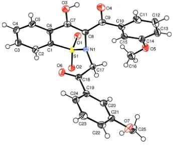
The structure of the title compound contains independent molecules separated by normal van der Waals distances (Fig. 1). The heterocyclic thiazine ring adopts a half-chair conformation, with atoms S1 and N1 displaced by -0.284 (3) and 0.411 (3) Å, respectively, from the plane formed by atoms C1/C6/C7/C8; the puckering parameters (Cremer & Pople, 1975) are: Q = 0.4576 (13) Å, θ = 58.6 (2)° and φ = 34.3 (3)°. Similar conformations of the corresponding rings have been reported in some closely related compounds (Siddiqui et al., 2008). The carbon fragments C1–C15 and C17–C24 are more or less planar individually and lie at an angle 77.17 (2)° with rest to each other.
The structure is devoid of any classical hydrogen bonds. However, intramolecular interactions C15—H15···N1 and O3 —H3O···O4 resulting in six membered rings and intermolecular interactions of the type C—H···O are present (Tab. 1).
S2. Experimental
4-Hydroxy-1,1-dioxido-2H-1,2-benzothiazin-3-yl)(3-methoxyphenyl) methanone (2 g, 6.0 mmol), K2CO3 (1.24 g, 9.0
mmol), 3-methoxyphenacyl bromide (1.42 g, 6.2 mmol) and acetonitrile (25 ml) were refluxed for 6 h. The completion of reaction was monitored by TLC. After cooling to room temperature, the reaction mixture was poured into ice cold water. Yellow precipitates obtained were filtered, washed with cold water and dried. The crystals suitable for crystallographic study were grown from a solution of methanol and chloroform (1:1).
S3. Refinement
The H-atoms were located from difference Fourier maps and were included in the refinement at geometrically idealized positions in riding-model approximation with O—H = 0.84 Å and C—H = 0.95–0.99 Å; the Uiso(H) were allowed at
Figure 1
The title molecule plotted with the displacement ellipsoids at 50% probability level (Farrugia, 1997).
4-Hydroxy-3-(3-methoxybenzoyl)-2-[(3-methoxybenzoyl)methyl]-2H-1,2- benzothiazine 1,1-dioxide
Crystal data C25H21NO7S
Mr = 479.49
Triclinic, P1 Hall symbol: -P 1 a = 10.3169 (2) Å b = 10.6923 (3) Å c = 11.6867 (3) Å α = 115.5965 (11)° β = 105.8041 (14)° γ = 97.6128 (13)° V = 1071.22 (5) Å3
Z = 2 F(000) = 500 Dx = 1.487 Mg m−3
Mo Kα radiation, λ = 0.71073 Å Cell parameters from 4699 reflections θ = 1.0–27.5°
µ = 0.20 mm−1
T = 173 K Prism, yellow
0.24 × 0.16 × 0.08 mm
Data collection Nonius KappaCCD
diffractometer
Radiation source: fine-focus sealed tube Graphite monochromator
ω and φ scans
Absorption correction: multi-scan (SORTAV; Blessing, 1997) Tmin = 0.953, Tmax = 0.984
9164 measured reflections 4860 independent reflections 4419 reflections with (I) > 2.0 σ(I) Rint = 0.021
θmax = 27.5°, θmin = 2.1°
supporting information
sup-3 Acta Cryst. (2010). E66, o2314–o2315
Refinement Refinement on F2
Least-squares matrix: full R[F2 > 2σ(F2)] = 0.038
wR(F2) = 0.098
S = 1.07 4860 reflections 310 parameters 0 restraints
Primary atom site location: structure-invariant direct methods
Secondary atom site location: difference Fourier map
Hydrogen site location: difference Fourier map H-atom parameters constrained
w = 1/[σ2(F
o2) + (0.0352P)2 + 0.7607P]
where P = (Fo2 + 2Fc2)/3
(Δ/σ)max < 0.001
Δρmax = 0.37 e Å−3
Δρmin = −0.42 e Å−3
Special details
Experimental. Yield: 2.44 g, 85%, m.p. 434–435 K, IR (KBr, νmax): 2972, 1708, 1331, 1172 cm-1, EI—MS (m/z): 479.0
Geometry. All e.s.d.'s (except the e.s.d. in the dihedral angle between two l.s. planes) are estimated using the full
covariance matrix. The cell e.s.d.'s are taken into account individually in the estimation of e.s.d.'s in distances, angles and torsion angles; correlations between e.s.d.'s in cell parameters are only used when they are defined by crystal symmetry. An approximate (isotropic) treatment of cell e.s.d.'s is used for estimating e.s.d.'s involving l.s. planes.
Refinement. Refinement of F2 against ALL reflections. The weighted R-factor wR and goodness of fit S are based on F2,
conventional R-factors R are based on F, with F set to zero for negative F2. The threshold expression of F2 > σ(F2) is used
only for calculating R-factors(gt) etc. and is not relevant to the choice of reflections for refinement. R-factors based on F2
are statistically about twice as large as those based on F, and R- factors based on ALL data will be even larger.
Fractional atomic coordinates and isotropic or equivalent isotropic displacement parameters (Å2)
x y z Uiso*/Ueq
C11 0.65958 (18) −0.09010 (18) 0.24566 (18) 0.0278 (3) H11 0.6521 −0.1530 0.1552 0.033* C12 0.76153 (19) −0.0826 (2) 0.35613 (19) 0.0337 (4) H12 0.8241 −0.1403 0.3408 0.040* C13 0.77297 (18) 0.0075 (2) 0.48786 (18) 0.0311 (4) H13 0.8431 0.0116 0.5626 0.037* C14 0.68140 (17) 0.09261 (17) 0.51133 (16) 0.0245 (3) C15 0.57875 (16) 0.08652 (16) 0.40222 (16) 0.0223 (3) H15 0.5159 0.1438 0.4180 0.027* C16 0.6187 (2) 0.2765 (2) 0.67580 (18) 0.0354 (4) H16A 0.6443 0.3321 0.7749 0.043* H16B 0.5186 0.2226 0.6324 0.043* H16C 0.6370 0.3429 0.6410 0.043* C17 0.54988 (15) 0.32707 (16) 0.29135 (15) 0.0194 (3) H17A 0.6254 0.2789 0.2869 0.023* H17B 0.5774 0.4045 0.3870 0.023* C18 0.54062 (15) 0.39652 (16) 0.20087 (15) 0.0194 (3) C19 0.67638 (15) 0.48054 (16) 0.21338 (15) 0.0199 (3) C20 0.79776 (16) 0.53606 (16) 0.33060 (15) 0.0216 (3) H20 0.7942 0.5258 0.4066 0.026* C21 0.92400 (15) 0.60661 (16) 0.33434 (16) 0.0222 (3) C22 0.92881 (17) 0.62253 (17) 0.22334 (17) 0.0250 (3) H22 1.0154 0.6691 0.2257 0.030* C23 0.80723 (17) 0.57047 (18) 0.10920 (17) 0.0263 (3) H23 0.8104 0.5839 0.0347 0.032* C24 0.68086 (16) 0.49892 (17) 0.10313 (16) 0.0233 (3) H24 0.5980 0.4627 0.0245 0.028* C25 1.04318 (18) 0.7108 (2) 0.57431 (17) 0.0323 (4) H25A 1.1368 0.7708 0.6436 0.039* H25B 1.0120 0.6279 0.5859 0.039* H25C 0.9767 0.7687 0.5845 0.039*
Atomic displacement parameters (Å2)
U11 U22 U33 U12 U13 U23
supporting information
sup-5 Acta Cryst. (2010). E66, o2314–o2315
C5 0.0225 (7) 0.0236 (8) 0.0176 (7) −0.0006 (6) 0.0041 (6) 0.0093 (6) C6 0.0187 (7) 0.0188 (7) 0.0179 (7) 0.0007 (5) 0.0052 (5) 0.0091 (6) C8 0.0202 (7) 0.0164 (7) 0.0174 (7) 0.0035 (5) 0.0080 (5) 0.0068 (6) C7 0.0231 (7) 0.0180 (7) 0.0169 (7) 0.0028 (6) 0.0085 (6) 0.0077 (6) C9 0.0241 (7) 0.0189 (7) 0.0212 (7) 0.0043 (6) 0.0093 (6) 0.0084 (6) C10 0.0236 (7) 0.0190 (7) 0.0257 (8) 0.0066 (6) 0.0101 (6) 0.0126 (6) C11 0.0347 (9) 0.0244 (8) 0.0300 (8) 0.0136 (7) 0.0163 (7) 0.0141 (7) C12 0.0360 (9) 0.0369 (10) 0.0411 (10) 0.0229 (8) 0.0188 (8) 0.0237 (8) C13 0.0301 (9) 0.0352 (9) 0.0335 (9) 0.0148 (7) 0.0087 (7) 0.0218 (8) C14 0.0261 (8) 0.0243 (8) 0.0262 (8) 0.0070 (6) 0.0095 (6) 0.0151 (7) C15 0.0236 (7) 0.0208 (7) 0.0260 (8) 0.0081 (6) 0.0095 (6) 0.0137 (6) C16 0.0469 (11) 0.0305 (9) 0.0256 (8) 0.0165 (8) 0.0121 (8) 0.0103 (7) C17 0.0167 (6) 0.0182 (7) 0.0192 (7) 0.0012 (5) 0.0037 (5) 0.0085 (6) C18 0.0193 (7) 0.0175 (7) 0.0190 (7) 0.0049 (5) 0.0060 (5) 0.0076 (6) C19 0.0197 (7) 0.0177 (7) 0.0227 (7) 0.0056 (5) 0.0085 (6) 0.0099 (6) C20 0.0217 (7) 0.0208 (7) 0.0210 (7) 0.0048 (6) 0.0074 (6) 0.0099 (6) C21 0.0191 (7) 0.0197 (7) 0.0238 (7) 0.0046 (6) 0.0065 (6) 0.0086 (6) C22 0.0244 (8) 0.0224 (7) 0.0302 (8) 0.0053 (6) 0.0129 (6) 0.0134 (7) C23 0.0306 (8) 0.0274 (8) 0.0276 (8) 0.0084 (7) 0.0144 (7) 0.0169 (7) C24 0.0236 (7) 0.0245 (8) 0.0230 (7) 0.0071 (6) 0.0074 (6) 0.0133 (6) C25 0.0263 (8) 0.0363 (9) 0.0233 (8) 0.0068 (7) 0.0041 (6) 0.0090 (7)
Geometric parameters (Å, º)
C6—C7 1.472 (2) C23—H23 0.9500 C8—C7 1.409 (2) C24—H24 0.9500 C8—C9 1.425 (2) C25—H25A 0.9800 C9—C10 1.488 (2) C25—H25B 0.9800 C10—C11 1.396 (2) C25—H25C 0.9800
supporting information
sup-7 Acta Cryst. (2010). E66, o2314–o2315
C12—C11—C10 119.58 (16) O7—C25—H25A 109.5 C12—C11—H11 120.2 O7—C25—H25B 109.5 C10—C11—H11 120.2 H25A—C25—H25B 109.5 C13—C12—C11 120.78 (16) O7—C25—H25C 109.5 C13—C12—H12 119.6 H25A—C25—H25C 109.5 C11—C12—H12 119.6 H25B—C25—H25C 109.5
Hydrogen-bond geometry (Å, º)
D—H···A D—H H···A D···A D—H···A C25—H25C···O1i 0.98 2.57 3.438 (2) 147
C17—H17B···O2i 0.99 2.26 3.244 (2) 174
C17—H17B···O2 0.99 2.51 2.844 (2) 100 C15—H15···N1 0.95 2.41 2.986 (2) 119 O3—H3O···O4 0.84 1.67 2.428 (2) 149

![Crystal structure of methyl 1 allyl 4 methyl 1H benzo[c][1,2]thiazine 3 carboxylate 2,2 dioxide](data:image/gif;base64,R0lGODlhAQABAIAAAP///wAAACH5BAEAAAAALAAAAAABAAEAAAICRAEAOw==)