Cyclohexane-1,2,3,4,5-pentol
G. Ganesh,aC. Sivaraj,b P. S. Kannan,aN. Raamanb and A. SubbiahPandic*
a
Department of Physics, SMK Fomra Institute of Technology, Thaiyur, Chennai 603 103, India,bCAS in Botany, University of Madras, Guindy Campus, Chennai 600 025, India, andcDepartment of Physics, Presidency College (Autonomous),
Chennai 600 005, India
Correspondence e-mail: a_spandian@yahoo.com
Received 10 March 2009; accepted 15 April 2009
Key indicators: single-crystal X-ray study;T= 293 K; mean(C–C) = 0.001 A˚; Rfactor = 0.033;wRfactor = 0.086; data-to-parameter ratio = 23.0.
In the title compound, C6H12O5, the cyclohexane ring adopts a
chair conformation. The absolute configuration is not defined. However, the relative configuration can be assigned as 1R*,3R*,4S*,S*. In the crystal structure, molecules are linked by strong intermolecular O—H O hydrogen bonds, produ-cing a three-dimensional network.
Related literature
For details of the biological activity and applications of cyclohexane derivatives, see: Eddingtonet al.(2000); Padma-vathi et al.(2000, 2001); Li & Strobel (2001). For puckering parameters and displacement asymmetric parameters, see: Cremer & Pople (1975); Nardelli (1983).
Experimental
Crystal data
C6H12O5 Mr= 164.16
Monoclinic, P21 a= 6.4727 (5) A˚ b= 8.4851 (6) A˚ c= 6.8249 (5) A˚
= 110.796 (2)
V= 350.41 (5) A˚3 Z= 2
MoKradiation
= 0.14 mm1 T= 293 K
0.210.190.17 mm
Bruker Kappa APEXII CCD diffractometer
Absorption correction: multi-scan (SADABS; Sheldrick, 1996) Tmin= 0.972,Tmax= 0.977
5019 measured reflections 2418 independent reflections 2314 reflections withI> 2(I) Rint= 0.020
Refinement
R[F2> 2(F2)] = 0.033 wR(F2) = 0.086 S= 1.06 2418 reflections 105 parameters 1 restraint
H-atom parameters constrained
max= 0.39 e A˚
3
min=0.19 e A˚
3
Absolute structure: Flack (1983), 994 Friedel pairs
Flack parameter: 0.7 (6)
Table 1
Hydrogen-bond geometry (A˚ ,).
D—H A D—H H A D A D—H A
O1—H1A O3i
0.82 1.94 2.7347 (11) 164 O2—H2A O4ii
0.82 1.96 2.7761 (12) 170 O3—H3A O1iii
0.82 2.02 2.8417 (11) 177 O4—H4A O5iv
0.82 1.91 2.7067 (12) 165 O5—H5 O2v
0.82 2.00 2.8036 (12) 166
Symmetry codes: (i)xþ1;y;z; (ii)xþ1;yþ1
2;zþ1; (iii)xþ1;y 1
2;zþ1; (iv)
x;y1
2;z; (v)x;y;z1.
Data collection:APEX2(Bruker, 2004); cell refinement:APEX2; data reduction: SAINT (Bruker, 2004); program(s) used to solve structure:SHELXS97(Sheldrick, 2008); program(s) used to refine structure: SHELXL97 (Sheldrick, 2008); molecular graphics: ORTEP-3(Farrugia, 1997); software used to prepare material for publication:SHELXL97andPLATON(Spek, 2009).
GG and ASP thank Dr Babu Varghese, SAIF, IIT, Chennai, India, for the X-ray intensity data collection.
Supplementary data and figures for this paper are available from the IUCr electronic archives (Reference: KP2211).
References
Bruker (2004).APEX2andSAINT. Bruker AXS Inc., Madison, Wisconsin, USA.
Cremer, D. & Pople, J. A. (1975).J. Am. Chem. Soc.97, 1354–1358. Eddington, N. D., Cox, D. S., Roberts, R. R., Stables, J. P., Powell, C. B. & Scott,
A. R. (2000).Curr. Med. Chem.7, 417–436. Farrugia, L. J. (1997).J. Appl. Cryst.30, 565. Flack, H. D. (1983).Acta Cryst.A39, 876–881.
Li, J. Y. & Strobel, G. A. (2001).Phytochemistry,57, 261–265. Nardelli, M. (1983).Acta Cryst.C39, 1141–1142.
Padmavathi, V., Reddy, B. J. M., Balaiah, A., Reddy, K. V. & Reddy, D. B. (2000).Molecules,5, 1281–1286.
Padmavathi, V., Sharmila, K., Reddy, A. S. & Reddy, D. B. (2001).Indian J. Chem. Sect. B,40, 11–14.
Sheldrick, G. M. (1996).SADABS. University of Go¨ttingen, Germany. Sheldrick, G. M. (2008).Acta Cryst.A64, 112–122.
Spek, A. L. (2009).Acta Cryst.D65, 148–155.
Structure Reports Online
supporting information
Acta Cryst. (2009). E65, o1114 [doi:10.1107/S1600536809014135]
Cyclohexane-1,2,3,4,5-pentol
G. Ganesh, C. Sivaraj, P. S. Kannan, N. Raaman and A. SubbiahPandi
S1. Comment
Cyclohexanes are either prepared from natural sources or entirely via synthetic routes. The reason for their preparation is
a variety of medical effects. The molecules provide anticonvulsant, antimalarial, antiinflamatory and cardiovascular
effects (Eddington et al., 2000). Cyclohexanes are also important intermediates for many biologically active compounds
(Padmavathi et al., 2001; Padmavathi et al., 2000). A number of their derivatives have fungicidal and antitumor activities
(Li & Strobel, 2001). Taking into consideration of these aspects, and in order to obtain a detailed information on the
molecular structure in the solid state, the X-ray study of the title compound has been carried out.
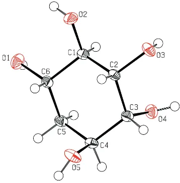
X-Ray analysis confirms the molecular structure and atom connectivity for (I) (Fig. 1).The cyclohexane ring adopts the
chair conformation with the puckering parameters q2 and φ (Cremer & Pople, 1975) and the smallest displacement
asymmetric parameters, Δ, (Nardelli, 1983) as follows: q2=0.0673 (11) Å, φ=111.3 (9)°, Δs(C1)= 1.27 (8).
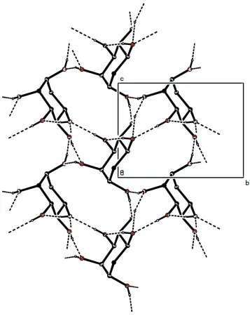
The atom O1 acts as a donor to the atom O3 of the neighbouring molecule. This hydrogen bond is involved in a motif
C(6) forming an infinite chain along a axis, and also the atom O5 acts as a donor to the atom O2. This hydrogen bond is
involved in a motif C(7) forming an infinite chain along c axis. The crystal packing is defined by O–H···O hydrogen
bonds (Table 1, Fig. 2)
S2. Experimental
The compound was isolated from Manilkara zapota(L) Van Royan leaves of ethyl acetate fraction by column
chromatography. Single crystals of the title compound suitable for X-ray diffraction were obtained from a mixture of
ethyl acetate and methanol (3:1) by slow evaporation.
S3. Refinement
All the H atoms were positioned geometrically, with O—H = 0.82 Å and C—H = 0.93 - 0.98 Å and constrained to ride on
Figure 1
Figure 2
The packing diagram of the title compound, view along the a axis forming a three dimensional network.
Cyclohexane-1,2,3,4,5-pentol
Crystal data
C6H12O5 Mr = 164.16 Monoclinic, P21 Hall symbol: P 2yb
a = 6.4727 (5) Å
b = 8.4851 (6) Å
c = 6.8249 (5) Å
β = 110.796 (2)°
V = 350.41 (5) Å3 Z = 2
F(000) = 176
Dx = 1.556 Mg m−3
Mo Kα radiation, λ = 0.71073 Å Cell parameters from 2418 reflections
θ = 3.2–33.3°
Bruker Kappa APEXII CCD diffractometer
Radiation source: fine-focus sealed tube Graphite monochromator
ω scans
Absorption correction: multi-scan (SADABS; Sheldrick, 1996)
Tmin = 0.972, Tmax = 0.977
5019 measured reflections 2418 independent reflections 2314 reflections with I > 2σ(I)
Rint = 0.020
θmax = 33.3°, θmin = 3.2° h = −9→9
k = −12→12
l = −9→10
Refinement
Refinement on F2 Least-squares matrix: full
R[F2 > 2σ(F2)] = 0.033 wR(F2) = 0.086 S = 1.06 2418 reflections 105 parameters 1 restraint
Primary atom site location: structure-invariant direct methods
Secondary atom site location: difference Fourier map
Hydrogen site location: inferred from neighbouring sites
H-atom parameters constrained
w = 1/[σ2(F
o2) + (0.0548P)2 + 0.0106P] where P = (Fo2 + 2Fc2)/3
(Δ/σ)max < 0.001 Δρmax = 0.39 e Å−3 Δρmin = −0.19 e Å−3
Absolute structure: Flack (1983), 994 Friedel pairs
Absolute structure parameter: 0.7 (6)
Special details
Geometry. All e.s.d.'s (except the e.s.d. in the dihedral angle between two l.s. planes) are estimated using the full covariance matrix. The cell e.s.d.'s are taken into account individually in the estimation of e.s.d.'s in distances, angles and torsion angles; correlations between e.s.d.'s in cell parameters are only used when they are defined by crystal symmetry. An approximate (isotropic) treatment of cell e.s.d.'s is used for estimating e.s.d.'s involving l.s. planes.
Refinement. Refinement of F2 against ALL reflections. The weighted R-factor wR and goodness of fit S are based on F2, conventional R-factors R are based on F, with F set to zero for negative F2. The threshold expression of F2 > σ(F2) is used only for calculating R-factors(gt) etc. and is not relevant to the choice of reflections for refinement. R-factors based on F2 are statistically about twice as large as those based on F, and R- factors based on ALL data will be even larger.
Fractional atomic coordinates and isotropic or equivalent isotropic displacement parameters (Å2)
x y z Uiso*/Ueq
C1 0.39464 (15) 0.00491 (11) 0.41069 (16) 0.01595 (17)
H1 0.4688 −0.0926 0.4757 0.019*
C2 0.15563 (14) −0.03135 (11) 0.27557 (15) 0.01522 (17)
H2 0.0821 0.0694 0.2247 0.018*
C3 0.13572 (14) −0.13189 (11) 0.08297 (15) 0.01626 (17)
H3 −0.0198 −0.1352 −0.0093 0.020*
C4 0.27248 (16) −0.06837 (11) −0.04069 (15) 0.01663 (17)
H4 0.2741 −0.1468 −0.1456 0.020*
C5 0.51009 (15) −0.03378 (11) 0.10101 (16) 0.01768 (18)
H5A 0.5837 −0.1318 0.1590 0.021*
H5B 0.5889 0.0137 0.0188 0.021*
C6 0.51703 (15) 0.07685 (11) 0.27875 (16) 0.01628 (17)
H6 0.4414 0.1747 0.2168 0.020*
O1 0.73862 (11) 0.11569 (9) 0.40896 (14) 0.02328 (17)
O2 0.38759 (13) 0.11155 (10) 0.57023 (13) 0.02410 (17)
H2A 0.5140 0.1337 0.6464 0.036*
O3 0.04224 (12) −0.10449 (9) 0.39647 (13) 0.02046 (16)
H3A 0.1094 −0.1839 0.4529 0.031*
O4 0.20891 (12) −0.28909 (9) 0.14488 (13) 0.02034 (16)
H4A 0.1094 −0.3390 0.1642 0.031*
O5 0.16705 (13) 0.07199 (9) −0.14612 (13) 0.02343 (17)
H5 0.2119 0.0922 −0.2416 0.035*
Atomic displacement parameters (Å2)
U11 U22 U33 U12 U13 U23
C1 0.0173 (4) 0.0179 (4) 0.0146 (4) −0.0003 (3) 0.0080 (3) −0.0012 (3)
C2 0.0156 (3) 0.0175 (4) 0.0152 (4) 0.0008 (3) 0.0088 (3) 0.0005 (3)
C3 0.0166 (3) 0.0182 (4) 0.0147 (4) 0.0003 (3) 0.0065 (3) −0.0001 (3)
C4 0.0192 (4) 0.0187 (4) 0.0136 (4) 0.0020 (3) 0.0078 (3) 0.0000 (3)
C5 0.0179 (4) 0.0215 (4) 0.0167 (4) 0.0012 (3) 0.0099 (3) −0.0006 (3)
C6 0.0159 (3) 0.0180 (4) 0.0173 (4) −0.0007 (3) 0.0088 (3) −0.0006 (3)
O1 0.0168 (3) 0.0249 (3) 0.0288 (4) −0.0031 (3) 0.0089 (3) −0.0074 (3)
O2 0.0253 (3) 0.0310 (4) 0.0205 (4) −0.0075 (3) 0.0136 (3) −0.0112 (3)
O3 0.0215 (3) 0.0226 (3) 0.0233 (4) 0.0010 (3) 0.0155 (3) 0.0027 (3)
O4 0.0231 (3) 0.0162 (3) 0.0237 (4) −0.0008 (2) 0.0107 (3) 0.0000 (3)
O5 0.0265 (3) 0.0278 (4) 0.0195 (4) 0.0093 (3) 0.0125 (3) 0.0094 (3)
Geometric parameters (Å, º)
C1—O2 1.4289 (13) C4—H4 0.9800
C1—C6 1.5223 (14) C5—C6 1.5218 (14)
C1—C2 1.5249 (12) C5—H5A 0.9700
C1—H1 0.9800 C5—H5B 0.9700
C2—O3 1.4264 (12) C6—O1 1.4319 (11)
C2—C3 1.5337 (14) C6—H6 0.9800
C2—H2 0.9800 O1—H1A 0.8200
C3—O4 1.4282 (12) O2—H2A 0.8200
C3—C4 1.5221 (14) O3—H3A 0.8200
C3—H3 0.9800 O4—H4A 0.8200
C4—O5 1.4328 (12) O5—H5 0.8200
C4—C5 1.5266 (13)
O2—C1—C6 111.16 (8) O5—C4—H4 108.9
O2—C1—C2 106.77 (7) C3—C4—H4 108.9
C6—C1—C2 110.64 (8) C5—C4—H4 108.9
O2—C1—H1 109.4 C6—C5—C4 111.20 (8)
C6—C1—H1 109.4 C6—C5—H5A 109.4
C2—C1—H1 109.4 C4—C5—H5A 109.4
O3—C2—C1 110.97 (8) C6—C5—H5B 109.4
O3—C2—C3 110.48 (7) C4—C5—H5B 109.4
C1—C2—H2 107.4 O1—C6—C1 110.01 (8)
C3—C2—H2 107.4 C5—C6—C1 110.48 (8)
O4—C3—C4 107.32 (8) O1—C6—H6 108.0
O4—C3—C2 110.35 (8) C5—C6—H6 108.0
C4—C3—C2 112.94 (7) C1—C6—H6 108.0
O4—C3—H3 108.7 C6—O1—H1A 109.5
C4—C3—H3 108.7 C1—O2—H2A 109.5
C2—C3—H3 108.7 C2—O3—H3A 109.5
O5—C4—C3 107.80 (8) C3—O4—H4A 109.5
O5—C4—C5 110.57 (8) C4—O5—H5 109.5
C3—C4—C5 111.73 (8)
O2—C1—C2—O3 −61.20 (10) O4—C3—C4—C5 −72.15 (10)
C6—C1—C2—O3 177.71 (7) C2—C3—C4—C5 49.69 (10)
O2—C1—C2—C3 174.15 (8) O5—C4—C5—C6 65.25 (11)
C6—C1—C2—C3 53.07 (10) C3—C4—C5—C6 −54.82 (11)
O3—C2—C3—O4 −54.15 (10) C4—C5—C6—O1 −177.75 (8)
C1—C2—C3—O4 70.77 (10) C4—C5—C6—C1 59.17 (10)
O3—C2—C3—C4 −174.27 (7) O2—C1—C6—O1 59.39 (10)
C1—C2—C3—C4 −49.35 (10) C2—C1—C6—O1 177.84 (8)
O4—C3—C4—O5 166.16 (7) O2—C1—C6—C5 −176.32 (7)
C2—C3—C4—O5 −71.99 (10) C2—C1—C6—C5 −57.87 (9)
Hydrogen-bond geometry (Å, º)
D—H···A D—H H···A D···A D—H···A
O1—H1A···O3i 0.82 1.94 2.7347 (11) 164
O2—H2A···O4ii 0.82 1.96 2.7761 (12) 170
O3—H3A···O1iii 0.82 2.02 2.8417 (11) 177
O4—H4A···O5iv 0.82 1.91 2.7067 (12) 165
O5—H5···O2v 0.82 2.00 2.8036 (12) 166