The principal function of the cardiopulmonary system is the matching of oxygen and carbon dioxide transport to the metabolic requirements of different tissues. Increased oxygen demands (V.O∑), for example during physical activity, result in a rapid compensatory increase in cardiac output and the redistribution of blood flow to the appropriate skeletal muscles, matched by suitable ventilatory increments. This matching of cardiopulmonary performance and metabolic rate during activity seems to be universal among vertebrates and has been demonstrated in a number of different taxa (for a review, see Jones, 1994).
In some animals, large increments in aerobic metabolic rate are also associated with physiological states other than activity. In particular, V.O∑ increases following feeding because of the energy-requiring processes associated with prey handling, digestion and the ensuing protein synthesis (Andrade et al., 1997; Brody, 1945; Cruz-Neto et al., 1999; Houlihan, 1991;
Secor and Diamond, 1995, 1997a,b; Wang et al., 1995). This large increase in V.O∑is referred to as ‘specific dynamic action’ (SDA) (Brody, 1945). In relative terms, it is more pronounced in ectothermic vertebrates than in endothermic birds and mammals; the increase in V.O∑ during SDA may resemble or even exceed that during physical activity. For example, in the Burmese python Python molurus, V.O∑reaches values as high as 20 ml kg−1min−1, 24 h post-feeding, which is six times greater than the values measured while crawling at 0.5 km h−1 (Secor and Diamond, 1997a,b). Furthermore, in contrast to the relatively short duration (minutes) of physical activity observed in reptiles, the elevated metabolic rates during SDA are sustained for several days (Secor and Diamond, 1997a,b). The physiological processes underlying the increase in metabolic rate resulting from physical activity and digestion are similar in some ways and dissimilar in others. Both states require increased convection of air and blood to permit oxygen
JEB2804
The principal function of the cardiopulmonary system is the precise matching of O2 and CO2 transport to the metabolic requirements of different tissues. In some ecothermic vertebrates (amphibians and reptiles), V.O∑ increases dramatically following feeding. Factorial increments in V.O∑range from 1.7 to 44 times above resting rates, and in some cases V.O∑approaches or even exceeds values measured during physical activity. There is virtually no information on the cardiopulmonary response during the postprandial period in these animals or how the pattern of cardiopulmonary support compares with that during activity. In our experiments, pulmonary ventilation (V.E), heart rate (fH), systemic blood flow (Q.sys), rate of oxygen consumption (V.O∑) and rate of carbon dioxide production (V.CO∑) were measured at 35 °C in the lizard Varanus exanthematicus for 24 h prior to the ingestion of meals of various sizes and measured continuously for up to 72 h during the postprandial period. The results of this study were compared with previously published values for treadmill exercise in the same experimental animals. The change in fHand stroke
volume (VS) for a given increment in V .
O∑ did not differ during exercise and digestion. In contrast, the ventilatory response was very dependent on the nature of the elevated metabolic state. During digestion, an increase in V.O∑ resulted in a relative hypoventilation in comparison with resting values, whereas hyperventilation characterized the response during activity. During exercise, breathing frequency (f) increased 10- to 40-fold above resting values accompanied by large reductions in tidal volume (VT). In contrast, postprandial increases in V.O∑ resulted in relatively minor changes in f and VT almost doubled. These results indicate that, in these lizards, the cardiac response to elevated V.O∑is stereotyped, the response being predictable irrespective of the source of the metabolic increment. In contrast, the ventilatory response is flexible and state-dependent, not only in pattern but also in its frequency and volume components.
Key words: cardiac output, digestion, exercise, lizard, oxygen consumption, postprandial, reptile, specific dynamic action, Varanus
exanthematicus, ventilation. Summary
Introduction
PATTERNS OF CARDIOVASCULAR AND VENTILATORY RESPONSE TO ELEVATED
METABOLIC STATES IN THE LIZARD VARANUS EXANTHEMATICUS
JAMES W. HICKS1,*, TOBIAS WANG2 ANDALBERT F. BENNETT1
1Department of Ecology and Evolutionary Biology, University of California at Irvine, Irvine, CA 92697, USA and
2School of Biological Sciences, University of Birmingham, Birmingham B15 2TT, UK
*e-mail: [email protected]
uptake, carbon dioxide release and the uptake and delivery of nutrients. However, the physiological processes underlying these metabolic increments are very different in tissue location (skeletal muscle versus gastrointestinal tract), metabolic pattern (catabolism versus anabolism), activating system (somatic motor and sympathetic nervous systems versus parasympathetic nervous system), acid–base status (acidosis versus alkalosis) and time course (minutes versus hours or days).
It is currently not known whether the cardiopulmonary response to similar metabolic demands during these different physiological states (exercise or digestion) is stereotyped or flexible. In the former case, equal metabolic increments (e.g. ml O2or J) in either the skeletal muscle or the intestine would
elicit an equal increment in the cardiac response (heart rate and stroke volume, with appropriate redistribution of blood flow) and ventilatory response (breathing frequency and tidal volume) regardless of the physiological state generating the demand. In the latter case, the cardiopulmonary system might have a variety of state-dependent and appropriate responses that result in the same level of gas exchange. In such a flexible system, the convective components (either cardiac output or lung ventilation) might differ markedly, depending on the location and type of metabolic increment, appropriate to matching more subtle regulatory factors other than simple energetic demands.
In spite of the interesting perspectives regarding patterns of cardiopulmonary performance during the postprandial period and its comparison with other physiological states (e.g. exercise), most studies of SDA in ectothermic vertebrates (for example, Benedict, 1932; Preest, 1991; Secor and Diamond, 1997a,b) have, for the most part, been restricted to measuring changes in metabolic rate, while changes in ventilation and blood flows have received limited attention (see Wang et al., 1995). In the present study, we report on the patterns of gas exchange, ventilation and systemic blood flow following voluntary feeding in the monitor lizard Varanus exanthematicus. All cardiopulmonary variables were measured simultaneously and thereby provide the first complete data set on cardiopulmonary performance during the postprandial period. In addition, the results of this study were compared with previously published values for treadmill exercise obtained in the same experimental animals (Wang et al., 1997).
Materials and methods Animals
Five savannah monitor lizards (Varanus exanthematicus Bosc) with body mass ranging from 260 to 660 g were purchased from a licensed supplier (Glades Herp Inc., Florida, USA) and transported by air to Irvine, California. Upon arrival, the lizards were kept in a large terrarium with free access to heating lamps and other heat sources, allowing behavioral thermoregulation. A 12 h:12 h light:dark cycle was maintained, and water was always available ad libitum. Food was withheld for 3–4 days before surgery.
Measurement of ventilation, gas exchange and blood flow Ventilation and gas exchange were measured using a modification of the experimental apparatus described by Wang and Warburton (1995). Briefly, short pieces of flexible gas-tight tubing were glued into both nostrils, fused on top of the head of the lizard and connected to a T-piece. This T-piece was, in turn, attached to gas-tight Tygon tubing at both ends. One end fed into Applied Electrochemistry O2 and CO2 analyzers (S-3A and CD-3A, respectively) connected in series, while the other served as a reservoir. A flow pump (Applied Electrochemistry), connected in series with the gas analyzers, maintained a constant gas flow between the T-piece and the gas analyzers and thus provided continuous delivery of room air to the lizard. A pneumotachograph (8421 series, 0-5 LPM, H. Rudolph, Inc.) was connected ‘up-stream’ relative to the T -piece. At this position, airflow decreased during exhalations, while inhalations caused increases in airflow. A Valendyne (MP-45-1-871) differential pressure transducer continuously monitored the resulting changes in pressure gradients across the pneumotachograph. At any given breath, the signal from the differential pressure transducer preceded that from the gas analyzers by approximately 2 s. Because the breathing pattern invariably consisted of single breaths with low frequency, this arrangement allowed breath-to-breath gas exchange and tidal volume to be measured.
V.O∑and V .
CO∑were determined as the area of the differential between the inspired and expired gas concentration for each breath. The relationship between this area and gas exchange was determined by simulating exhalations with known gas compositions through the T-piece (which was connected to the nostrils of the lizard during experiments). Similarly, the expired tidal volume of single breaths was determined from the integrated flow signal from the differential pressure transducer. Again, the relationship between this integral and tidal volume (in ml) was quantified by injection of a range of gas volumes through the T-piece. All calibration procedures produced very significant correlations between injected gas volumes and integrated flow signal (r2>0.98) and were reproducible before, during and after experiments.
subclavian arteries or their branches. In addition, our analysis assumes proportionate blood flow distribution to all major vessels with increases in metabolic rate during digestion and exercise. Although this arrangement cannot provide measures of total cardiac output, the resulting changes in blood flow would provide, at least, qualitative changes in cardiac output and its components, heart rate (fH) and stroke volume (VS). The
wires from the flow probes were exteriorized through the incision, which was closed with intermittent sutures and surgical glue. All lizards were allowed to recover for a minimum of 14 days before experimentation. During this period, water was available ad libitum; however, food was withheld. All animals remained in excellent health until killed several months after surgery.
The flow probe was connected to a Transonic flow meter (T201) for measurement of blood flow. Heart rate was calculated continuously from the instantaneous blood flow signal. All cardiovascular and respiratory measurements were recorded on a computer using the AcqKnowledge (version 3.2.4) data-acquisition system (Biopac Inc., Goleta, CA).
Experimental protocol
All experiments were conducted in a large climatic chamber maintained at 35 °C. At least 18 h before feeding, lizards were placed in a large plastic container (50 l) for recording pre-feeding levels of the measured variables. The container served to prevent visual and auditory stimuli and to restrain the animals from any gross movements. On the following day, dead prey (mice or rats) were presented to the lizard, which normally grabbed them directly from the hand of the investigator. The lizard was subsequently left undisturbed and allowed to swallow its meal. In no case was the lizard forced to eat. Two lizards refused to eat while connected to the Tygon tubing. In these two instances, we disconnected the animal from the experimental apparatus, allowed the animal to eat voluntarily and then reassembled the experimental apparatus after it had swallowed the prey. After feeding, all cardiopulmonary variables were continuously measured for at least 60 h.
Data analysis: cardiopulmonary changes during digestion The mean values for V.O∑, V
.
CO∑, breathing frequency (f), VT,
V.E, systemic blood flow (Q.sys), fHand VSwere determined for
10 min periods of each hour. All data were analyzed using AcqKnowledge (3.2.4) data-analysis software (Biopac system, Inc., CA, USA). The effects of digestion on the variables measured were tested by analyzing the values during fasting and 24 h post-feeding using a two-tailed paired t-test (P⭐0.05).
Cardiopulmonary responses: comparisons with exercise The results of this study were compared quantitatively with similar measurements made on the same experimental animals during treadmill exercise (Wang et al., 1997). This comparison was made to determine whether the cardiopulmonary response to increases in rates of oxygen consumption, generated during two different physiological states (exercise or digestion), was
the same (stereotyped) or different (flexible). In this analysis, for each animal, we regressed each cardiopulmonary variable (i.e. fH, VS, Q
.
sys, f, VT, V .
E, V.E/V.CO∑, V .
E/V.O∑) against V . O∑ measured during digestion and during fasting treadmill exercise. The slopes for each variable, under the two different physiological states (digestion versus exercise), were compared using a two-tailed paired t-test (P<0.05). If significant differences were determined between the slopes of a variable during exercise and digestion, the response was considered flexible and state-dependent. If the slopes were not significantly different, the response was considered to be stereotyped.
Results
Cardiopulmonary responses to feeding
Our experimental arrangement allowed for continuous measurements of ventilation, heart rate and systemic blood flow during fasting, feeding and digestion. In a representative trace (Fig. 1), a 360 g monitor lizard was presented with a dead 60 g rat. Initially, the lizard became agitated and grabbed and then vigorously shook the rat for several minutes before swallowing it head-first. As shown in Fig. 1, heart rate and systemic blood flow increased rapidly upon grabbing the rodent. As the monitor lizard shook the rat, heart rate, ventilation and systemic blood flow continued to increase, reaching values that were 3–4 times resting values. Heart rate and blood flow remained elevated throughout the entire eating period until the rat had been swallowed, and then both slowly returned towards resting values. Our experimental method could not be used to quantify ventilation and gas exchange during the act of feeding, because the mouth remains open. However, as the trace in Fig. 1 clearly shows, ventilatory frequency increased substantially during prey handling. During swallowing, costal ventilation was probably depressed, although the animal may have been breathing through its mouth during this period. Following the act of swallowing, breathing frequency and ventilation increased transiently and then slowly returned towards the values measured prior to eating.
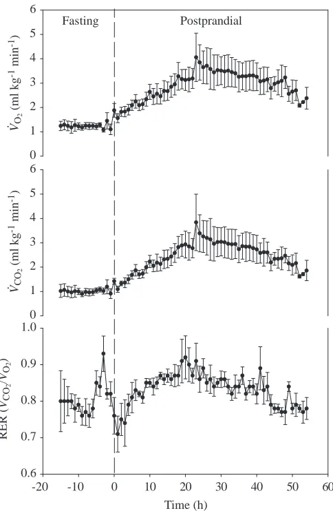
Gas exchange
The mean rates of gas exchange during fasting and for 54 h post-feeding are summarized for all animals in Fig. 2. V.O∑ increased gradually following feeding, reaching peak values at approximately 24 h post-feeding. Peak values ranged from two to six times fasting values (Table 1). The peak value of V.O∑ following feeding varied among individuals, but was significantly correlated with meal size (Fig. 3). With the exception of the first few hours following feeding, the elevated rate of oxygen uptake following ingestion was mirrored by an increased rate of CO2 excretion (Fig. 2) and, thus, the respiratory gas exchange ratio (RER; V.CO∑/V
.
Ventilatory and cardiovascular responses during digestion Prior to feeding, expired minute ventilation (V.E) averaged 20–25 mlBTPSmin−1kg−1, and 24 h after feeding this increased approximately 2.5-fold (Table 1; Fig. 4). This elevation in V.E
was accomplished primarily through increases in tidal volume (VT) (Table 1; Fig. 4). During digestion, all animals exhibited a
tendency for hypoventilation, with air convection requirements for V.O∑ (V
.
E/V.O∑) and V
.
CO∑ (V
.
E/V.CO∑) tending to decrease
compared with fasting rest values (Fig. 5; Table 1). At 24 h post-feeding, V.E/V.CO∑and V
.
E/V.O∑were reduced in some animals by
as much as 50 % of the fasting value. Although the difference in the mean postprandial and fasting values for V.E/V.CO∑ just
failed to be statistically significant (P=0.07), all individuals hypoventilated, and the failure of the statistical test reflects variability within those hypoventilatory states. Subsequent measurements on additional animals consuming a meal of 10 % of body mass (D. Crossley, J. W. Hicks and A. F. Bennett, unpublished data) confirm postprandial hypoventilation in this species (V.E/V.CO∑ fasting 25.2±2.1, V
.
E/V.CO∑ postprandial
21.8±1.4, P=0.047, paired t-test, N=5, means ±S.E.M.).
Qsys
(ml min
-1)
0 20 40 60
f
H
(min
-1)
0 25 50 75 100
Time (min)
0 20 40 60 80
Ventilation -2 0 2
Grab prey
Shaking prey Swallowing prey
Varanus exanthematicus
[image:4.609.108.486.73.328.2].
Fig. 1. A representative trace for a 360 g monitor lizard presented with a dead 60 g rat. Initially, the lizard became agitated, grabbed and vigorously shook the rat for several minutes before swallowing the rodent head-first. Systemic blood flow (Q.sys) and heart rate (fH) increased rapidly upon grabbing the rodent. As the monitor lizard shook the rat, heart rate, ventilation and systemic blood flow continued to increase, reaching values that were 3–4 times above resting values. Heart rate and blood flow remained elevated throughout the entire eating period, until the rat was swallowed, and then both slowly returned towards resting values. During swallowing, it appears that costal ventilation was probably depressed, although the animal may have been breathing through its mouth during this period; this would not have been detected by our respiratory arrangement.
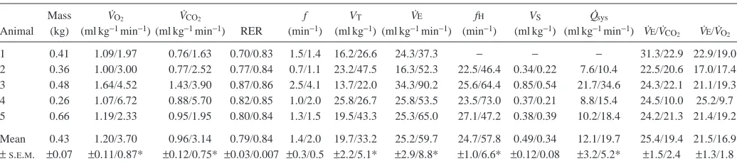
Table 1. Cardiopulmonary variables at rest and 24 h after feeding in Varanus exanthematicus at a body temperature of 35 °C
Mass V˙O2 V˙CO2 f VT V˙E fH VS Q
.
sys
Animal (kg) (ml kg−1min−1) (ml kg−1min−1) RER (min−1) (ml kg−1) (ml kg−1min−1) (min−1) (ml kg−1) (ml kg−1min−1) V˙E/V˙
CO2 V˙E/V˙O2
1 0.41 1.09/1.97 0.76/1.63 0.70/0.83 1.5/1.4 16.2/26.6 24.3/37.3 − − − 31.3/22.9 22.9/19.0 2 0.36 1.00/3.00 0.77/2.52 0.77/0.84 0.7/1.1 23.2/47.5 16.3/52.3 22.5/46.4 0.34/0.22 7.6/10.4 22.5/20.6 17.0/17.4 3 0.48 1.64/4.52 1.43/3.90 0.87/0.86 2.5/4.1 13.7/22.0 34.3/90.2 25.6/64.4 0.85/0.54 21.7/34.6 24.3/22.1 21.1/19.3 4 0.26 1.07/6.72 0.88/5.70 0.82/0.85 1.0/2.0 25.8/26.7 25.8/53.5 23.5/73.0 0.37/0.21 8.8/15.4 24.5/10.0 25.2/9.7 5 0.66 1.19/2.33 0.95/1.95 0.80/0.84 1.3/1.5 19.5/43.3 25.3/65.0 27.1/47.2 0.38/0.39 10.2/18.4 24.2/21.3 21.4/19.2
Mean 0.43 1.20/3.70 0.96/3.14 0.79/0.84 1.4/2.0 19.7/33.2 25.2/59.7 24.7/57.8 0.49/0.34 12.1/19.7 25.4/19.4 21.5/16.9
± S.E.M. ±0.07 ±0.11/0.87* ±0.12/0.75* ±0.03/0.007 ±0.3/0.5 ±2.2/5.1* ±2.9/8.8* ±1.0/6.6* ±0.12/0.08 ±3.2/5.2* ±1.5/2.4 ±1.3/1.8
Values are expressed as rest/24 h post-feeding.
An asterisk denotes a significant difference (P<0.05) between pre- and postprandial values.
[image:4.609.42.560.581.698.2]Systemic blood flow (Q.sys) increased approximately twofold
24 h after feeding (Table 1; Fig. 6). The increase in Q.sysduring
digestion resulted primarily from an increase in fH (Table 1; Fig. 6).
Ventilatory responses during digestion and exercise A comparison of the responses of these animals to fasting treadmill activity and digestion revealed that an increase in V.O∑during exercise was associated with a marked increase in
breathing frequency, while tidal volume decreased (Fig. 7). In contrast, during digestion, increased ventilation was accomplished primarily through increments in VT. V
.
E
increased dramatically during exercise, but showed only a modest increase during digestion (Fig. 7). A major difference in the ventilatory response to exercise and digestion is revealed in the air convection requirement for CO2(V
.
E/V.CO∑).
Over the entire range of V.O∑ measured during treadmill
exercise, varanid lizards exhibit a relative hyperventilation (Fig. 7). For example, a threefold increase in V.O∑ is
associated with a near doubling of V.E/V.CO∑ (Fig. 7). In VO
2
(ml kg
-1 min -1)
VCO
2
(ml kg
-1 min -1)
0 1 2 3 4 5 6
Fasting Postprandial
0 1 2 3 4 5 6
-20 -10 0 10 20 30 40 50 60
RER (
VCO
2
/V
O2
)
0.6 0.7 0.8 0.9 1.0
Time (h) .
.
.
.
Fig. 2. V.O∑, V
.
CO∑ and respiratory exchange ratio (RER; V
.
CO∑/V
.
O∑)
measured every hour in Varanus exanthematicus during fasting and during digestion. Values are means ±S.E.M., N=5.
Relative meal size (% of body mass)
3 6 9 12 15
Increment in
VO
2
(ml kg
-1 min -1)
0 1 2 3 4 5
[image:5.609.337.532.69.272.2].
Fig. 3. The effects of meal size, expressed as a percentage of body mass, on the incremental increase in V.O∑ during digestion. The
increment in V.O∑ represents the value measured 24 h after ingesting
the meal. The line is a first-order linear regression through the data points (r2=0.91, P=0.012).
f (min
-1)
0 1 2 3 4 5
Fasting Postprandial
VT
(ml kg
-1)
10 20 30 40 50
Time (h)
-20 -10 0 10 20 30 40 50 60
V
E
(ml kg
-1 min -1)
20 40 60 80
.
Fig. 4. Breathing frequency (f), tidal volume (VT) and minute
[image:5.609.53.295.70.441.2] [image:5.609.326.558.345.703.2]contrast, as V.O∑increases during digestion, V
.
E/V.CO∑decreases
(Fig. 7).
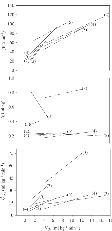
Cardiovascular responses during exercise and digestion The relationship between systemic blood flow and its components, fH and VS, did not differ as a function of V
.
O∑
during exercise and digestion (Fig. 8). In both physiological states, the increase in systemic blood flow resulted primarily from changes in fH. Thus, for a given increment in V.O∑,
irrespective of whether the increase was due to digestion or physical activity, fH increased by a similar amount. There was no consistent trend in VS. In some animals, VS tended
to increase as V.O∑ increased, both during exercise and
during digestion; however, in other animals, there was either a slight reduction or no change in VS as metabolic rate
increased.
Discussion
Specific dynamic action in varanids compared with other animals
The increased metabolic rate following feeding is consistent with the findings for both vertebrates and invertebrates (Benedict, 1932; Coulson et al., 1950; Preest, 1991; Lyndon et al., 1992; Secor et al., 1994; Wang et al., 1995; Secor and Diamond, 1995; Secor and Phillips, 1997). In Varanus exanthematicus, V.O∑ began to increase within several hours
following ingestion of the meal and reached a peak within 24–30 h, increasing approximately threefold (Fig. 2). This
factorial increase in V.O∑ is lower than the seven- to tenfold
increase reported in the closely related species Varanus albigularis (Secor and Phillips, 1997a,b) and far lower that the 44-fold increase measured in the python Python morulus (Secor and Diamond, 1997a,b).
The relative magnitude of specific dynamic action (SDA) is influenced by a number of factors, including the initial resting metabolic rate, differences in the cost of digestion among species, the length of the fasting period prior to feeding, the composition of the meal and the size of the meal (Secor and Phillips, 1997a,b; Secor and Diamond, 1997a,b). In our study, the monitor lizards fed voluntarily, consuming meals that ranged from 5 to 15 % of body mass. The increment in V.O∑at
24 h post-feeding was positively correlated with the relative meal size. A similar correlation has been reported in Python morulus and Varanus albigularis (Secor and Diamond, 1997a,b; Secor and Phillips, 1997). In spite of the many variables that potentially influence the magnitude of SDA, the absolute values for peak V.O∑ are strikingly similar among
different species after ingesting meals of similar size. In
V
E
/V
O2
V
E
/V
CO
2
10 20 30 40
Fasting Postprandial
Time (h)
-20 -10 0 10 20 30 40 50 60
10 20 30 40 50 . .
[image:6.609.316.556.72.441.2]. .
Fig. 5. Air convection requirements for O2 (V
.
E/V.O∑) and CO2
(V.E/V.CO∑), where V
.
Eis minute ventilation, measured every hour in Varanus exanthematicus during fasting and during digestion. Values are means ±S.E.M., N=5.
f
H
(min
-1)
20 30 40 50 60 70 80
Fasting Postprandial
VS
(ml kg
-1)
0.2 0.4 0.6 0.8
Relative change in
Qsys
0.5 1.0 1.5 2.0 .
Time (h)
[image:6.609.46.285.72.327.2]-20 -10 0 10 20 30 40 50 60
Fig. 6. Heart rate (fH), stroke volume (VS) and relative systemic
blood flow (Q.sys) measured every hour in Varanus exanthematicus
Varanus albigularis, peak V.O∑following meals that range from
6 to 9 % of body mass is 4.9 ml kg−1min−1(Secor and Phillips,
1997). In Python morulus, a meal of 5 % of body mass results in a peak V.O∑ of 3.2 ml kg−1min−1. In the present study, the
mean meal size was 9 % of body mass, resulting in a peak V.O∑
of 3.7 ml kg−1min−1.
Cardiopulmonary responses during digestion and exercise The results of this study were compared with previously published (Wang et al., 1997) values for treadmill exercise obtained in the same experimental animals. Although we cannot extrapolate the variables of interest during digestion to the high metabolic rates during activity, we have analyzed the data
0 200 400 600 800 1000 1200 (5) (4) (1) (2) (3) 0 20 40 60 80 100 (1) (1) (2) (2) (3) (3) (4) (4) (5) (5) f (min -1) 0 20 40 60
(1) (3) (4)
(2) (5)
VO2 (ml kg
-1 min-1)
0 3 6 9 12 15 18
VT (ml kg -1) 0 20 40 60 80 (3) (1) (4) (2)(5) (5) (1) (3) (2) (4) . (1) (2)(3) (4) (5) (1) (2)(3) (4) (5)
VO2 (ml kg-1 min-1)
0 3 6 9 12 15 18 .
V
E
(ml kg
[image:7.609.329.550.67.521.2]-1 min -1) V E /V CO 2 . . .
Fig. 7. The effects of an increase in metabolic rate (V.O∑) on breathing
frequency (f), tidal volume (VT), minute ventilation (V
.
E) and air convection requirement for CO2 (V
.
E/V.CO∑) in Varanus
exanthematicus at 35 °C. The dashed regression lines represent the relationship between V.O∑ and respiratory variables during treadmill
exercise (taken from Wang et al., 1997). The solid regression lines represent the response to increased V.O∑during digestion (this study).
The numbers in parentheses identify individual animals.
0 20 40 60 80 100 120 140 f H (min -1) (5) (2) (4) (3) (4) (5) (2) (3) 0.2 0.4 0.6 0.8 1.0 VS (ml kg -1) (2) (4) (5) (4) (2) (5) 0 15 30 45 60 75 Qsys (ml kg
-1 min -1)
(3)
(5) (5) (4)
(4) (2) (2) . (3) (3) (3)
VO2 (ml kg
-1 min-1)
0 2 4 6 8 10 12 14 16 18 .
Fig. 8. The effects of an increase in metabolic rate (V.O∑) on heart rate
(fH), stroke volume (VS) and systemic blood flow (Q
.
sys) in Varanus
exanthematicus at 35 °C. The dashed regression lines represent the response to increased V.O∑ during treadmill exercise (taken from
Wang et al., 1997). The solid regression lines represent the response to increased V.O∑ during digestion (this study). The numbers in
[image:7.609.74.264.73.570.2]encompassing an overlapping range of metabolic rates. The cardiovascular response in digesting and exercising animals was stereotyped. For a given increment in V.O∑, changes in fH, VSand
Q.sysdid not differ between exercise and digestion. In contrast,
the ventilatory response was flexible and was dependent on physiological state. During exercise, breathing frequency (f) increased 10- to 40-fold above resting values, accompanied by significant reductions in tidal volume (VT). In contrast,
postprandial increases in V.O∑ resulted in relatively minor
increments in f and VT. This pattern accords with that observed
in our companion paper examining cardiopulmonary responses to exercise and digestion in the snake Python molurus (Secor et al., 2000): the cardiovascular response is stereotyped, and the pulmonary response is flexible and state-dependent.
A major difference in the ventilatory response to exercise and digestion is revealed in the air convection requirement for CO2 (V
.
E/V.CO∑). During treadmill exercise in this lizard, the
increase in minute ventilation is disproportionately larger than that in metabolic rate, resulting in a relative hyperventilation (Mitchell et al., 1981a,b; Hopkins et al., 1995; Wang et al., 1997). V.E/V.CO∑can increase as much as two- to threefold above
the values measured at rest (Mitchell et al., 1981a,b; Wang et al., 1997). This relative hyperventilation is also demonstrated by an increase in end-tidal PO∑and a significant reduction in
end-tidal and arterial PCO∑ during exercise (Mitchell et al.,
1981a,b; Hopkins et al., 1995; Wang et al., 1997). A relative hyperventilation during exercise also occurs in Python molurus (Secor et al., 2000).
In contrast, as metabolic rate increases during digestion, V.E/V.CO∑ decreases. Postprandial hypoventilation was also
found in Python molurus (Secor et al., 2000). This hypoventilation leads to an increase in arterial PCO∑ and has
been shown to occur in the snake Python molurus (Overgaard et al., 1999), the lizard Varanus exanthematicus (Hartzler et al., 2000) and the alligator Alligator mississipiensis (M. Busk, J. Overgaard, J. W. Hicks, A. F. Bennett and T. Wang, unpublished observations). Because these reptiles ingest a relatively large meal with a subsequent distention of the stomach, the postprandial hypoventilation might result from a mechanical constraint on costal ventilation. Alternatively, it is more likely that the postprandial hypoventilation represents changes in ventilatory control, specifically resulting from the elevation of plasma [HCO3−] associated with digestion.
Perspectives
This study has demonstrated a stereotypy in the cardiovascular response to different elevated metabolic states, and a state-dependent and flexible response on the part of the pulmonary system. Specifically in regard to the latter, activity results in hyperventilation and digestion results in hypoventilation in comparison with fasting rest values. The generality of this pattern is currently unknown, but it also appears to be present in a snake and a crocodilian. What accounts for the state-dependent response of the pulmonary system? The physiological changes associated with activity in reptiles are dominated by a pronounced acidosis, resulting
from their reliance on anaerobic metabolism (Bennett, 1994). In contrast, digestion is dominated by an alkalosis, resulting from the secretion of H+into the stomach and accumulation of
HCO3−in the blood (Coulson et al., 1950). These differences
in acid–base status during activity and digestion are undoubtedly associated with the state-dependent responses of the pulmonary system. Current studies in our laboratory (e.g. Hartzler et al., 2000; Crossley et al., 2000) are investigating the interrelationships and consequences of these ventilatory patterns for blood gas and acid–base composition. The state-dependency of this response may also serve as a useful model system in which to investigate ventilatory control systems.
We thank Frank Powell and an anonymous reviewer for helpful comments on the manuscript. T.W. was supported by a postdoctoral fellowship from the Carlsberg Foundation (Denmark). This research was supported by NSF grant IBN-9727762 to A.F.B. and J.W.H.
References
Andrade, D. V., Cruz-Neto, A. P. and Abe, A. S. (1997). Meal size
and specific dynamic action in the rattlesnake Crotalus durissus (Serpentes: Viperidae). Herpetologica 53, 485–493.
Benedict, F. G. (1932). The Physiology of Large Reptiles with Special
Reference to the Heat Production of Snakes, Tortoises, Lizards and Alligators. Washington: Carnegie Institute Publication.
Bennett, A. F. (1994). Exercise performance of reptiles. In
Comparative Vertebrate Exercise Physiology: Phyletic Adaptations, vol. 38B (ed. J. H. Jones), pp. 113–138. San Diego: Academic Press, Inc.
Brody, S. (1945). Bioenergetics and Growth. New York: Hafner
Press.
Busk, M., Jensen, F. B. and Wang, T. (2000). The effects of
feeding on blood gases, acid–base parameters and selected metabolites in the bullfrog Rana catesbeiana. Am. J. Physiol. 278, R185–R195.
Coulson, R. A., Hernandez, T. and Dessauer, H. C. (1950).
Alkaline tide in alligators. Soc. Exp. Biol. Med. 74, 866–869.
Crossley, D., Hicks, J. W. and Bennett, A. F. (2000). Postprandial
changes in cardiopulmonary control in the savanna monitor lizard. FASEB J. 72, A46.
Cruz-Neto, A. P., Andrade, D. V. and Abe, A. S. (1999). Energetic
cost of predation: Aerobic metabolism during prey ingestion by juvenile rattlesnakes, Crotalus durissus. J. Herpetol. 33, 229–234.
Hartzler, L. K., Hicks, J. W. and Bennett, A. F. (2000).
Postprandial acid–base status in the savannah monitor lizard. Am. Zool. 39, 69A.
Hopkins, S., Hicks, J. W., Cooper, T. and Powell, F. (1995).
Ventilation–perfusion relationships in the savannah monitor during treadmill exercise. J. Exp. Biol. 198, 1783–1789.
Houlihan, D. F. (1991). Protein Turnover in Ectotherms and its
Relationships to Energetics (ed. D. F. Houlihan, D. R. Livingstone and R. F. Lee). Adv. Comp. Env. Physiol. 7, 1–213. Berlin: Springer-Verlag.
Jones, J. H. (1994). (ed.) Comparative Vertebrate Exercise
Physiology: Phyletic Adaptations, vol. 38B. San Diego: Academic Press.
of short-term fasting and a single meal on protein synthesis and oxygen consumption in cod, Gadus morhua. J. Comp. Physiol. B
162, 209–215.
Mitchell, G. S., Gleeson, T. T. and Bennett, A. F. (1981a).
Ventilation and acid–base balance during graded activity in lizards. Am. J. Physiol. 240, R29–R37.
Mitchell, G. S., Gleeson, T. T. and Bennett, A. F. (1981b).
Pulmonary O2transport during activity in lizards. Respir. Physiol.
43, 365–375.
Overgaard, J., Busk, M., Hicks, J. W., Jensen, F. B. and Wang, T. (1999). Acid–base status and arterial oxygen transport following
feeding in the snake Python molorus. Comp. Biochem. Physiol.
124A, 361–367.
Preest, M. R. (1991). Energetic costs of prey ingestion in a scincid
lizard, Scincella lateralis. J. Comp. Physiol. B 161, 327–332.
Secor, S. M. and Diamond, J. (1995). Adaptive responses to feeding
in Burmese pythons: pay before pumping. J. Exp. Biol. 198, 1313–1325.
Secor, S. M. and Diamond, J. (1997a). Determinants of the
postfeeding metabolic response of Burmese pythons, Python molurus. Physiol. Zool. 70, 202–212.
Secor, S. M. and Diamond, J. (1997b). Effects of meal size on
postprandial responses in juvenile Burmese pythons (Python molurus). Am. J. Physiol. 272, R902–R912.
Secor, S. M., Hicks, J. W. and Bennett, A. F. (2000). Ventilatory
and cardiovascular responses of a python (Python molurus) to exercise and digestion. J. Exp. Biol. 203, 2447–2454.
Secor, S. M. and Phillips, J. A. (1997). Specific dynamic action of
a large carnivorous lizard, Varanus albigularis. Comp. Biochem. Physiol. A 117, 515–522.
Secor, S. M., Stein, E. D. and Diamond, J. (1994). Rapid
upregulation of snake intestine in response to feeding: a new model of intestinal adaptation. Am. J. Physiol. 266, G695–G705.
Wang, T., Burggren, W. W. and Nobrega, E. (1995). Metabolic,
ventilatory and acid–base responses associated with specific dynamic action in the toad, Bufo marinus. Physiol. Zool. 68, 192–205.
Wang, T., Carrier, D. R. and Hicks, J. W. (1997). Ventilation and
gas exchange in lizards during treadmill exercise. J. Exp. Biol. 200, 2629–2639.
Wang, T. and Warburton, S. J. (1995). Breathing pattern and cost