With 5 text-figures Printed in Great Britain
CHANGES OF INTERNAL HYDROSTATIC PRESSURE AND
BODY SHAPE IN ACANTHOCEPHALUS RANAE
BY R. A. HAMMOND
Department of Zoology, University of Bristol*
(Received 25 January 1966)
Two hydraulic systems are primarily concerned in the movements of
Acantho-cephahis ranae (Schrank, 1788; Lvihe, 1911), that of the proboscis apparatus and that
of the trunk (Hammond, 1966a). The muscles associated with each system operate about a fluid skeleton of constant volume. Muscular activity in either system, and therefore change of body shape, is accompanied by a change of hydrostatic pressure within the skeletal fluid.
The present paper describes an investigation into the correlation between the move-ments of the animal and changes of hydrostatic pressure within it.
MATERIAL AND METHODS
The worms were obtained from parasitized common toads (Bufo bufo) and were maintained apart from the host in 35% sea water (Hammond, 1966a). Although the animals survived well in this medium experiments were carried out as soon as possible after their removal from the host.
Two indirect methods of recording changes of hydrostatic pressure were devised. Both allowed recordings to be made from intact animals. The first system is illu-strated in Fig. 1.
A fragment of coverslip connected to a light balsa-wood lever by a fine, stiff wire rested on the animal. The lever was adjusted by means of riders so that the body wall was slightly flattened.
A small mirror was cemented to one end of the lever. A narrow beam of light directed on to this from a spotlight was reflected to a slit camera. This consisted of a multi-speed Palmer kymograph with a 12 in. drum, around which was wrapped a strip of 4 in. wide photographic recording paper. The drum was mounted in a box in the front of which was a slit 1 mm. wide, and a window 3 cm. square.
Below the worm was a spotlight which was flashed every 30 sec. by an electronic timer. A series of profile images of the animal was projected, via a 2 in. lens and mirror, through the square window of the camera. From each image it was possible to deter-mine whether the proboscis was evaginated or invaginated. Experiments were carried out in a darkroom.
The degree of flattening of the body-wall by the coverslip resting on it depended on the weight of the system, the rigidity of the body-wall, and on the internal pressure. The animal became more flattened as its internal hydrostatic pressure decreased, and less so as the pressure increased. The lever moved in response to pressure changes and
• Present address: Department of Zoology, University College, Cardiff.
in response to the general changes of body shape of the active worm (Hammond, 1966a).
In these experiments the worm lay on its side because of the dorso-ventral curva-ture of the body. The degree of flattening was measured as the ratio of dorso-ventral
Spotlight
Mirror
Coversllp
[image:2.451.47.407.129.336.2]Worm
Fig. 1. Apparatus used for the first method of indirect pressure recording.
Dissecting microscope
Camera
Spotlight
Prism
lover
(a)
UjKworm
RiderWorm
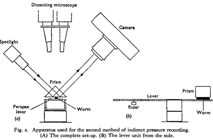
Fig. 2. Apparatus used for the second method of indirect pressure recording. (A) The complete set-up. (B) The lever unit from the side.
extension (given by the profile) to lateral extension (given by the distance between the coverslip and the microscope slide).
[image:2.451.53.399.361.586.2]Hydrostatic pressure and body shape in Acanthocephalus ranae 199
The worm was brought into contact with a prism by a Perspex lever. The force exerted by the prism against the animal could be varied by adjustment of the rider on the lever. A beam of light was directed on to the prism so that it underwent total internal reflexion at the air/glass interface. At the glass/animal interface, however, light was not reflected. The part of the worm in contact with the prism thus became visible and could be photographed. The area of contact between animal and prism at different phases of the animal's activity was measured from the photographs.
As the force exerted by the prism against the animal and the area of contact between the two were known, the force per unit area exerted at any phase of activity could be calculated. This gave a comparative measure of changes of hydrostatic pressure within the animal. Methods similar in principle have been used by Cole (1932) and Wilson (1962).
Stage I
4
15
1-0
g,
OS
-Stage III Stage II Stage IV
I 4 4
Stage V Stage VI
High
Low
10 15 20 25 30 35 40 45 50 55 60
[image:3.451.65.390.222.460.2]Time (sec.)
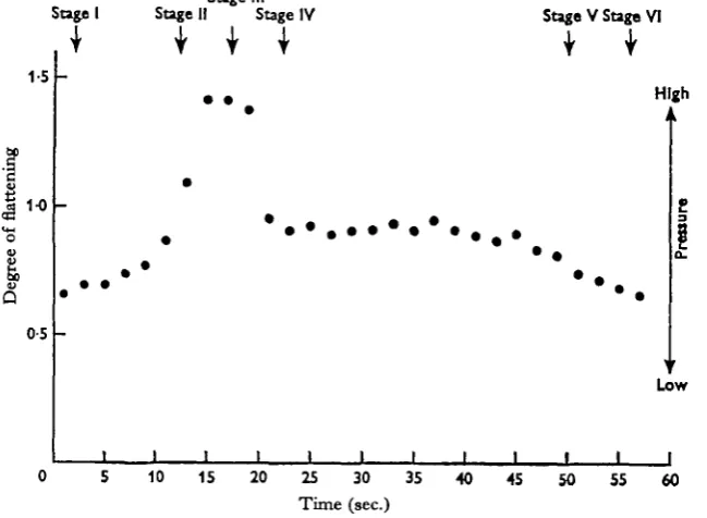
Fig. 3. Pressure changes within the trunk cavity of Acanthocephalus ranae recorded by the first method. Relative pressure changes are expressed in terms of the degree of flattening of the body.
RESULTS
Both methods gave readily repeatable results. With Method 1 (Fig. 3) changes of internal pressure are expressed in terms of the degree of flattening of the body. In Fig. 4 (Method 2), a compilation of results obtained with ten worms, pressure is expressed in cm. Hg. As the apparatus was not calibrated, and the intrinsic rigidity of the body-wall remained an unknown factor, this figure is not absolute.
With Method 2 (Fig. 4) it was found convenient to record pressure at the following points in each cycle of activity (see Hammond, 1966a):
Stage I. Trunk maximally short and thick, proboscis invaginated.
Stage II. Trunk fully elongated after contraction of the circular muscles, proboscis invaginated.
Stage III. Trunk elongated, as in Stage II, but proboscis evaginated.
Stage IV. Proboscis evaginated but partially ensheathed by the fore-trunk, because of the contraction of the neck retractor muscles.
0-6 -• Stage I
0 5
0 4
0 3
0 2
0 1
-Stagell Stage I Stage IV Stage V Stage VI
• • I •
•. . . t . » . . . ' . . i i i i L i i i . . i r i i i i i i i i i i i i i i i ' i i i I i ' t I i t I i i t I
1 3 5 7 9 1 3 5 7 9 1 3 5 7 9 1 3 5 7 9 1 3 S 7 9 1 3 5 7 9 2 4 6 8 1 0 2 4 6 8 1 0 2 4 6 8 1 0 2 4 6 8 1 0 2 4 6 8 1 0 2 4 6 8 1 0
Animal number
Fig. 4. Pressure changes within the trunk cavity of Acanthocephahu ranae recorded by the second method. A compilation of results from ten worms is shown.
Stage V. Proboscis evaginated but completely ensheathed within the fore-trunk because of the complete relaxation of the trunk wall circular muscles.
Stage VI. Proboscis in the process of invagination, trunk shortening and thickening. These points are indicated for reference in Figs. 3-5.
Hydrostatic pressure within the trunk is lowest when the trunk is contracted and the proboscis invaginated (Stage I). At this stage the circular muscles are relaxed and the longitudinal muscles contracted. As the circular muscles contract, and the trunk elongates in consequence (Stage II), pressure increases. The pressure is greatest either at (Fig. 4), or just before (Fig. 3), the time of proboscis evagination (Stage III). At Stage IV, at which the proboscis remains evaginated but full elongation of the trunk is not maintained, the internal pressure falls, but not to the lowest value. The neck retractor muscles are contracted at this time.
Hydrostatic pressure and body shape in Acanthocephalus ranae 201
From Method 2 it appears that overall pressure changes of up to 0-5 cm. Hg occur within the trunk during a cycle of activity.
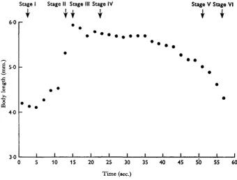
Changes of body length of an active worm are illustrated in Fig. 5. As the circular muscles of the trunk wall contract the body rapidly extends in length, becoming longest between Stages II and III. However, even when fully extended the body length of
A. ranae is only 40-50% greater than when fully contracted.
At Stage IV the body shortens and remains for a while at an intermediate length. The circular trunk muscles become fully relaxed at Stage V, after which the longi-tudinal muscles contract, the trunk shortening as they do so.
Stage I Stage II Stage III Stage IV
U I
Stage V Stage VI60 r
5 0
4 0
3 0
10 15 20 25 30 35 4 0 45 50 55 60
[image:5.451.54.395.196.454.2]Time (sec.)
Fig. 5. Changes of body length of Acanthocephalus ranae.
DISCUSSION
It is clear that the fluid filling the central cavity of the trunk of A. ranae functions as a simple hydrostatic skeleton. Changes of pressure within the fluid accompany contraction of the muscles operating about it. There is a clear correlation between internal fluid pressure and all the major phases of the animal's activity.
It has been emphasized by Batham & Pantin (1950) that the 'functional objective' of an animal with a hydrostatic skeleton may be achieved as an indirect result of muscular contraction. In A. ranae the direct effect of contraction of the circular muscles of the trunk wall is to lengthen the animal. The result of primary significance to the animal, however, is that the proboscis apparatus is forced to the tip of the animal by displacement of body fluid.
considerable amount of work that these muscles perform at this stage. They act antagonistically to the longitudinal trunk muscles, to the neck retractor muscles, and to the receptacle retractor muscle, all of which are extended as the trunk elongates.
During the major phases of trunk elongation and shortening the circular and longitudinal musculature of the trunk contract and relax as entire units. This results in simple uniform changes of body shape from short-and-thick to long-and-narrow. Throughout Stage IV, however, local contractions occur in isolated regions of the trunk musculature. These bring about small local changes of body shape, causing the trunk to bend from side to side (Hammond, 1966 a), and are reflected in small fluctua-tions of internal pressure (Fig. 3).
The extensibility of the trunk of A. ranae is low compared with that of many nemerteans and turbellarians (Clark, 1964, table 1). Worms have been observed to extend by a maximum of 40-50%. Further, part of this extension is accounted for by the untucking of the fore-trunk (Hammond, 1966a). The cuticle, which consists of a homogeneous material perforated by pores, and the basement membrane, a homo-geneous matrix containing small fibres (Hammond, 19666), almost certainly impose restraint on the extensibility of the worm. Stretching of the body surface might be disadvantageous to an acanthocephalan if it led to distortion or occlusion of the cuticular pores through which nutrients enter. Change of body shape seems primarily to be accommodated by wrinkling of the body-wall when the animal contracts, rather than by stretching when it elongates.
SUMMARY
1. Two indirect methods for recording changes of hydrostatic pressure within the trunk of Acanthocephalus ranae have been described.
2. Internal pressure has been shown to be lowest when the trunk is fully con-tracted and the proboscis invaginated, and highest when the trunk is fully elongated. 3. A rapid rise of internal pressure occurs when the circular trunk muscles contract. 4. Overall internal pressure changes of up to 0*5 cm. Hg have been shown to occur in active specimens.
5. The body length when fully extended is only 40-50% greater than when contracted.
6. The correlation between muscular activity, body shape, and internal hydro-static pressure in A. ranae is discussed
I wish to thank Prof. J. E. Harris for the provision of laboratory facilities and Dr H. D. Crofton for his advice and criticism during the course of this work. I am grateful to Prof. James Brough and Dr D. A. Erasmus for reading and criticizing the manuscript.
REFERENCES
BATHAM, E. J. & PANTIN, C. F. A. (1950). Muscular and hydrostatic action in the sea-anemone
Metri-ddum senile (L.). J. Exp. Biol. 27, 264-89.
CLARK, R. B. (1964). Dynamics in Metazoan Evolution, pp. x + 313. Oxford. COLE, K. S. (1932). Surface forces of the Arbacia egg. J. Cell. Comp. Pkysiol. 1, 1-9.
HAMMOND, R. A. (1966a). The proboscis mechanism of Acanthocephalus ranae. J. Exp. Biol. 45, 203—13. HAMMOND, R. A. (19666). The fine structure of the trunk and praesoma wall of Acanthocephalus ranae
(Schrank, 1788), Llihe, 1911. To be published.