THE RELATIONSHIP BETWEEN LUNG VOLUME,
RESPIRATORY DRIVE AND BREATHING PATTERN IN THE
TURTLE, CHRYSEMYS PICTA
BY W. K. MILSOM AND P. CHAN
Department of Zoology, University of British Columbia, Vancouver, B.C., Canada, V6T2A9
Accepted 24 June 1985
SUMMARY
Induced changes in resting lung volume (VLR) in the turtle Chrysemys picta (Schneider) had no effect on resting levels of minute ventilation in animals breathing room air but did change their breathing pattern. Increasing VLR caused an increase in the number of breaths in each episode (burst) of breathing but a reduction in the incidence of such breathing bursts and thus an increase in the length of periods of breath-holding. The data indicate that these effects were largely the consequence of changes in lung volume per se rather than changes in lung gas stores.
Although both hypoxia and hypercapnia stimulated ventilation via increases in tidal volume and breathing frequency, they produced distinct changes in breathing
pattern. While hypoxia (3% O2) caused an increase in the number of bursts of
breathing (B/min) and reduced the number of breaths (b) in each burst (b/B),
hypercapnia (5% CO2) increased both B/min and b/B. These data suggest that
the size and incidence of bursts of breathing must be under separate control. One consequence of the different effects of hypoxia and hypercapnia on breaths per burst
(b/B) was that hypoxic-hypercapnic gas mixtures (3% O2+5% CO2) failed to
stimulate ventilation as much as hypercapnia alone.
Administration of hypoxic, hypercapnic and hypoxic-hypercapnic gas mixtures to elevate respiratory drive eliminated the effects of changes in VLR on breathing pattern. Thus, although changes in VLR are important in the control of breath-holding in animals breathing air, their effect decreases as respiratory drive increases.
INTRODUCTION
The pattern of lung ventilation is periodic in most reptiles. Many species exhibit episodes (bursts) of continuous breathing separated by a highly variable period of holding, while others take only single breaths which each end with a breath-hold. In all cases studied thus far, these periods of breath-holding are the major controlled variable determining breathing frequency (Glass & Johansen, 1976; Milsom & Jones, 1980; Benchetrit & Dejours, 1980; Ballam, 1984). Although it was expected that fluctuations in lung and/or blood gas composition would be instru-mental in the control of this portion of the breathing pattern, studies have failed to
establish the existence of any clear 'thresholds' for initiating or terminating breath-holding (Lumsden, 1923; Lenfant, Johansen, Petersen & Schmidt-Nielsen, 1970; Burggren & Shelton, 1979; Ackerman & White, 1979; Glass, Boutilier & Heisler, 1983). Some general trends emerged from these studies but large fluctuations in alveolar and arterial PQ and, to a lesser extent Pco , frequently occurred at both the onset and termination of breath-holding.
The role of lung volume in the determination of breath-hold length also remains unclear. In marine turtles, there is a good correlation between breath-hold length and end inspiratory lung volume over a range of ± 35 % of resting lung volume (Jacobs, 1939; Milsom & Johansen, 1975). Furthermore, as a consequence of a progressive decline in respiratory quotient (R) in the lungs of many species during breath-holding due to CO2 storage and/or CO2 excretion via extrapulmonary routes
(Burggren & Shelton, 1979; Ackerman & White, 1979), lung volume declines during breath-holding as a function of the end-inspiratory lung volume and the rate and extent of oxygen consumption. Since this fall in lung volume should stimulate ventilation, it has been suggested that this could provide a correlation between breathing frequency and metabolic rate (Johansen, 1970). Despite these corre-lations, however, there was still a high degree of variability in the breath-hold lengths measured at any given resting lung volume (Milsom & Johansen, 1975; Burggren & Shelton, 1979; Ackerman & White, 1979).
The relationships reported between lung volume, lung gas stores, blood gas composition and metabolic rate suggest that breath-hold length may be controlled by an interaction between afferent inputs from pulmonary stretch receptors and peripheral and central chemoreceptors rather than by any one input alone. Given this, the goal of the present study was to examine the effect of changes in resting lung volume, with and without changes in chemoreceptor drive, on the ventilation pattern of the western painted turtle, Chrysemys picta. This species exhibits a burst-breathing pattern and routinely undergoes large changes in lung volume associated with buoyancy control (Milsom, 1975; Milsom & Jones, 1980).
MATERIALS AND METHODS
Specimens of Chrysemys picta, ranging from 300 to llOOg, were housed in large Plexiglas tanks, 45x60cm, filled to a depth of 8cm with water at 22-23 °C. They were provided with heat lamps and dry areas for basking, maintained on a 12 h light: 12 h dark photoperiod and were not fed for at least 3 days before any measurements were made.
Changes in lung volume
weights and styrofoam floats equal to ± 2 and ± 4% of the animal's body weight. The addition of weights and floats altered the animal's buoyancy state inducing lung volume changes which returned specific gravity to a state of neutral buoyancy, usually within 24 to 48 h (Table 1). Following compensation, end-inspiratory lung volume remained constant as long as specific gravity remained constant.
Protocol and measurements
At the beginning of each experiment, the turtle was transferred to a tank, similar to the holding tank but covered just below water level with a grid containing an 8 cm diameter breathing hole. This hole was covered with a ventilation chamber as described by Glass et al. (1983) and, as in that study, ventilation was measured by pneumotachography using a Fleisch model 00 pneumotachograph, Validyne DP 103—18 differential pressure transducer and Gould integrating amplifier. Each ani-mal was allowed 12—24 h to adjust to this set-up before any measurements were made.
Ventilation was first measured while animals breathed air and then during random administration of 3% O2 in N2 (hypoxia), 5% CO2 in air (hypercapnia) and 3 %
O2+5% CO2 in N2 (hypoxic hypercapnia). Animals were returned to breathing air
for at least 1 h between each test gas. Gas flow rates were maintained throughout at either 500 or lOOOmlmin"1 and gas composition was monitored using a Beckman OM-11 oxygen analyser and LB-2 CO2 analyser calibrated with commercially
purchased, analysed, gas mixtures. The zero balance of the pneumotachograph was adjusted to cancel the constant signal resulting from gas flow through the system. Each gas mixture was flushed through the chamber for at least 1 h or until a steady response was achieved and measurements were made from traces obtained following this period. All measurements were made during daytime (08.00 to 20.00 h) and are taken to represent steady-state conditions in animals equilibrated to the various gas mixtures.
At the end of each experimental run, body weight was measured and lung volume determined by whole body plethysmography as described previously (Milsom, 1975). Each turtle was placed in an aluminium chamber filled with water and fitted with a dome-shaped lid. The turtle could breathe freely through a small air space at the top of the lid. When the turtle was resting quietly near the hole and not breathing, the air space was quickly filled with water, the hole sealed, and lung volume determination performed. As a consequence, all lung volumes were determined for turtles with buoyancy states adjusted for floating at the surface, the condition under which all ventilation measurements were made during these studies. It is assumed that turtles rested quietly at the surface with similar lung volumes in both the experimental tank and the plethysmograph.
RESULTS
Effect of changes in resting lung volume (VLR) on breathing pattern
The normal breathing pattern of C. picta consists of breathing episodes separated by highly variable periods of breath-holding. Such a breathing pattern can be analysed in terms of the number of breaths in each burst of breathing (breaths/burst), the number of episodes of breathing in each minute (burst/min), the length of breath-holding between breathing episodes (Tnon.ventil,tory period or TNVP), the overall breathing frequency (f = total breaths/min), the tidal volume of each breath (VT) and the overall level of minute ventilation (VE = f X VT).
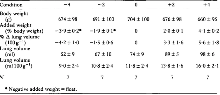
The effect of adding weights and floats to turtles to induce changes in VLR for buoyancy compensation is shown in Table 1 and Fig. 1. The induced changes in lung volume (in ml) were roughly equal to the changes in body weight (in g) and thus neutral buoyancy was maintained, providing a range of VLR from 9-0 to 160ml
100 g"1, almost a doubling of VLR.
Changes in VLR had no effect on the size of each breath (VT) in absolute terms (ANOVA; F(4,16) = 0-47, P*»0-l), although because of the changes in VLR, the
amount of lung volume turned over with each breath (VT/l00ml VLR) decreased as VLR increased (Fig. 1) (ANOVA; F(4>16) = 2-91, 0-05 < P < 0 - l ) .
Overall breathing frequency (f) is a function of the number of breaths/burst multiplied by the number of bursts/min. The number of bursts per minute is in-versely related to the length of the non-ventilatory period (TNVP) between breathing episodes. As can be seen from Fig. 2, as VLR increased animals exhibited fewer breathing episodes, (ANOVA, F(416) = 3-09, P < 0 0 5 ) hence longer breath-hold
periods (ANOVA; F(4il6) = 3-40, P< 0-05), but took more breaths during each bout
of breathing (ANOVA: F( 4 1 6 ) = 3-37, P < 0 0 5 ) . The overall effect was to produce
very little change in overall breathing frequency (f) (ANOVA; F(4#i6) = 1-62,
P > 0" 1), except at the lowest values of VLR (but not significant), and hence very little
[image:4.451.39.416.486.649.2]change in VE (Fig. 1) (ANOVA; F(4,16) = 0-23, P > 0-1).
Table 1. Effect of changes in body weight, by the addition of weights or floats, on lung
•volume Condition
Body weight (g)
Added weight (% body weight) % A lung volume
(lOOg-1) Lung volume
(ml) Lung volume
(mllOOg"1)
N
* Negative added
- 4
674 ± 98
-3-9 ±0-2*
- 4 - 2 ± l - 0
52 ± 9
9-0 ±2-4
7
weight = float.
- 2
691 ± 100
— 1-9 ± 0-1*
-1-5 ±0-6
67 ± 1 0
10-8 ±2-4 7 0 704 ±100 0 0
74 ± 9
ll-8±2-4
7
+2
676 ± 98
2-0 ±0-1
3-3 ±1-6
89 ± 5
13-8 ±1-6 7 +4 660 ±95 4-1 ±0-2 5-6 ±1-8
98 ± 6
160±2-l
20
E 10
00
8
i
H
20
ri
>
E8
s
- 4 - 2 0+2+4 -4 -2 0 +2+4
[image:5.451.93.333.37.391.2]Weight condition
Fig. 1. The relationships between resting lung volume (VLR), minute ventilation ( V E ) , and tidal volume [VT both in absolute (ml lOOg"1) and relative (ml 100 ml V L R "1) units] and the various weight conditions listed in Table 1.
Effect of changes in respiratory drive on breathing pattern under control conditions
Under control conditions (i.e. no weights or floats added; V L R = ll-8ml lOOg"1) the inspiration of 3 % O2 provided a mild respiratory stimulation while inhalation of 5% CO2 (with or without concomitant hypoxia) greatly excited ventilation (Fig. 3). The hypoxic stimulus (3 % O2) caused small increases in both VT and f while the hypercapnic stimulus (5% CO2) caused substantial increases in both these values (only the increases in f, however, were significant atP < 0-05). When 3 % O2 and 5 %
CO2 were administered together they caused a greater increase in VT than either gas alone but failed to stimulate f as much as the hypercapnic gas alone and thus, overall, had slightly less effect on VE than 5 % CO2 inhalation alone. It can be seen from Fig.
4 that all gases produced similar increases in the number of bursts/min, and hence similar decreases in TNVP, and that all differences in breathing frequency seen in
significant at the 005 <P< 0-1 level under control conditions but were more significant, statistically, at other lung volumes. In the case of the gas concentrations used in this study, these opposing effects offset one another and thus no change was seen in the number of breaths/burst when 3 % O2 and 5 % CO2 were administered
together. This accounts for the inability of hypoxic hypercapnia to stimulate breathing frequency as much as hypercapnia alone.
Effect of changes in VLRon the ventilatory responses to changes in respiratory drive
Once ventilation was stimulated by changing the inspired gas composition, all effects of changes in VLR on breathing pattern disappeared. As can be seen from Figs 3 and 4, all respiratory variables were independent of VLR on each of the hypoxic, hypercapnic and hypoxic-hypercapnic gas mixtures (ANOVA; F(4i6)<2-33, PS>0-l).
The relationship between changes in VLR and respiratory drive on breathing pattern can best be seen in Figs 5 and 6. The 'Hey Plot' in Fig. 5 shows the relative
- 4 - 2 0 +2 +4 - 4 - 2 0 +2 +4
Weight condition
:ontributions of changes in f and VT to changes in VE resulting from alterations in VLR and inspired gas composition. Whereas the hypoxic gas mixture led to a doubling of both VT and f, the hypercapnic gas mixture led to a three-fold increase in VT and a five-fold increase in f. Changes in VLR can, again, be seen to have had little effect on the overall responses to each gas. Thus each gas produced a characteristic breathing pattern which was independent of lung volume. The plot in Fig. 6 illustrates the contribution of the components of breathing frequency to the overall anges in f. Two important trends can be seen from this figure. First, although changes in VLR can significantly alter the breathing pattern in animals breathing air, without altering overall breathing frequency substantially, such changes have no effect on the breathing patterns of animals breathing hypoxic or hypercapnic gas mixtures. Secondly, if values recorded for animals with the largest VLR are compared (dashed lines, Fig. 6), it can be seen that hypercapnia results only in a shortening of
TNVP (i.e. an increase in bursts/min) while both hypoxia and a reduction in lung gas
stores (i.e. a reduction in VLR) produce an increase in the number of breathing
40
00
8
I 20
u
4-00
8
i 2
10 12 14 16 10 12 14 16 VutOnllOOg"1)
0 Air
• 3 % O2
A 5 % C O2
V 3 % O2+ 5 % C O2
10 12 14 16
[image:7.451.93.356.281.586.2]VLR (ml 100 g"1)
episodes (a reduction in TNVP) and a reduction in the number of breaths in each breathing episode.
DISCUSSION
Effect of changes in resting lung volume on breathing pattern
Chronic changes in resting lung volume over the range of 9-0 to 160 ml lOOg"1 had no overall effect on resting levels of breathing frequency, tidal volume or their product, minute ventilation. They did, however, alter the breathing pattern. As VLR increased, the periods of breath-holding between breathing episodes increased, the number of breaths in each episode increased and the relative tidal volume (VT 100 ml
V L R "1) decreased. The simplest interpretation of these results is that the increase in lung gas stores associated with increasing VLR permits longer periods of breath-holding but to replenish this enhanced gas store with a relatively smaller VT requires a greater number of breaths.
20
3
% 10
s
oa|
200
2 100
10 12 14 16 10 12 14 16
VLR (ml 100 g"1)
0 Air • 3%O2 A 5%CO2 V 3%O2+5%CO2
10 12 14 16 VLR (ml 100 g"1)
40
30
on 8
20
10
f =
• 90 O 10-8
• 11-8 A 13-8
• 160
Fig. 5. The 'Hey Plot' demonstrating the relationship between minute ventilation (VE) and its components, breathing frequency (f) and tidal volume (VT), for animals with various resting lung volumes (VLR) breathing different gas mixtures. Solid lines connect values recorded at increasing levels of VLR in animals breathing each gas mixture.
In the only other study of this nature, performed on the Atlantic loggerhead turtle
(Caretta caretta, Milsom & Johansen, 1975), increases in VLR also lead to increases
in TNVP. Since this species only takes single breaths, however, this necessarily caused a decrease in breathing frequency. Under these conditions, either VT must have increased or VE would also have been reduced. Unfortunately, this was not measured.
In mammals (primarily cats; Woldring, 1965; Grunstein, Wyszogrodski & Milic-Emili, 1975; D'Angelo & Agostoni, 1975; Finkler & Iscoe, 1984) acute (lOmin) increases in functional residual capacity (FRC = end-expiratory lung volume;
VLR = end-inspiratory lung volume, i.e. VLR = F R C + V T ) also lead to decreases in f
reduction in f (Finkler & Iscoe, 1984). Increases in FRC in mammals also elicit a Breuer-Hering, inspiratory-inhibitory reflex which should shorten inspiration. This would act to offset the prolongation of expiration and thus maintain the total breath length and breathing frequency constant. It has been shown, however, that both adaptation of pulmonary stretch receptors and central habituation of pulmonary afferent input occur in less than lOmin and act to offset completely the inhibition of inspiration (D'Angelo & Agostoni, 1975; Muza & Frazier, 1983; Finkler & Iscoe, 1984). Thus, under steady-state conditions, only a prolongation of expiration and a depression in breathing frequency are seen. The fall in VE which follows causes an increased chemoreflex drive which acts to increase VT, but this compensatory effect is only sufficient to restore VE to resting levels if the increase in FRC is small (Finkler & Iscoe, 1984).
It would appear, therefore, that increasing lung volume acts to prolong the end-inspiratory breath-hold in turtles while it prolongs expiration in mammals. It has no
[image:10.451.46.380.275.592.2]10
overall effect on VE in the western painted turtle but generally decreases VE in cats. These differences are consistent with previous reports that the central integration of lung volume information, under resting conditions, is quite different in turtles from that described in mammals (Milsom & Jones, 1980). There are insufficient data, however, to determine whether these differences are simply a function of differences in the control of periodic versus continuous breathing patterns, true differences between the control of breathing in reptiles and mammals, or whether they are specific to this one species of turtle.
Effect of changes in respiratory drive on breathing pattern under control conditions
Although no blood samples were taken during these experiments to verify that steady-state levels of blood gases had been achieved during exposure to the various inspired gases, care was taken to ensure that ample time was allowed for stable responses to develop. The responses we recorded under control conditions (VLR = 11'8 ml 100 g"1) correspond well with those recorded by other investigators. Table 2 lists, for comparison, values recorded in other studies of turtles exhibiting burst-breathing patterns at similar temperatures and exposed to similar gases. The slight hypocapnia which will have developed during the hypoxic hyperventilation and the mild hyperoxia which will have developed during the hypercapnic hyperventilation have not been corrected for and thus the responses reported here are probably slight underestimates of the true ventilatory responses. These effects will be small, how-ever, and common to all the studies reported in Table 2. Consistent with these other studies, severe levels of hypoxia were required to produce even a modest ventilatory response, whereas mild hypercapnia produced a vigorous response.
Inhalation of both hypoxic and hypercapnic gas mixtures led to increases in VT and overall breathing frequency. An analysis of the components of f, however, shows that although both mixtures led to an increase in the number of breathing episodes per minute, hypoxia produced a decrease in the number of breaths in each burst of breathing, whereas hypercapnia produced an increase in this number. These variables are reported infrequently and while one study on Pseudemys scripta failed to show this difference in response to hypoxia and hypercapnia (Frankel, Spitzer, Blaine & Schoener, 1969), another on Chrysemyspicta, the same species which was used in the present study, is consistent with our results (Glass et al. 1983). Also consistent with these results was the lack of change in the number of breaths taken in each breathing episode when animals were exposed to hypoxic-hypercapnic gas mixtures. Under these conditions, the opposing effects of hypoxia and hypercapnia on the size of each burst of breathing just offset one another. As a consequence, even though one effect of administering both gases together was to increase VT in an additive fashion, both f and VE increased less than when animals were exposed to only the hypercapnic mixture.
Table 2. Respiratmy vatiables for species of burst-breathing turtles exposed to van'ous gas mixtures Gas VE % Time Temp. Fd (rnlkg;' VT fTNVP B/nun achvely Species PC) Flrh min- ) d' mi) (8) b/~ (rnin-') breath- Rcfcrence ~e&dusa subtufa Chqwmys picta Ch ysemys picta Pehdusa subrufa Chtymys picta Calculated from original papers (aenumca T, = 5 s). t98% NZ in air for 10-15 min. 110-12% COz in air for 10-15 rnin. Air
1.4 1.4
83
2.3
1.6 1.4
following vagotomy in turtles while the number of breaths in each burst remains unchanged (Frankel et al. 1969; Milsom & Jones, 1980).
Effect of changes in resting lung volume on the ventilatory response to changes in respiratory drive
There were no effects of changing VLR on any of the respiratory variables measured during exposure to hypoxic, hypercapnic or hypoxic-hypercapnic gas mixtures. Stated otherwise, the effects of VLR on TNVP, b/B and B/min in animals breathing air were eliminated by increasing respiratory drive with any of these gas mixtures.
In cats, increasing FRC acts to reduce transiently the ventilatory response to hypoxic or hypercapnic stimuli. Decreasing FRC has the opposite effect, but neither response is seen under steady-state conditions (Woldring, 1965; Cherniack et al. 1973), which is consistent with the data presented here. Interestingly, however, exposure to hypoxic and hypercapnic gases has been shown to have steady-state effects on FRC. Hypoxia increases FRC in dogs (Bouverot & Fitzgerald, 1969) and guinea pigs (Lechner & Banchero, 1980) and VLR in snakes (Glass & Johansen, 1976; Bartlett & Birchard, 1983) and tadpoles (Burggren & Mwalukomo, 1983), and hypercapnia has similar effects in Ii2ards (Ballam, 1984). In the present study, VLR was only measured under control conditions and any such effects would not have been recorded. Given the magnitude of our experimental changes in VLR, however, these additional effects should have been relatively small and, as already noted, would not have had any effect on the ventilatory responses that were recorded.
The effects of reducing VLR on breathing pattern are extremely similar to the effects of hypoxia but not of hypercapnia. This raises the possibility that the changes seen in breathing pattern following changes in VLR, in animals breathing air, could arise from changes in lung O2 stores rather than changes in lung volume perse. That is, stimulation of systemic chemoreceptors rather than pulmonary mechanoreceptors could underlie these changes in breathing pattern. Unfortunately, this possibility could not be tested directly. The changes in VLR elicited by reflex changes in specific gravity in our study are under pulmonary vagal control. As a consequence, pul-monary stretch receptor denervation would have eliminated the changes in VLR. At present, too little is known about the presence and innervation of systemic chemoreceptors in turtles to make chemodenervation a viable alternative (Ishii, Ishii &Kusakabe, 1985).
On the basis of published values for the O2 concentration in the luminal gas of the lungs of Chrysemys (Pseudemys) scripta breathing air (Po = 120 mmHg; Burggren, Glass & Johansen, 1978), the lung O2 stores of a turtle with a VLR of 16-0 ml lOOg"1
would be estimated at approximately 2-5 ml 100 g"1 (16 ml 100 g~*X 120 mmHg/ 760 mmHg). If VLR is reduced to 9 0 ml 100 g"1, lung O2 stores would be reduced to 1-4ml 100g"1 (9 ml 100 g~xX 120 mmHg/760 mmHg). Although this represents a significant fall in lung O2 stores, it falls far short of the effect of reducing lung O2 concentration to 3 %. Such a change in O2 concentration would lower lung O2 stores
simple calculations serve to indicate that the effects of changing VLR on breathing pattern can at most be only partially explained on the basis of changing lung O2 stores, and thus changes in lung volume per se - probably acting via pulmonary afferent input — must account for most of the observed changes.
In conclusion, this study has shown that changes in lung volume play an important role in the control of the breathing pattern in turtles under resting conditions. Although they do not contribute to the control of the overall level of minute ventilation, they strongly influence the size and incidence of breathing bursts and, therefore, the length of the periods of breath-holding. This influence is probably reduced as respiratory drive increases and is definitely absent at the levels of hypoxia and hypercapnia used in this study. Under these conditions, breathing pattern appears to be solely under chemoreceptor influence but the effects of hypoxia and hypercapnia on breathing pattern (specifically on the number of breaths per burst) are distinct.
This work was supported by the NSERC of Canada.
REFERENCES
ACKERMAN, R. A. & WHITE, F. N . (1979). Cyclic carbon dioxide exchange in the turtle Pseudemys
scripta. Physiol. Zool. 52, 378-389.
BALLAM, G. 0 . (1984). Ventilatory response to inspired CO2 in the lizard, Tupinambis
mgropunctatus. Comp. Biochem. Physiol. 78A, 757-762.
BARTLETT, D . , JR. & BlRCHARD, G. F. (1983). Effects of hypoxia on lung volume in the garter snake. Respir. Physiol. 53, 63-70.
BENCHETRIT, G. & DEJOURS, P. (1980). Ventilatory CO2 drive in the tortoise Testudo horsefeldi.
J. exp. Biol. 87, 229-236.
BOUVEROT, P. & FITZGERALD, R. S. (1969). Role of the arterial chemoreceptors in controlling lung volume in the dog. Respir. Physiol. 7, 203-215.
BURGGREN, W. W., GLASS, M. L. &JOHANSEN, K. (1978). Intrapulmonary variation of gas partial
pressures and ventilation inequalities in chelonian reptiles. J. comp. Physiol. 126, 203-209. BURGGREN, W. & MWALUKOMO, A. (1983). Respiration during chronic hypoxia and hyperoxia in
larval and adult bullfrogs (Rana catesbeiana). I. Morphological responses of lungs, skin and gills. J . exp. Biol. 105, 191-204.
BURGGREN, W. & SHELTON, G. (1979). Gas exchange and transport during intermittent breathing in chelonian reptiles. J. exp. Biol. 82, 75-92.
CHERNIACK, N . S., STANLEY, N. N., TUTEUR, P. G., ALTOSE, M. D. & FISHMAN, A. P. (1973). Effects of lung volume changes on respiratory drive during hypoxia and hypercapnia. J. appl.
Physiol. 35, 635-641.
D'ANGELO, E. & AGOSTONI, E. (1975). Tonic vagal influences on inspiratory duration. Respir.
Physiol. 24, 287-302.
FrNKLER, J. & ISCOE, S. (1984). Control of breathing at elevated lung volumes in anaesthetized cats. J. appl. Physiol. 56, 839-844.
FRANKEL, H. M., SPITZER, A., BLATNE, J. & SCHOENER, E. P. (1969). Respiratory response of
turtles (Pseudemys scripta) to changes in arterial blood gas composition. Comp. Biochem.
Physiol. 31, 535-546.
GLASS, M., BOUTTLDER, R. G. & HEISLEK, N . (1983). Ventilatory control of arterial Po2 in the turtle Chrysemyspicta bellh: effects of temperature and hypoxia. J. comp. Physiol. 151, 145-153.
GLASS, M., BURGGREN, W. W. & JOHANSEN, K. (1978). Ventilation in an aquatic and a terrestrial
chelonian reptile. J . exp. Biol. 72, 165-179.
GRUNSTEIN, M. M., WYSZOGRODSKI, I. & M H I C - E M U , J. (1975). Regulation of frequency and
depth of breathing during expiratory threshold breathing in cats. J. appl. Physiol. 38, 869—874. ISHU, K., ISHU, K. & KUSAKABE, T . (1985). Electrophysiological aspects of reflexogenic area in
the chelonian, Geoclemmys reevesn. Respir. Physiol. 59, 45-54.
JACKSON, D . C. (1969). Buoyancy control in the freshwater turtle, Pseudemys scripta elegans.
Science, N.Y. 166, 1649-1651.
JACKSON, D . C. (1971). Mechanical basis for lung volume variability in the turtle Pseudemys
scripta elegans. Am. J. Physiol. 22J0, 754-758.
JACKSON, D. C. (1973). Ventilatory response to hypoxia in turtles at various temperatures. Respir.
Physiol. 18, 178-187.
JACKSON, D. C , PALMER, S. E. & MEADOW, W. L. (1974). The effects of temperature and carbon
dioxide breathing on ventilation and acid-base status of turtles. Respir. Physiol. 20, 131—146. JACOBS, W. (1939). Die Lunge der Seeschildkrote Caretta caretta (L.) als Schwebeorgan. Z. vergl.
Physiol. 27, 1-28.
JOHANSEN, K. (1970). Air breathing in fishes. In Fish Physiology, Vol. 4, (eds W. S. Hoar & D. J. Randall). New York: Academic Press.
LECHNER, A. J. & BANCHERO, N. (1980). Lung morphometry in guinea pigs acclimated to hypoxia during growth. Respir. Physiol. 42, 155-169.
LENFANT, C , JOHANSEN, K., PETERSEN, J. A. & SCHMIDT-NIELSEN, K. (1970). Respiration in the fresh water turtle, Chelys fimbriata. Respir. Physiol. 8, 261-275.
LUMSDEN, T . (1923). The regulation of respiration. Part l.J. Physiol., Land. 58, 82-91.
MlLSOM, W. K. (1975). Development of buoyancy control in juvenile Atlantic loggerhead turtles,
Caretta c. caretta. Copeia 1975, 758-762.
MILSOM, W. K. & JOHANSEN, K. (1975). The effect of buoyancy induced lung volume changes on respiratory frequency in a Chelonian (Caretta caretta). J. comp. Physiol. 98, 157-160.
MlLSOM, W. K. & JONES, D . R. (1980). The role of vagal afferent information and hypercapnia in control of the breathing pattern in Chelonia. J. exp. Biol. 87, 53—63.
MUZA, S. R. & FRAZIER, D . T . (1983). Response of pulmonary stretch receptors to shifts of functional residual capacity. Respir. Physiol. 52, 371-386.
WALLENSTEIN, S., ZUCKER, C. L. & FLEISS, J. L. (1980). Some statistical methods useful in circulation research. Circulation Res. 47, 1-9.
![Fig. 1. The relationships between resting lung volume (VLR),and tidal volume minute ventilation (VE), [VT both in absolute (ml lOOg"1) and relative (ml 100 ml VLR" 1) units]and the various weight conditions listed in Table 1.](https://thumb-us.123doks.com/thumbv2/123dok_us/1179322.640604/5.451.93.333.37.391/relationships-resting-ventilation-absolute-relative-various-weight-conditions.webp)


