CT-Guided Aspirations in the Head and Neck:
Assessment of the First 216 Cases
Paul M. Sherman, David M. Yousem, and Laurie A. Loevner
BACKGROUND AND PURPOSE: The growth of cross-sectional imaging has increased the detection of nonpalpable head and neck masses. We sought to determine the reliability of CT-guided fine-needle aspiration (FNA) over 216 consecutive cases.
METHODS: We retrospectively reviewed histopathologic findings and notes from 216 con-secutive head and neck CT-guided FNA procedures performed between 1993 and 2003. Types of needles used, passes required, lesion location, initial cytologic diagnosis, and final histopatho-logic or clinical diagnosis were reviewed.
RESULTS: Diagnostic samples were obtained in 195 (90.3%) of the lesions, with 21 (9.7%) inadequate samples. A correct diagnosis was made in 191 cases (88.4%). Final FNA diagnosis was discordant in four (1.9%) specimens, with the parapharyngeal space and parotid gland having the highest rate of inaccuracy. The range in number of passes required for final diagnosis was one to six (mode, 2.0 passes per specimen; median, 2.0; mean, 2.6; standard deviation, 1.13). In 135 (63%) of 216 cases, definitive surgical pathologic results, including findings for the four discordant specimens, were obtained. Of the nondiagnostic specimens, six (29%) of 21 went to surgery, five (83%) of six were neoplastic, and one (17%) of six was fibrous tissue. The remainder underwent clinical and imaging follow-up.
CONCLUSION: CT-guided FNA is a safe, well tolerated, and accurate for the diagnosis of head and neck lesions. In our series, the percentage of diagnostic samples obtained improved compared with prior reports. The low diagnostic error rate overall was possibly related to onsite evaluation by the cytopathologist and to improved FNA technique.
The growing quantity of cross-sectional imaging stud-ies has led to the detection of an increasing number of nonpalpable head and neck masses. Many of these masses are not clinically suspected and asymptomatic. They may be found serendipitously during a search for degenerative disease of the cervical spine or other primary head and neck lesions.
Although palpable masses are usually aspirated by otorhinolaryngologists, endocrinologists, or cyto-pathologists, nonpalpable lesions or lesions in regions adjacent to critical structures (e.g., carotid arteries, vertebral arteries, or cranial nerves) are often re-ferred to the radiologist for diagnostic intervention. Postoperative or previously irradiated sites are
an-other area where physical examination may be unin-formative, even when recurrence is suspected.
Sonographic guidance has been generally advo-cated for biopsy of most head and neck lesions be-cause of its real-time capability. However, CT guid-ance has benefit in that it has less image degradation by the air-containing aerodigestive system structures and the bony content of the head and neck. Although bony structures may cause some beam-hardening ar-tifact, this has been substantially reduced by thin-section collimation. Accordingly, CT can be used for accurate assessment of adjacent anatomy. With the advent of multidetector-row CT scanning, more cases of CT-guided aspirations are being requested. In our experience, the time it takes to perform these inter-ventions is decreasing. We sought to determine the reliability of head and neck CT-guided aspiration over an experience of 216 consecutive cases.
Methods
We retrospectively reviewed the histopathologic findings, clinical records, and notes from 216 consecutive head and neck CT-guided fine-needle aspiration (FNA) procedures in 212 patients (110 women and 102 men). One neuroradiologist (D.M.Y.) and his trainees performed the procedure at two institutions between 1993 and 2003. All patients had previously Received December 11, 2003; accepted February 10, 2004.
From the Russell H. Morgan Department of Radiology and Radiological Sciences, Division of Neuroradiology, Johns Hopkins Medical Institution, Baltimore, MD (P.M.S., D.M.Y.), and the Department of Radiology, Neuroradiology Section, Hospital of the University of Pennsylvania, University of Pennsylvania School of Medicine, Philadelphia (L.A.L.).
Address reprint requests to Paul M. Sherman, MD, 600 N. Wolfe Street, Phipps B-112, Baltimore, MD 21287.
©American Society of Neuroradiology
undergone a contrast-enhanced CT or MR imaging. Limited axial nonenhanced CT images were obtained through the le-sion at the start of the procedure; these allowed adequate delineation between the lesion and the surrounding tissues. In cases in which the lesion (e.g., tumor recurrence within scar tissue) was difficult to visualize on nonenhanced CT scans, the prior imaging study was used to determine appropriate land-marks for tissue sampling. Intravenous contrast material was not used at the time of FNA. Local anesthesia with bupivacaine was administered generously. Intravenous sedation was not used in any case.
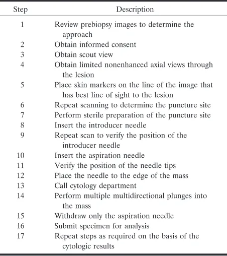
All aspirations were performed by using a coaxial technique. In most cases, a 19-gauge introducer needle was passed to the margin of the lesion, and subsequent aspirations were accomplished with coaxially placed 22-gauge lumbar puncture spinal needles (Becton Dickinson, Franklin Lakes, NJ). The cytopathologist determined the required number of passes. Table 1 provides the step-by-step approach to obtaining an FNA sample.
Radiologists acquired all FNA samples, and a cytopatholo-gist was present for all aspirations. The aspirated material was processed as air-dried direct smears for Giemsa-type staining (Diff-Quick; EM Diagnostic Systems, Gibbstown, NJ) for im-mediate evaluation and as wet-fixed smears for Papanicolaou staining. The aspiration procedure was repeated until diagnos-tic material was obtained. At the time of immediate cytologic evaluation, a preliminary diagnosis was rendered. Special an-cillary studies such as flow cytometry or cell block preparation for immunohistochemical staining were performed at the re-quest of the cytopathologist as needed to facilitate diagnosis. In cases in which core biopsy was required, a 20-gauge device (Biopty gun; Bard Radiologic, Covington, GA) was used. Com-plications were assessed at the time of the procedure. In addi-tion, patients were provided with the phone numbers for the neuroradiology department and the neuroradiologist perform-ing the FNA procedure in the event of any adverse outcomes, which were discussed with the patients at the time of informed consent and before their leaving the CT suite.
The types and sizes of needles used, number of passes required, locations of the lesion, initial cytologic diagnoses, and final histopathologic diagnoses or final clinical diagnoses were retrospectively obtained. Base-of-the-skull lesions and cervical
paraspinal lesions were included as part of the head and neck sample. The lesions were categorized as in the thyroid or parotid glands; in the parapharyngeal space; in paraspinal ar-eas, which included cervical spinal lesions and lesions in the anterior and posterior paraspinous soft tissues; and in other locations, such as the oral cavity, supraglottic larynx, carotid space, paraesophageal and paratracheal regions, masticator space, supraclavicular space, brachial plexus, and sternocleido-mastoid muscle.
Results
Of the 216 head and neck FNA procedures, 27 were involved the parapharyngeal space; 53, the thy-roid gland; 25, the skull base or a paraspinal location; 36, the parotid glands; and 75, other locations.
A diagnostic sample was obtained in 195 (90.3%) of the 216 lesions, with a comparable percentage of diagnostic specimens in all of the locations stratified except for the parotid gland, where the rate was 83% (Table 2). Inaccurate samples in which the final di-agnosis was discordant with the FNA results occurred in four (1.9%) of 216 specimens, with the parapha-ryngeal space and the parotid gland having the high-est rate. A correct diagnosis was made in 191 cases (88.4%). The sample was nondiagnostic in 21(9.7%) cases. The range in number of passes required to make a final diagnosis was one to six, with a mode of two passes per specimen, a median of 2.0, and a mean of 2.6. (standard deviation ⫽1.13).
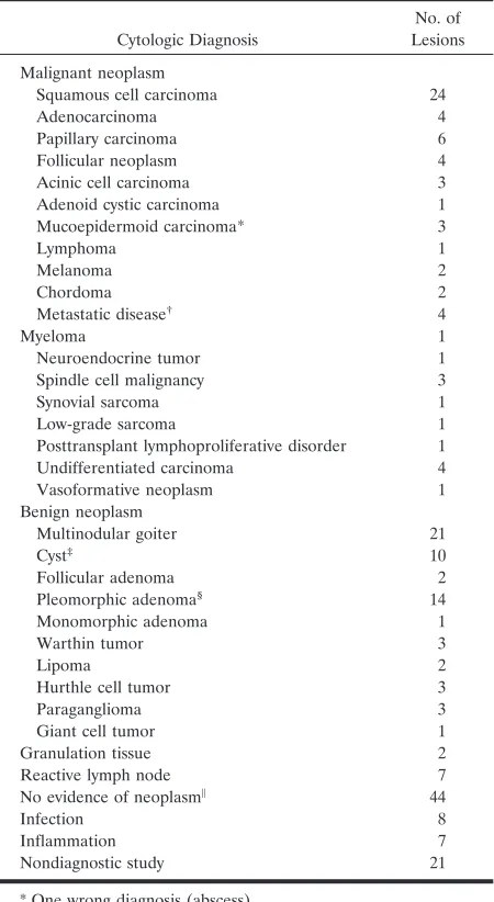
In 135 (63%) of 216 cases, definitive surgical pa-thology results were obtained, including those for the four specimens with wrong diagnoses. Among the nondiagnostic specimens, six (29%) of 21 went to surgery, five (83%) of six were neoplastic, and one (17%) of six was fibrous tissue. In the remainder, clinical or imaging follow-up was used to arrive at a final diagnosis (e.g., no recurrent tumor, inflamma-tion, scar material, multinodular goiter). Table 3 shows the cytologic results with FNA.
[image:2.585.54.280.70.324.2]In 14 instances, the cytopathologist requested core biopsy for histologic evaluation at the time of FNA. Core biopsy was deemed necessary when nonspecific neoplastic cells, such as spindle cells, were found on cytology or when supplemental material was required for more definitive diagnosis. Of the 14 histologic spec-imens, five were paraspinal, two were parapharyngeal, two were in a postsurgical site, and five were in other locations. A correct diagnosis was made in 11 (78.6%) of cases. The core sample was nondiagnostic in two (14.3%), and the wrong diagnosis was made in one (7.1%). The range in number of passes was one to two. Of the 216 FNA procedures, 196 were performed TABLE 1: Steps in performing aspiration
Step Description
1 Review prebiopsy images to determine the approach
2 Obtain informed consent 3 Obtain scout view
4 Obtain limited nonenhanced axial views through the lesion
5 Place skin markers on the line of the image that has best line of sight to the lesion
6 Repeat scanning to determine the puncture site 7 Perform sterile preparation of the puncture site 8 Insert the introducer needle
9 Repeat scan to verify the position of the introducer needle
10 Insert the aspiration needle 11 Verify the position of the needle tips 12 Place the needle to the edge of the mass 13 Call cytology department
14 Perform multiple multidirectional plunges into the mass
15 Withdraw only the aspiration needle 16 Submit specimen for analysis
[image:2.585.306.533.71.144.2]17 Repeat steps as required on the basis of the cytologic results
TABLE 2: Accuracy of CT-guided FNA
Diagnosis
Parapharyngeal
Space Thyroid Parotid Paraspinal Other Total
Correct 23 (85) 49 (92) 29 (81) 25 (100) 65 (87) 191 None 3 (11) 4 (8) 6 (17) 0 8 (11) 21
Wrong 1 (4) 0 1 (2) 0 2 (2) 4
Total 27 53 36 25 75 216
with 22-gauge spinal needles, 10 were performed with 25-gauge needles, and 10 were performed with 20-gauge or larger needles. Although few patients un-derwent biopsy with 25- or 20-gauge needles, no dra-matic differences were noted in the success rate for diagnostic samples. (The sample size was inadequate for statistical comparison.) Most the aspirations done with large-gauge needles involved paraspinal samples. No postprocedural complications occurred except for one facial hematoma, which required observation in the emergency department for 6 hours. Table 2 sum-marizes the overall results in the 216 samples.
Discussion
FNA is an effective means for determining the histology of most head and neck lesions. The value of
FNA includes its low invasiveness, the use of small needles in an area where several vascular structures are present, and the lack of a tell-tale scar from the procedure. The pain associated with the procedure is usually minimal when adequate local anesthesia is used. Neither sedation nor general anesthesia is re-quired. We inform our patients that the aspiration needle is smaller than the needle used for drawing blood samples and that whatever discomfort may oc-cur resolves after the needle is removed. We also inform our patients that the risk of an adverse reac-tion to anesthesia is greater than the risk of the procedure itself. We use a coaxial technique, as it requires only one pass through the skin, and it allows the radiologist to achieve different angles for aspira-tion in a specific lesion. In general, this approach is less traumatic to the patient, though it does require an introducer needle of a slightly larger gauge. In our experience, CT-guided FNA in the head and neck is a well-tolerated procedure devoid of notable complica-tions, and it is an effective alternative to a more invasive and risky open surgical biopsy.
Non–image-guided FNA of palpable lesions is well established as an accurate diagnostic tool in the head and neck, particularly in the salivary glands (1– 4). FNA via a transoral approach for visible parapharyn-geal space lesions is an option with an accuracy of 78 – 86% (5, 6), but a false-negative rate as high as 19% secondary to inadequate stabilization of the le-sion (lele-sions moves away from the needle, possibly due to a shallow depth from the needle entry site or loose surrounding connective tissue) at the time of aspiration, limits of the intraoral angles available to puncture the mass, and an inability to make deep blind passes secondary to the underlying vessels (7). Percutaneous image-guided FNA is not constrained by these limitations. Image-guided FNA can accu-rately evaluate nonpalpable lesions, particularly those in a poorly accessible or deep location in the head and neck. sonography-guided FNA is an established tech-nique for lesion localization, particularly in the eval-uation of the thyroid gland and superficial cervical lymph nodes (8 –14). However, lesions deep to the bony structures of the face and air-containing spaces are not well visualized during sonography (10, 15, 16). These can accurately and readily be localized and aspirated under CT guidance (15–23).
MR-guided FNA has been performed with good re-sults and minimal morbidity (24 –31). MR imaging pro-vides excellent soft-tissue resolution, but it does not provide detail of bony structures. Although MR imaging has the advantage of the lack of ionizing radiation, it has the disadvantages of limited availability (particularly with open MR imaging systems), longer acquisition times, and the need for instruments compatible with a magnetic field. While MR imaging guidance will likely have an expanded role in the future, especially with open higher-field-strength magnets, CT is currently the imaging technique of choice for biopsy of deep-seated or poorly localized head and neck lesions when sonog-raphy is inadequate.
[image:3.585.57.282.71.482.2]The risks of CT-guided FNA are usually minor and TABLE 3: Cytohistologic findings with CT-guided FNA
Cytologic Diagnosis
No. of Lesions
Malignant neoplasm
Squamous cell carcinoma 24
Adenocarcinoma 4
Papillary carcinoma 6
Follicular neoplasm 4
Acinic cell carcinoma 3
Adenoid cystic carcinoma 1
Mucoepidermoid carcinoma* 3
Lymphoma 1
Melanoma 2
Chordoma 2
Metastatic disease† 4
Myeloma 1
Neuroendocrine tumor 1
Spindle cell malignancy 3
Synovial sarcoma 1
Low-grade sarcoma 1
Posttransplant lymphoproliferative disorder 1 Undifferentiated carcinoma 4
Vasoformative neoplasm 1
Benign neoplasm
Multinodular goiter 21
Cyst‡ 10
Follicular adenoma 2
Pleomorphic adenoma§ 14
Monomorphic adenoma 1
Warthin tumor 3
Lipoma 2
Hurthle cell tumor 3
Paraganglioma 3
Giant cell tumor 1
Granulation tissue 2
Reactive lymph node 7
No evidence of neoplasm㛳 44
Infection 8
Inflammation 7
Nondiagnostic study 21
* One wrong diagnosis (abscess).
†Renal cell, breast, or prostate carcinoma. ‡Colloid, hemorrhagic, or branchial cleft cyst. §One wrong diagnosis (mucoepidermoid carcinoma).
㛳Radiation change or fibrous, glandular, adipose, or muscular tissue
include pain, vasovagal reaction, minor infection, and minor hemorrhage (8). Notable complications, such as seeding of the biopsy tract, pneumothorax, severe hemorrhage, and death have been estimated to occur in only 0.003– 0.031% of patients; these have been directly correlated to the size of the needle (11, 17, 32). It is common opinion that, with needles smaller than 18 gauge, this risk is infinitesimal. Injury to the adjacent nerves is possible but usually transient. There is a potential for a reaction to local anesthesia or the sterile preparation agent, typically povidone iodine. Walker et al (32) described a case of pseudo-aneurysm formation in an internal maxillary artery manifesting with transoral hemorrhage 3 months af-ter CT-guided biopsy in the masticator space. They believe that the potential for vascular complications may be increased in patients who have undergone prior radical neck surgery and radiation therapy. Por-cellini et al (33) describe a complication of pseudo-aneurysm rupture in the internal carotid artery. Con-trast-enhanced CT had failed to depict the lesion, which was not pulsatile and thought to be a neoplastic cervical lymph node. After FNA and leakage of the aneurysm, color Doppler scanning and MR-imaging showed aneurysmal changes of the carotid artery wall with an intraluminal thrombus. Successful emergency resection with a polytetrafluoroethylene interposition graft was performed. We did not have any notable complications. All patients reported resolution of procedure-related pain within 24 hours. In a recent 5-year series of CT-guided FNA procedures of head and neck lesions, DelGaudio et al (23) also reported no significant complications.
The results of this study compare favorably to those of a previously reported 5-year experience (22). We obtained a diagnostic sample in 195 (90.3%) of 216 cases, which compares positively with the rate from a series of 111 cases (83.8%). A correct diagnosis was made in 191 (88.4%) of our 216 cases compared with the prior study result of 83.4%. The improved sam-pling rate is likely due to a combination of improve-ment in FNA technique and cytopathologist tech-nique or skill.
DelGaudio et al (23) suggested a learning curve on the part of the radiologist using the CT-guided nee-dle-biopsy technique. They reported an improvement from 20% to 6% in the nondiagnostic rate in the first 10 biopsy procedures compared with the subsequent 32. Improved collection likely results from several factors including 1) more vigorous aspiration as ex-perience increases, 2) more effective approaches to the lesions, 3) better feedback from the cytologists, and 4) correct choices of needle size. Improved cyto-pathologic techniques and cytopathologist skill are also likely contributing factors.
The critical value of onsite cytology must be high-lighted. In our study, there were few instances in which a single FNA pass was considered diagnostic. This was partly because the cytologist often requests aspirations of different portions of the lesions to re-duce sampling error. It may also be that initial passes through the lesions have more contamination with
adjacent tissue than subsequent passes for which a track has already cleared a path in the underlining subcutaneous fat and deep muscle. Austin and Cohen (34) showed a significant increase from 80% to 100% in obtaining diagnostic specimens with a cytopatholo-gist present. FNA is most accurate when immediate assessment for specimen adequacy is performed and a differential diagnosis constructed so that additional material can be acquired for special studies (including conversion to core biopsy) when needed (35). The cytopathologist also allows for termination of the pro-cedure once diagnostic material is obtained; this in-creases the safety of the procedure.
In no instance did a sample obtained after the fourth pass yield diagnostic material. This observation suggests that some lesions are less readily sampled than others. In this case, it is our policy to attempt a true cut biopsy; however, the overall yield on performing this study is intermediate, as only 11 of 14 true cut histologic samples yielded the correct diagnosis.
We perform a higher rate of thyroid biopsy proce-dures under CT guidance than clinicians at many other institutions. This is because of a high rate of referrals from the otorhinolaryngologists directly to our neurora-diology department. Our neuroradiologists have more experience performing CT-guided FNA, whereas our body-imaging radiologists use sonography.
The 90% yield of diagnostic samples reported in this study is commensurate with that of other studies of FNA in the head and neck and other parts of the body. A relatively low rate of incorrect diagnoses (1.9%) was attributable to FNA in this study, possibly because of the multiple aspirations with onsite eval-uations by the cytology team.
Conclusion
CT-guided FNA is a safe, well tolerated, and accu-rate tool in diagnosis of head and neck lesions. Our series demonstrates an overall low diagnostic error rate, which may have been secondary to improved cytopathology and neuroradiologist FNA technique. The presence of a cytopathologist during CT-guided FNA is critical to maintaining high diagnostic sam-pling rates, achieving low rates of incorrect diagnoses, and providing the best possible care to the patient.
References
1. Meyers DS, Templer J, Davis WE, Balch JA. Aspiration cytology for diagnosis of head and neck masses. Otolaryngology 1978;86:650 – 655
2. Layfield LJ, Glasgow BJ.Diagnosis of salivary gland tumors by fine-needle aspiration cytology: a review of clinical utility and pitfalls.Diagn Cytopathol1991;7:267–272
3. Frable MA, Frable WJ.Fine-needle aspiration biopsy of salivary glands.Laryngoscope1991;101:245–249
4. Cohen MB, Reznicek MJ, Miller TR.Fine-needle aspiration biopsy of the salivary glands.Pathol Ann1992;27:213–245
5. Das DK, Gulati A, Bhatt NC, Mandal AK, Khan VA, Bhambhani S.Fine needle aspiration cytology of oral and pharyngeal lesions. A study of 45 cases.Acta Cytologica1993;37:333–342
biopsy of intraoral and parapharyngeal lesions. Acta Cytol 1993;37:448 – 450
8. Charboneau JW, Reading CC, Welch TJ.CT and sonographically guided needle biopsy: current techniques and new innovations.AJR Am J Roentgenol1990;154:1–10
9. van den Brekel MWM, Castelijns JA, Stel HV, Golding RP, Meyer CJL, Snow GB. Modern imaging techniques and ultrasound-guided aspiration cytology for the assessment of neck node metas-tases: a prospective comparative study.Eur Arch Otorhinolaryngol 1993;250:11–17
10. McIvor NP, Freeman JL, Salem S, Elden L, Noyek AM, Bedard YC. Ultrasonography and ultrasound-guided fine-needle aspira-tion biopsy of head and neck lesions: a surgical perspective. La-ryngoscope1994;104:669 – 674
11. Smith EH. The hazards of fine-needle aspiration biopsy. Ultra-sound Med Biol1994;10:629 – 634
12. Yokozaw T, Fukata S, Kuma K, et al.Thyroid cancer detected by ultrasound-guided fine needle aspiration biopsy. World J Surg 1996;20:848 – 853
13. Screaton NJ, Berman LH, Grant JW.Head and neck lymphade-nopathy: evaluation with US-guided cutting-needle biopsy. Radiol-ogy2002;224:75– 81
14. Titton RL, Gervais DA, Boland GW, Maher MM, Mueller PR.
Sonography and sonographically guided fine-needle aspiration bi-opsy of the thyroid gland: indications and techniques, pearls and pitfalls.AJR Am J Roentgenol2003;181:267–271
15. Robbins KT, vanSonnenberg E, Casola G, Varney RR. Image-guided needle biopsy of inaccessible head and neck lesions.Arch Otolaryngol Head Neck Surg1990;116:957–961
16. Yousem DM, Sack MJ, Scanlan KA. Biopsy of parapharyngeal space lesions.Radiology1994;193:619 – 622
17. Welch TJ, Sheedy PF II, Johnson CM, Stephens DH.CT-guided biopsy: prospective analysis of 1,000 procedures. Radiology 1989;171:493– 496
18. Barakos JA, Dillon WP.Lesions of the foramen ovale: CT-guided fine-needle aspiration.Radiology1992;182:573–575
19. Yousem DM, Sack MJ, Weinstein GS, Hayden RE. Computed tomography-guided aspirations of parapharyngeal and skull base masses.Skull Base Surg1995;5:131–136
20. Esposito MB, Arrington JA, Murtagh FR, Ridley MB, Endicott JN, Silbiger ML.Anterior approach for CT-guided biopsy of skull base and parapharyngeal space lesions. J Comput Assist Tomogr 1996;20:739 –741
21. Abrahams JJ.Mandibular sigmoid notch: a window for CT-guided biopsies of lesions in the peripharyngeal and skull base regions. Radiology1998;208:695– 699
22. Sack MJ. Weber RS, Weinstein GS, Chalian AA, Nisenbaum HL, Yousem DM.Image-guided fine-needle aspiration of the head and neck: five years’ experience. Arch Otolaryngol Head Neck Surg 1998;124:1155–1161
23. DelGaudio JM, Dillard DG, Albritton FD, Hudgins P, Wallace VC, Lewis MM.Computed tomography– guided needle biopsy of head and neck lesions.Arch Otolaryngol Head Neck Surg2000;126:366 –370 24. Duckwiler G, Lufkin RB, Teresi L, et al.Head and neck lesions:
MR-guided aspiration biopsy.Radiology1989;170:519 –522 25. Lufkin RB, Robinson JD, Castro DJ, et al.Interventional magnetic
resonance imaging in the head and neck.Top Magn Reson Imaging 1990;2:76 – 80
26. Hathout G, Lufkin RB, Jabour B, Andrews J, Castro D.MR-guided aspiration cytology in the head and neck at high field strength.J Magn Reson Imaging1992;2:93–94
27. Lee MH, Lufkin RB, Borges A, et al.MR-guided procedures using contemporaneous imaging frameless stereotaxis in an open-config-uration system.J Comput Assist Tomogr1998;22:998 –1005 28. Lewin JS, Petersilge CA, Hatem SF, et al.Interactive MR
imaging-guided biopsy and aspiration with a modified clinical C-arm sys-tem.AJR Am J Roentgenol1998;170:1593–1601
29. Kacl GM, Carls FR, Moll C, Debatin JF.Interactive MR-guided biopsies of maxillary and skull-base lesions in an open-MR system: first clinical results.Eur Radiol1999;9:487– 492
30. Wang SJ, Sercarz JA, Lufkin RB, Borges A, Wang MB. MRI-guided needle localization in the head and neck using contempo-raneous imaging in an open configuration system. Head Neck 2000;22:355–359
31. Merkle EM, Lewin JS, Aschoff AJ, et al.Percutaneous magnetic resonance image-guided biopsy and aspiration in the head and neck.Laryngoscope2000;110:382–385
32. Walker AT, Chaloupka JC, Putman CM, Abrahams JJ, Ross DA.
Sentinel transoral hemorrhage from a pseudoaneurysm of the internal maxillary artery: a complication of CT-guided biopsy of the masticator space.AJNR Am J Neuroradiol1996;17:377–381 33. Porcellini M, Bernardo B, Perretti B, Bauleo A.Iatrogenic rupture
of internal carotid artery aneurysm: a complication of CT-guided needle biopsy of the neck.Giornale Chirurgia1996;17:531–533 34. Austin JH, Cohen MB.Value of having a cytopathologist present
during percutaneous fine-needle aspiration biopsy of lung: report of 55 cancer patients and metaanalysis of the literature.AJR Am J Roentgenol1993;160:175–177