Great Britain © The Company of Biologists Limited 1984
DEVELOPMENT OF SWIMMING MOVEMENTS AND
MUSCULATURE OF LARVAL HERRING
(CLUPEA HARENGUS)
BY R. S. BATTY
Scottish Marine Biological Association, Dunstaffnage Marine Research Laboratory, P.O. Box 3, Oban, Argyll
Accepted 20 December 1983
SUMMARY
A kinematic analysis was made of swimming of larval herring Clupea harengus L. Swimming style was found to change with growth and develop-ment; the amplitude of swimming movements of early post-yolk-sac larvae increases linearly towards the tail, a style of swimming which relies mainly on resistive forces for propulsion. Later, after the caudal and dorsal fins have developed, the swimming style changes, in response to an increase in Rey-nold's Number, such that inertial forces are more important. In this type of swimming the amplitude increases more rapidly than linearly towards the tail.
The distribution of red and white muscle fibre types was studied in rela-tion to development. On hatching, red muscle fibres were found to be arranged as a single layer on the outside of the myotomes. They develop into the adult distribution, concentrated at the midline of the flank near the skin, only after the gills and circulation become fully functional.
INTRODUCTION
On hatching, larvae of herring (Clupea harengus) have elongate eel-like bodies and are 6-8 mm long. During the yolk-sac stage, swimming is probably for depth control (Weihs, 19806) and the amount of time herring larvae spend swimming upwards is correlated with their sinking rate. Once the yolk-sac has been resorbed, larval herring are observed to swim almost continuously at light intensities adequate for feeding (Blaxter & Staines, 1971). This search-swimming has been analysed in anchovy by Hunter (1972) and in plaice by Batty (1981).
There is a very marked scale effect during the growth of a herring larva from 8 mm at hatching to near 40 mm at metamorphosis. Since swimming speed is related to length, Reynold's Number (Re, which is proportional to the product of length and swimming speed) increases substantially during larval life. The present study examines the changes which occur in swimming style of herring larvae as they grow, relating this to increasing Re which indicates changes in the importance of inertial over viscous forces.
A method of describing and understanding the swimming movement of a particular
fish is the two-wave model of Gray (1933), further developed by Videler & W ^ (1978). In this model, undulatory swimming is described as a propulsive wave travel-ling backwards along the body at a velocity v, wavelength A&, and period T. A further stationary wave with wavelength A, describes the track in space followed by a point on the body. The parameters of these two waves can be found by examining the lateral and longitudinal movements of a number of fixed points on the body of the fish (Videler & Wardle, 1978).
Examination of the swimming of plaice larvae (Batty, 1981) shows that larval fish swimming has many similarities to adult swimming previously examined by the model. The main differences are that the swimming speed to wave speed ratio u/v is lower in larvae than adults and that, at the tail, the maximum value of 6, the angle of the body to the direction of motion, is higher and can exceed 90°. The two-wave model does not however consider this angle 6, which is a very important factor in the generation of thrust force by the fish's body (Lighthill, 1971).
Search-swimming is powered by aerobic (red) muscles. The amount and location of these aerobic muscles is important in relation to the swimming ability of the fish and the development of its respiration. Walker & Pull (1975) made a survey of the relative proportions of red and white muscle in adult fish but little work has been done on fish larvae. The present study examines the distribution of red muscle of herring larvae from hatching to metamorphosis.
MATERIALS AND METHODS
Source of animals
Spring-spawning herring were caught on Ballantrae Bank, in the Clyde, Scotland, during early March and the gonads were transported to the laboratory where artificial fertilizations were made. The hatched larvae were reared in circular black plastic tanks of 120-2401 capacity, temperatures rising from about 8 °C to 14 °C during larval life.
Body dimensions
Body dimensions were measured from living anaesthetized larvae using a silhouette photography technique (Edgerton, 1977; Neave & Batty, 1982). The larvae were placed in a container 75 X 40 X 25 mm made of microscope slides glued with 'Aral-dite'. It was filled with a filtered anaesthetic-seawater solution (1: 40000 benzocaine) rested on a strip of Kodak fine grain release positive film in a dark room. An electronic flash 2 m from the film was used to make a 1/1000 s exposure. The developed films were projected and measurements made of body dimensions at various stages of larval development.
Szuimming movements
Swimming movements of larval herring 219
kground beneath the tank with a 15 W strobe light synchronized with the television eld pulse. Light from the strobe lamp was reflected off a semi-silvered mirror set at an angle of 45 ° in front of the camera lens so that the camera lens and illumination shared the same optical axis (see Fig. 1). Video tape recordings were replayed frame-by-frame and analysable sequences copied onto 35 mm film by photographing the individual frames on the monitor screen. These photographs were projected onto a digitizing tablet which was linked to a Hewlett Packard 9825 computer. The outline of each image of a larva from a series of frames (Fig. 2) was entered into the computer and stored as x, z coordinates, x being the mean path of motion and z normal to it in the horizontal plane.
The outline information was transformed, using the method described by Batty (1981), to a series of points on a line along the centre of the larva's back which has values, a, from 0 at the snout to 1 at the tip of the caudal fin. This co-ordinate system, which defines any point on the centre line of the larva by the three co-ordinates a, x, z is shown in Fig. 3. A set of 11 points of fixed position on a were calculated using
interpolation.
Histology
Herring larvae were sampled and killed by an overdose of anaesthetic (Benzocaine 1:10 000). Frozen transverse sections were cut in a cryostat and subsequently stained for succinic dehydrogenase (SDHase) to demonstrate the occurrence and distribution of different fibre types. A positive reaction should occur in red aerobic muscle and a negative reaction in white anaerobic muscle. Since the standard method for SDHase staining (Pearse, 1968) failed to demonstrate known red muscle in adult herring satisfactorily, a modified technique was used based on earlier work by Aronson & Pharmakis (1962). This involved using nitroblue tetrazolium with cyanide.
[image:3.451.165.295.411.607.2]M
3 mm
[image:4.451.40.420.55.342.2]5 mm
Fig. 2. Outlines of herring larvae swimming. (A) An 11 mm larva; (B) a 22 mm larva; note difference in scale. Frames are 20 ms intervals.
Fig. 3. The three co-ordinates x, a and a used to describe the movement of any point along the length of the larval body (i.e. along a) in terms of x and z. Note that a increases from 0 at the head to 1 at the tail.
[image:4.451.133.320.390.504.2]J P tetrazolium is reduced by SDHase, via phenazine methosulphate, to form an insoluble formazan which is deposited in the tissue at the site of the reaction and so gives a dark blue stain in the vicinity of the enzyme. Sections were preserved by fixing in 10% phosphate-buffered formalin for lOmin, counter-stained with nuclear fast red, and mounted in an aqueous medium.
RESULTS
Swimming movements
Swimming speeds of 5—20 mm s"1 were observed in first feeding larvae about 10 mm long. Swimming speed increased with length, larvae 22 mm long having swim-ming speeds in the range 5-60 mm s~'. Two examples of herring larvae swimswim-ming are shown in Fig. 2. The figure shows the outlines of two larvae, of 11 mm and 22 mm length, from successive 20-ms frames of the TV recording. Top views are shown drawn to different scales in order to compare the details of movement. This figure clearly shows apparent similarities between the two larvae. For example there is slightly more than one wavelength on the body at any time, the wave moving along the body with increasing amplitude towards the tail.
Further analysis shows differences in detail between the two sizes of larva. In Fig. 4 amplitude versus length curves are shown for the 11 mm and 22 mm larva and also for a 60 mm juvenile herring. The amplitudes of lateral movements, z, and the maximum value of angle 6 between the body and the direction of motion, which is the x axis, are plotted. The shape of both curves is different in the two sizes of larvae. For the 11 mm larva, amplitude and #m»x increase almost linearly from head to tail. In contrast, amplitude and 0m« in the 22 mm larva increase at a steadily increasing rate from head to tail, and the amplitude is lower at the head and in the anterior part of the body compared with the 11 mm larva. Note that the angle ft™* at the tail is less than 90° for the 11 mm larvae but reaches 100° for the 22 mm larva.
In the 22 mm larva the anterior part of the body, for about one-third of its length, is almost rigid. The amplitude versus length curve for a juvenile herring (Fig. 4C) shows that the non-linear increase in amplitude is retained after metamorphosis. There is now a considerably greater amplitude at the head however, and the position of minimum amplitude has moved rearwards to between 0 2 and 0-4L (where L is the body length). This indicates that yaw of the head due to lateral recoil is much greater in the juvenile herring than in the larva.
222 R. S. BATTY
2 "5.
1 0
0-8
0-6
0-4:
0-2
0
- c
-, 1
y
W——K * ^
l 1 1 1 1 1 l
0-2 0-4 0-6
Distance along body a (L)
[image:6.451.84.361.56.465.2]0-8 1-0
Fig. 4. Maximum relative amplitudes for each length coordinate a (x), and maximum angle 0 of each interval between the points shown by the symbol O, (A) for an 11 mm larva; (B) for a 22mm larva; (C) for a 60mm juvenile (with the adult form).
The ratio of swimming speed to wave speed u/vp is a measure of the slippage of the
wave speed in relation to the water. The ratio lies between 0*2 and 0*4 (Fig. 6) which is much lower than that found in adult fish. There is apparently a slightly higher efficiency at faster swimming speeds.
Body dimensions and svnmming
Swimming movements of larval herring
223
(7 ) c oI
>
a c ti o Pos i 10 0-8 0-6 0-4 0-2 0 A -p . .d
1 /P
rp
h
P 9
_r
Tr P
if
D/
/
/
i i
J J
I
/
$
r / a
4
r ( i i0-2 0-4 0-6
15
10
10
2 0'8 o ! 0-6
s
%
°'
4
*
1 0-2
£ o
20
0 0-2 0-4 0-6
IS
10
0-2 0-4 Time (s)
0-6 0-2 0-4
Time (s)
[image:7.451.50.399.56.392.2]0-6
Fig. S. Wave crest positions against time for an 11 mm larva (A and B) and a 22 mm larva ( C a n d D ) . Position is shown on the body coordinate a (A and C) and on the* coordinate, i.e. direction of motion (B and D).
l-0r 0-8 0-6 0-4 0-2 10 20 Swimming speed u (mms"')
30
[image:7.451.93.350.435.620.2]224 R. S. BATTY
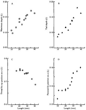
expressed as proportions of length, are shown in Fig. 8. Body depth/L (Fig. ^ P increases as length increases, and the position of maximum height moves forward as the need for a streamlined shape increases. The position of the pectoral fins (Fig. 8D) is initially well forward. The pectoral fins generate lift, initially well ahead of the centre of mass, giving a head-up attitude which has been noted in behavioural ob-servations (Rosenthal, 1968). The head-up attitude is lost as the fish grows and the pectoral fins move relatively rearwards.
The dorsal (Fig. 8C) and anal fins move forward during growth. The dorsal fin develops very early (at about 15 mm length) and is relatively large during larval life. The dorsal fin is too close to the tail to be of use in making turns (Aleev, 1958, 1963). This fin, in fact, increases the lateral area behind the centre of mass so tending to increase directional stability (Aleev, 1963). The dorsal fin of larval herring is also likely to be important in propulsion. At the end of the caudal fin stroke thrust is low (Wardle & Reid, 1977) and lateral forces relatively large due to a low angle 8. The action of the trailing edge of the dorsal fin will tend to smooth the thrust and coun-teract the recoil produced by the caudal fin, as a result of its opposite lateral movement at the time when the caudal fin produces the maximum lateral force. The effectiveness
11
15
20
27
[image:8.451.82.331.293.612.2]35
Swimming movements of larval herring 225
s arrangement is apparent when comparing graphs showing amplitude and body position for a 22mm larva and for a 60mm juvenile herring (Fig. 4A,B,C). The 22 mm larva is successful in minimizing amplitude near the head caused by lateral recoil. Plaice larvae have similarly small lateral movements of the head (Batty, 1981) but in this species recoil is opposed by pectoral fin movements.
Muscle fibres
Transverse sections of herring larvae show two distinct areas in the body muscle,
0-20
0 1 5
0-10 0-05 o. •a E 3 e
5
s a a D D10 20 30 40
0-20
„ 015
0 1 0
0-05
10 20 30 40
0-8 8. 0-7 a.
1
o Q 0-68
10 20 30 Length (mm) 40 0-18 0-14
g
o 010• • 8
10 20 30 Length (mm)
[image:9.451.57.389.186.610.2]40
one of white anaerobic muscle, and the other of red aerobic muscle staining ^ for SDHase (Fig. 9). In the yolk-sac larva (10 mm length, see Fig. 9A), red muscle appears as a band of fibres, one fibre thick, around the external surface of the myotome. This arrangement persists through most of larval life (Fig. 9B,C).
At a length of around 27 mm, the distribution of red muscle begins to change (Fig. 9D). Between the band of red muscle and the skin are a number of new muscle fibres which stain well for SDHase but do not exhibit such a strong reaction as the original red fibres. As these fibres increase in proportion the staining reaction becomes more uniform. The red and white muscle distribution typical of adults is observed in the 35 mm fish (Fig. 9E) and here, on the basis of SDHase staining, only these two types of muscle can be recognized.
The change in the relative proportions of red and white muscle during growth of herring larvae was measured by finding their relative areas in camera lucida drawings of transverse sections. To make comparisons with the work of Walker & Pull (1975) all the sections used for this were taken from a point one-third of the length along the body from the tail (a — 0-67). The graph of red muscle area in transverse section (Fig. 10) shows a decline in the proportion of red muscle from 13 % at the end of the yolk-sac stage to a minimum of about 8 % at a length of about 30 mm. In the juvenile fish, after the full development of gills and circulation, it has increased to about 16% at 70 mm length, the same proportion found by Walker & Pull (1975) for adult herring. The minimum in red muscle bulk nearly coincides with the changeover in the distribution of the red muscle from the larval single fibre band to the adult arrange-ment concentrated about the centre of the myotomes near the skin.
DISCUSSION
During growth a fish larva experiences a great change in the nature of the water flow about its body whilst swimming. This is due to a scale effect of growth and increase in the swimming speed used for searching. In larval herring the swimming movements change as Reynolds Number (Re) increases during growth. The shape of the am-plitude against length curve (Fig. 4) changes as herring larvae grow. Initially (at 11 mm length) it is linear, but once larvae have grown to 22 mm it becomes non-linear. Amplitude then increases more rapidly as the tail is approached. The shape of this curve is characteristic of the type of swimming motion employed (Grillner & Kashin, 1976; Videler, 1981). A linear increase in amplitude along the body, giving a large amplitude over most of the length of the body, is characteristic of a style of swimming in which lateral acceleration of water is constant along the body, indicating that resistive forces are important. The non-linear increase in amplitude seen in a 22 mm larva (Fig. 4B) is typical of a style of swimming which is often called carangiform or subcarangiform; in this mode lateral acceleration of water by the fish body increases rapidly towards the tail, so that in this type of swimming reactive (inertial) forces predominate.
Journal of Experimental Biology, Vol. 110
B
D
Fie. 9
R. S. BATTY
Swimming movements of larval herring
111
2 0r
15
S 10
•8 06
D
D °
1
10
A A ,
r
1-2
0-8
O 0-4
6
20 40
Length (mm)
60
o
o
80
Weihs (1980a) showed theoretically that the swimming style of anchovy la changed as they grew, due to the increase of Re, so that intermittent swimming became energetically more advantageous as viscous forces became less important. Weihs (1980a) denned three regimes: Re < 10, where velocity is much more impor-tant than inertial force effects, 10 <Re < 200, which is a region of transition where the flow changes from the viscous regime to the inertial regime, where Re > 200. In low Re regimes, drag coefficient is inversely related to Re which makes swimming at low speeds very inefficient. This will affect herring larvae in the yolk-sac stage and during the early stages of feeding. Until the larva exceeds a length of 15 mm and the Re at the searching speed of the larva has exceeded 200, swimming using reactive forces would be very inefficient. It is therefore advantageous for herring larvae less than 15 mm to use resistive thrust production. The appearance of the caudal and dorsal fins when the larvae are between 15 mm and 20 mm in length increases the effectiveness of swimming in this mode and the change in swimming style occurs at the same size.
A distribution of red aerobic muscle fibres similar to that reported here has been described in larval zebra fish, Brachydanio rerio, by Waterman (1969) using SDHase staining and by Van Raamsdonk, Pool & te Kronnie (1978) using immunological techniques. Both studies showed a single layer of red fibres in newly hatched larvae, developing to an adult distribution similar to that of adult herring.
The development of respiration in herring larvae was studied by de Silva (1973, 1974). Her results for gill and body surface area are plotted in Fig. 10, together with the percentage of red muscle. A vertical dashed line indicates the body length at which the adult distribution of red muscle fibres starts to develop. The gills develop very rapidly from a length of 22 mm onwards, the most rapid development occurring at around 30 mm. The maximum gill area to body weight ratio attained was 1 -2 mm2 mg"1 at metamorphosis (length 35—40 mm). It thus appears that cutaneous respiration is more important than gill respiration during most of the larval stage. Gill respiration only becomes important when the gill area has increased at a body length of 25 mm. Up to this length, the red muscle is distributed peripherally, enabling it to utilize cutaneous respiration. Subsequently, new red fibres appear away from the periphery but by this time gill respiration has been established.
I thank J. H. S. Blaxter, D. A. Booth, R. Harvey, J. J. Videler and C. S. Wardle for their comments on the manuscript. The work was carried out while I was in receipt of a NERC research studentship.
R E F E R E N C E S
ALEEV, YU. G. (1958). Adaptation for movement and the turning ability of fish. Dokl. Biol. Sci. (Trarwl.) 120, 299-302.
ALEEV, YU. G. (1963). Function and Gross Morphology in Fish. Moscow. (Transl. from Russian, Isr. Program. Sci. Transl. No. 1773, Jerusalem, 1969).
ARONSON, W. & PHARMAKIS, T. (1962). Enhancement of neotetrazoleum staining for succinic dehydrogenase activity with cyanide. Stain Technology 37, 321.
BATTY, R. S. (1981). Locomotion of plaice larvae. Symp. zool. Soc. bond. 48, 53-69.
BLAXTER, J. H. S. &STAINES, M. E. (1971). Food searching potential in marine fish larvae. In Fourth Eur
Swimming movements of larval herring 229
SILVA, C. D. (1973). The ontogeny of respiration in herring and plaice larvae. Ph.D. thesis. Stirling
y
DE SILVA, C. D. (1974). Development of the respiratory system in herring and plaice larvae. In The Early Life
History of Fish, (ed. J. H. S. Blaxter), pp. 465-485. Berlin: Springer Verlag.
EDGERTON, H. E. (1977). Silhouette photography of small active subjects. J . Microscopie 110, 79-81. GRAY, J. (1933). The movement of fish with special reference to the eel. J. exp. Biol. 10, 88-104.
GRILLNER, S. &KASHIN, S. (1976). On the generation and performance of swimming in fish. In Neural control
of locomotion, (eds R. M. Herman, S. Grillner, P. S. G. Stein & D. G. Stuart), pp. 181-201. New York:
Plenum Press.
HUNTER, J. R. (1972). Swimming and feeding behaviour of larval anchovy Engraulismordax. Fishery Bull. Fish
Wildl. Serv. U.S. 70, 821-838.
LIGHTHILL, M. J. (1971). Large-amplitude elongated-body theory of fish locomotion. Proc. R. Soc. B. 179, 125-138.
NEAVE, D. A. & BATTY, R. S. (1982). A simple method for measuring fish larvae using silhouette photography.
Aquaculture 29, 165-168.
PEARSE, A. G. E. (1968). Histochemistry, Theoretical and Applied. Vol. 1. 759pp. London: Churchill. ROSENTHAL, H. (1968). Schwimmverhalten undschwimmgeschwindigkeit bei den Larven desHerringsClupea
harengus. Helgoldnder viiss. Meeresunters 18, 453-486.
VAN RAAMSDONK, W., POOL, C. W. & TE KRONNIE, G. (1978). Differentiation of muscle fiber types in the teleost Brachydanio rerio. Anat. Embryol. 153, 137—155.
VIDELER, J. J. (1981). Swimming movements, body structure and propulsion in cod Gadus morhua. Symp. Zool.
Soc. bond. 48, 1-27.
VIDELER, J. J. & WARDLE, C. S. (1978). New kinematic data from high speed cine film recordings of swimming cod (Gadus morhua). Neth.jf. Zool. 28, 465-484.
WALKER, M. G. & PULL, G. A. (1975). A survey of red and white muscle in marine fish..7. Fish Biol. 7, 295-300. WARDLE, C. S. tc REID, A. (1977). The application of large amplitude elongated body theory to measure swimming power in fish. In Fisheries Mathematics, (ed. J. H. Steele), pp. 171-191. London: Academic Press.
WATERMAN, R. E. (1969). Development of the lateral musculature in the teleost, Brachydanio rerio: A fine structural study. Am.jf. Anat. 125, 457-494.
WEIHS, D. (1980a). Energetic significance of changes in swimming modes during growth of larval anchovy
Engraulis mordax. Fishery Bull. Fish Wildl. Serv. U.S. 77, 597-604.
WEIHS, D. (19806). Respiration and depth control as possible reasons for swimming of northern anchovy,