Charles F. Lanzieri1 Mark Larkins2 Andrew Mancall1
Ronald Lorig 1 Paul M. Duchesneau 1 Scott A. Rosenbloom 1 Meredith A. Weinstein 1
Received April 14, 1987; accepted after revision July 29, 1987.
Presented at the annual meeting of the American Society of Neuroradiology, New York, May 1987.
, Department of Diagnostic Radiology, Cleveland Clinic Foundation, 9500 Euclid Ave., Cleveland, OH 44106. Address reprint requests to C. F. Lanzieri.
2 Division of Neurosurgery, Cleveland Clinic Foundation, Cleveland, OH 44106.
AJNR 9:27-34, January/February 1988
0195-6108/88/0901-0027
© American Society of Neuroradiology
27
Cranial Postoperative Site:
MR Imaging Appearance
The ability to diagnose adverse postcraniotomy or postcraniectomy events is essential for proper postoperative care. The importance of identifying postoperative changes on CT has previously been shown. The purpose of this study is to assess the normal and abnormal MR changes that may be seen in the postcraniotomy/postcraniectomy period. The postoperative MR, CT, and medical records of 41 postcraniotomy patients and 26 postcraniectomy patients were reviewed. Reasons for choosing craniectomy over craniotomy included decompression, infected flap, bony involvement by tumor, and posttraumatic skull. In general, the postoperative normal anatomy was better seen with MR. Postoperative events included hemorrhage (two), infection (five), cyst formation (10), and recurrent tumor (five).
In general, MR was found to be more useful than CT for the detection of hemorrhage and infection after craniotomy or craniectomy and for the proper localization of postop-erative cysts. MR proved to be a useful method for following postoperative sites in the skull.
The ability to recognize and localize correctly CSF collections, hematomas, infections, and recurrent tumors after a craniotomy or craniectomy is essential for proper postoperative care. The ability to diagnose these entities early in the postoperative period and to distinguish them from one another would enable the clinician to make more accurate treatment decisions regarding reexploration, shunt placement, initiation of antibiotic therapy, or simple observation during the critical
early postoperative period [1 , 2].
The purpose of this investigation was threefold: (1) to gain a better understanding of the MR imaging findings in the postcraniotomy/postcraniectomy period; (2) to use the normal MR appearance at the uncomplicated postoperative site to develop criteria for the early recognition of an abnormal postoperative appearance; and (3) to compare the MR and CT findings in an effort to determine which of the two methods would be most useful in a given location or clinical situation.
Materials and Methods
The hospital records and CT and MR scans of 67 postcraniotomy or postcraniectomy patients were reviewed retrospectively. In 34 uncomplicated craniotomies and 12 uncompli-cated craniectomies, CT and MR scans had been obtained 2 days to 2 months after surgery because of clinical suspicion of one of several potential postoperative events or for routine follow-up. These combined studies were used to establish criteria for normal postoperative appearances. The remaining 17 patients were considered to have abnormal MR or CT scans. The abnormal group included 10 postoperative fluid collections, five postoperative infections, two postoperative hematomas, and four recurrent tumors. The most frequent reasons for requesting CT or MR in these patients were alteration of mental status, seizure, and fever.
Results
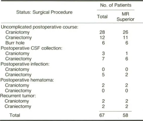
28
LANZIERI ET AL. AJNR:9, January/February 1988TABLE 1: Summary of Postcraniotomy and Postcraniectomy Patients in Whom MR Provided Additional or More Correct Information Than CT
No. of Patients Status: Surgical Procedure MR
Total
Superior Uncomplicated postoperative course:
Craniotomy 28 26
Craniectomy 12 11
Burr hole 6 6
Postoperative CSF collection:
Craniotomy 3 1
Craniectomy 7 6
Postoperative infection:
Craniotomy 0 0
Craniectomy 5 2
Postoperative hematoma:
Craniotomy 2 2
Craniectomy 0 0
Recurrent tumor:
Craniotomy 2 2
Craniectomy 2 2
Total 67 58
performed in the posterior fossa (21 of 27) as part of an effort to achieve decompression of the brainstem. The normal post-operative anatomy after craniectomy and the resulting CT findings have been described [2]. Nonspecific findings are encountered with some frequency, especially in the first few postoperative days. These include intracranial air, parenchy-mal and intraventricular hemorrhage, and mass effect. Edema and superficial cortical hemorrhages may be seen within the margins of the operative bed and are believed to be from manipulation of the brain or pressure from retractors. This is the subject of a future investigation. A relatively thin and regular enhancing meningogaleal complex (MGC) is usually seen bridging the craniectomy site. Thickening or interruption of the membrane may indicate infection, recurrent tumor, or pseudomeningocele formation.
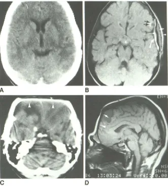
Uncomplicated craniotomies were encountered most often in our series. In most instances, a single broad zone of signal void was seen at the craniotomy site on spin-density images, which presumably represented the combined bone flap and dura with a subjacent zone of bright signal corresponding to the underlying subarachnoid space. In several clinically un-complicated craniotomies, up to four clearly identifiable layers could be seen (Fig. 1). Most superficially on spin-density images with a repetition time (TR) of 2000 msec and echo time (TE) of 32 msec, a relatively bright band of signal could be seen representing subcutaneous fat and skin. A zone of moderate signal intensity was then frequently encountered, which may have represented subcutaneous edema, tempo-ralis muscle, or scarring. Then, a zone of signal void was seen that represented the bone flap. Immediately subjacent to the bone flap, a high-signal layer could be seen representing CSF-containing blood or its breakdown products. The dura was then seen as a thin band of signal void. At the anterior and posterior margins of the craniotomy, suture artifacts were
often seen. To prevent the spread of postoperative extraaxial collections such as blood, pus, or CSF, the epidural and subdural spaces were often obliterated by suturing the dura to the edges of the craniotomy. This effectively isolated the epidural space at the operative site.
Only six burr holes were encountered (Fig. 2). Their char-acteristic MR appearance included intraosseous, subcuta-neous, and epidural collections. In two instances of seemingly normal postoperative burr holes on CT, MR revealed a pe-culiar region of high signal intenSity on T2-weighted images (TR = 2000 msec, TE = 64 msec). Both these patients were slow to regain consciousness after the procedure. This char-acteristic "mushroom sign" may represent edema secondary to inadvertent deep migration of surgical instruments while forming the burr hole.
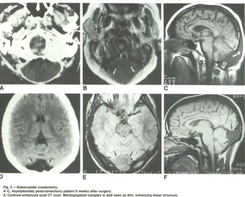
Twelve patients with clinically uncomplicated postoperative courses after suboccipital craniectomies formed the next largest group of normal patients (Fig. 3). While CT provided easier localization of the bony margins of the craniectomy, MR demonstrated the normal meningocutaneous complex equally well and showed its relationship to the resulting CSF collection to advantage. Seven patients had asymptomatic suboccipital pseudomeningoceles (Fig. 3). This common oc-currence is related to the practice of suturing the dural mar-gins to the bony marmar-gins after suboccipital craniectomies, thus leaving the dura open to avoid possible brainstem compression in the postoperative period. MR was more useful in judging the extent of these collections, especially at the level of the foramen magnum, where the potential for brain-stem compression is greatest because of the shortage of space. Dissection of pseudomeningoceles into the soft-tissue planes at this level may result in blind pockets, which from time to time may release their contents into the subarachnoid space proper and lead to episodes of aseptic meningitis. This condition is also called the postcraniectomy dumping syn-drome. Metallic surgical sutures are usually placed at the lateral inferior margins of suboccipital craniectomies. These produce characteristic artifacts at the foramen magnum. Strik-ingly similar findings are often seen in the posterior fossa in association with an enlarged cisterna magna.
[image:2.614.54.297.122.329.2]AJNR:9, January/February 1988 MR OF POSTOPERATIVE CRANIUM 29
Fig. 1.-A and B, Uncomplicated craniotomy. Contrast-enhanced CT scan (A) and MR image
(TR = 2000 msec, TE 32 msec) (B) at typical supratentorial craniotomy site 1 month after tem-poral craniotomy. MR reveals four distinct layers of postoperative site: subcutaneous fat and skin (arrowhead), temporalis muscle or galea (short straight white arrow), bone flap or dura (long straight arrow), and isolated epidural space, which may contain blood or its breakdown prod-ucts (curved arrow). At anterior margin of crani-otomy, artifact produced by one of the wire dural
tacking sutures is seen (black arrows). C and D, Uncomplicated craniectomy.
Con-trast-enhanced CT scan (C) and MR image (TR
=
2000 msec, TE=
32 msec) (D) 5 weeks after craniectomy reveal slightly different appear-ance. Meningogaleal complex (arrowheads). Wire dural tacking suture (arrow).A
c
of infection not requiring a baseline study. In one patient a sterile granuloma developed at one of the dural tacking su-tures. This phenomenon and the associated CT appearance has been described [2, 3]. The MR appearance is shown in Figure 5.
[image:3.612.229.560.84.449.2]Postoperative collections with signal characteristics similar to CSF were encountered with some frequency (10 of 68) (Fig. 6). Postoperative pseudomeningocele is anticipated and in some instances even a desirable occurrence, especially after a suboccipital craniectomy. These had a typical appear-ance related to the manner of dural closure. Hypertrophic scar formation may alter the appearance of suboccipital cran-iectomies and mimic recurrent meningiomas. Postoperative fluid collections were less common in the supratentorial com-partment, probably because of the manner of dural closure [1, 2]. In clinically uncomplicated cases, an intradural CSF collection is commonly encountered after the removal of a mass lesion. This represents the tumor site or operative bed and usually has relatively straight margins rather than the curvilinear margins seen with cysts or recurrent tumors. In the first few postoperative days these may be accompanied by surrounding edema or mass effect caused by surgical manipulation. The shape of such a collection appears to be more important than its size, although this specific issue has
B
o
not been addressed fully. MR was most helpful in differen-tiating the cyst associated with a recurrent tumor from the normal expected extraaxial collection (Fig. 7). Fifteen CSF-compatible or cystic structures were encountered. The ma-jority of these (10 of 15) represented the normal extraaxial CSF collections commonly encountered in the postoperative period. MR successfully separated the five cases of cysts
representing recurrent tumor from this total. CT in these patients was not definitive for correctly placing these st
ruc-tures, agreeing with MR in a minority of cases (seven of 15). In one case in which a recurrent tumor was adherent to the overlying dura, both CT and MR suggested this phenomenon. The superior sensitivity of MR in the detection of intracranial hemorrhage has been shown. MR was more sensitive in one of our two cases of postoperative hemorrhage (Fig. 8). More importantly, MR was more precise in defining the exact
com-partment into which a hemorrhage had occurred. This is of particular importance in guiding the neurosurgeon contem-plating reexploration.
Discussion
30 LANZIERI ET AL. AJNR:9, January/February 1988
A
8
c
o
E
F
of the normal changes that can be produced by either
pro-cedure as well as the appearances caused by postoperative complications. The reasons for choosing to perform a crani-ectomy include decompression and bone involvement by the underlying disease process. Whether or not the bone flap has
been discarded, the manner of dural closure and the surgical
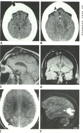
Fig. 2.-Burr holes.
A and B, CT scans of clinically uncomplicated burr holes typically show bone defects in outer
(A) and inner (B) tables.
C and D, Same patient. Sagittal (TR = 2000 msec, TE 32 msec) (C) and coronal (TR
=
2000 msec, TE=
64 msec) (D) MR images 2 days after surgery show postoperative subgaleal (ar·rows) and epidural (arrowheads) collections.
E and F, Contrast-enhanced CT scan (E) and
sagittal MR image (TR
=
2000 msec, TE=
120 msec) (F) 3 days after surgery in patient slow to regain full neurologic function. CT shows poorly defined region of decreased attenuation imme-diately subjacent to burr hole; "mushroom-shaped" region of increased signal (arrow) may result from edema secondary to deep migration of surgical instruments.isolation of the epidural space at the operative site are
impor-tant. For example, many surgeons prefer to leave the dura
open and may even suture it to the margins of the bone
defect after a craniotomy. This helps prevent brainstem compression caused by edema or hematoma after a
[image:4.612.53.404.76.639.2]AJNR:9. January/February 1988 MR OF POSTOPERATIVE CRANIUM 31
B
C
[image:5.614.58.559.77.479.2]E
F
Fig. 3.-Suboccipital craniectomy.
A-C, Asymptomatic postcraniectomy patient 6 weeks after surgery.
A, Contrast-enhanced axial CT scan. Meningogaleal complex is well seen as thin, enhancing linear structure.
B, Companion axial MR image (TR
=
2000 msec, TE=
64 msec). Dura has been left open and sutured to craniectomy margins and, therefore, is not seen (arrows).C, Sagittal MR image (TR = 500 msec, TE = 30 msec) clearly shows superior margin of craniectomy (arrowhead) and pseudomeningocele beginning
to dissect superiorly behind occipital bone (curved arrow) and inferiorly posterior to C2 (straight arrows). Dural line is not seen on MR as it has not been sutured closed. This common practice is used to avoid posterior fossa compression.
D, Unenhanced CT scan in patient without surgery shows incidental prominent cisterna magna.
E and F, Axial (TR
=
2000 msec, TE=
64 msec) and sagittal (TR=
500 msec, TE=
32 msec) (F) MR images show that this structure (arrows) resemblespostoperative pseudomeningocele. Suboccipital craniectomy may be mimicked by prominent foramen magnum or arachnoid cyst (arrows, D-F).
subdural and subarachnoid spaces at the craniectomy site,
the spread of postoperative blood, pus, or CSF is limited. Half of all postcraniotomy or postcraniectomy seizures are related to some local organic lesion such as postoperative hemorrhage, cyst, or fluid collection. Fulcamachi et al. [4] found that in 36 of 44 patients with postoperative seizures,
CT showed minimal postoperative changes compatible with normal appearances. In many cases, postoperative collec-tions can be missed easily on CT because of adjacent over-lying bone or, in the case of hematomas, because of low hematocrit. Our present series includes a relatively small number of patients with postoperative seizures; however, MR may prove to be more sensitive than CT in this situation [5]. Postsurgical arachnoid cysts may cause delayed seizures
[6]. Differentiating postoperative fluid collections from cystic recurrent tumors is a common, difficult, and important clinical problem. MR appears to enjoy some advantage over CT in this area because of its ability to identify more accurately a cystic collection as either intra- or extraaxial. Extraaxial col-lections are more likely to represent postoperative cysts, while intraaxial collections are more likely cystic tumors. Although none of the patients in our series had postoperative sonog-raphy, it has been suggested for following these patients [7].
Sonography can only be performed in the postcraniectomy patient, and even then is limited by overlying edema at the operative site. In addition, postoperative hemorrhage may be indistinguishable from residual tumor.
32
LANZIERI ET AL. AJNR:9, January/February 1988c
o
following patients after craniotomy or craniectomy. CT may
still be preferable in patients who are unable to cooperate
during the MR procedure or in whom recurrent meningioma
must be differentiated from hypertrophic scarring (Table 2).
Fig. 4.- lnfected craniectomy site.
A and B, Baseline contrast-enhanced CT scan
(A) and companion MR image (TR = 2000 msec,
TE = 64 msec) (B) show normal-appearing
post-operative findings 3 weeks after surgery. Men
-ingogaleal complex is seen as enhancing linear
structure on CT (arrow). Dura is seen on MR as linear structure of low or no signal intensity (ar·
rows).
C, Within 48 hr infected craniectomy site ex
-hibits slight thickening of meningogaleal com-plex (arrows).
D, Accompanying MR image shows dura as
poorly defined structure of mixed signal intensity
(arrows), possibly from edema and inflammatory reaction in spaces subjacent to and superficial to dura and possibly within dura itself.
Fig. 5.-Sterile granuloma. These uncommon occurrences at operative margin may be asso-ciated with wire dural tacking sutures.
A, Contrast-enhanced CT scan 7 weeks after
surgery shows nodular enhancement at margin of craniotomy (arrow).
B, MR image (TR = 2000 msec, TE = 64 msec),
shows that this surgically proved sterile granu-loma was associated with a dural tacking suture
(arrow).
[image:6.612.65.540.73.675.2]AJNR:9, January/February 1988
Fig. 6.-Postoperative cysts in patient with persistent seizures several months after
tem-porallobectomy.
A, CT scan shows apparent postoperative
ex-traaxial collection.
B, MR image (TR = 2000 msec, TE = 32 msec) reveals that this is actually an intraaxial cyst, which is separate from temporal horn (arrow).
Suboccipital pseudomeningoceles are common and expected after craniectomy.
C and D, Hypertrophic scarring within these
may mimic recurrent extraaxial tumor (arrows) on MR (TR = 500 msec, TE = 32 msec, C; TR =
2000 msec, TE = 32 msec, D).
E, Contrast-enhanced CT scan was able to
differentiate pseudomeningocele from surgically proved hypertrophic scar (arrows), while MR suggested recurrent tumor.
MR OF POSTOPERATIVE CRANIUM
B
o
E
Fig. 7.-Cysts associated with recurrent tumor 6 months after left frontal craniotomy in patient with known oligodendroglioma.
33
A, Follow-up CT study. Partially cystic, partially solid mass in left frontal region appears to be surrounded by thin rim of brain (arrows) and was
interpreted as intraaxial and part of recurrent tumor.
B, MR image (TR
=
2000 msec, TE=
32 msec) reveals that cyst is extraaxial. [image:7.612.56.567.78.682.2]34
LANZIERI ET AL. AJNR:9, January/February 1988A
B
TABLE 2: Summary of Postcraniotomy and Postcraniectomy
Patients in Whom CT Provided Additional or More Correct
Information Than MR
Status
Suboptimal MR study Recurrent tumor Hypertrophic scar
Total
No. of Patients (n = 68)
7 1
2 10
able to localize collections relative to the dura, an important
factor for the neurosurgeon contemplating intervention. A prospective evaluation of postneurosurgical patients with
sei-zures is needed to compare CT, sonography, and MR.
REFERENCES
Fig. 8.-Postoperative hematomas.
A and B, Patient who failed to regain con-sciousness after craniotomy.
A, CT scan shows large, extraaxial collection
of blood (arrow) with significant midline shift.
B, MR image shows additional large, intraaxial
hematomas (arrowheads). Importantly for sur-gical planning, MR clearly shows relationship of both collections to dura (arrows).
C and 0, Ability of MR to show relationship of postoperative hematomas and other collections to dura (black arrows) is evident on CT (C) and
MR (TR
=
500 msec, TE=
32 msec) (0) images.Extradural collection (while arrows); subdural collection (arrowheads).
1. Horowitz NH, Rizzoli HU. Post-operative complications of intracranial
neu-rological surgery. Baltimore: Williams & Wilkins, 1982:20-365
2. Lanzieri CF, Som PM, Sacher M, Solodnik P, Moore F. The
post-craniec-tomy site: CT appearance. Radiology 1986;159:165-170
3. Epstein AJ, Russell EJ, Berlin L, et al. Suture granuloma: an unusual case
of enhancing ring lesion in the post-operative brain. J Comput Assist Tomogr 1982;6(4):815-817
4. Fulcamachi A, Koizumi H, Nuleui H. Immediate post-operative seizures: incidence and computed tomographic findings. Surg Neuro/1985 ;24:671-676
5. Brant-Zawadzki M, Norman D, Newton TH, et al. Magnetic resonance of
the brain: the optimal screening technique. Radiology 1984;152:71-77
6. Luerssen TG, Kalsbeck JE. Post-surgical arachnoid cyst: report of two
cases. Neurosurgery 1983;13:438-440
7. De Slegte RGM, Kaiser MC. Sonography of the post-operative brain: a
[image:8.612.54.398.99.467.2] [image:8.612.50.294.527.595.2]