Malformations: A Clinical, Radiologic, and Histologic Study
Andreas Gruber, Peter R. Mazal, Gerhard Bavinzski, Monika Killer, Herbert Budka, and Bernd Richling
PURPOSE: To describe the pattern and time course of embolization-related tissue lesions and repermeation of the intranidal cast after endovascular embolization of cerebral arteriovenous malformations (AVMs) withN-butyl cyanoacrylate (NBCA). METHODS: We retrospectively re-viewed the records of 26 patients who were treated by endovascular embolization with NBCA and subsequent surgical extirpation to look for embolization-related tissue lesions and repermeation of the cast. A residual flow through the malformation was identified on preoperative angiograms in every case. RESULTS: Pattern and time course of embolization-related tissue lesions were typical. Until 3 months after embolization, repermeation of embolized structures did not occur. In contrast, repermeation was found in every patient who had surgery later than 3 months after the first embolization (n513; 50%). In these cases, histologic examination of the resected nidus disclosed capillary structures inside the lumen of embolized vessels. Capillaries were traced immunohisto-chemically with antibodies against membrane-bound factor VIII. No parameter other than the interval between the first embolization and surgery was found to relate to the repermeation of the cast. CONCLUSION: Intranidal recapillarization can occur later than 3 months after the first embolization with NBCA if total and solid casting of the nidus was not accomplished.
Index terms: Arteriovenous malformations, cerebral; Arteriovenous malformations, embolization
AJNR Am J Neuroradiol17:1323–1331, August 1996
Endovascular embolization has become a valuable tool in the treatment of cerebral arte-riovenous malformations (AVMs) as a result of improvements in endovascular techniques. Embolization of AVMs can be performed as de-finitive treatment (1) or as one step in a com-bined treatment with surgery (2– 4) or radiosur-gery (5). The decision as to which therapeutic tool should be used is guided by a comparison of the risks represented by the natural course of the malformation against the risks of treatment (6). For small malformations in highly func-tional areas of the brain, radiosurgery may be offered. However, at our clinic, endovascular embolization is the first-line treatment for most
patients with cerebral AVMs. It is our therapeu-tic strategy to attain a total and solid casting of the nidus, and hence a definitive treatment of the malformation, by endovascular emboliza-tion. However, definitive cure was accom-plished in only 15% of our patients by emboli-zation alone. In the remaining cases, further therapy (ie, radiosurgery or surgery) was nec-essary to obtain a definitive cure. Neurologically intact patients with lesions located in highly functional areas of the brain, however, may rep-resent an exception, as subtotal results of em-bolization can be acceptable (1). Residual he-modynamic stress, generated by remaining arteriovenous shunts, may trigger the reperme-ation of the nidus.
A residual flow through the malformation was identified on preoperative angiograms in every case in the present study. Our intention was therefore to describe the pattern and time course of embolization-related tissue lesions and repermeation of the intranidal cast after incomplete endovascular embolization with N -butyl cyanoacrylate (NBCA).
Received July 26, 1995; accepted after revision February 2, 1996. From the Department of Neurosurgery (A.G., G.B., M.K., B.R.) and the Institute of Neurology (P.R.M., H.B.), University of Vienna (Austria).
Address reprint requests to Andreas Gruber, MD, Department of Neu-rosurgery, University of Vienna Medical School, Wa¨hringer Gu¨rtel 18 –20, A-1090 Vienna, Austria.
AJNR 17:1323–1331, Aug 1996 0195-6108/96/1707–1323
qAmerican Society of Neuroradiology
Materials and Methods
Over a period of 9 years, 186 patients were treated for cerebral AVMs in our department. Of these, 96 patients were scheduled for further treatment, as nidus obliteration was still incomplete, and 90 were definitively cured by surgery, embolization, radiosurgery, or a combination of these tools. These patients remained stable at angio-graphic follow-up. Fifty-three of the cured patients had treatment consisting of endovascular embolization fol-lowed by surgical extirpation of the nidus. Of these, 27 cases were excluded from the study because either embo-lization was performed with materials other than NBCA or the whole surgical specimen was not available for histo-logic processing. Twenty-six cases in which a single or staged embolization with NBCA was done before surgery were thus available for analysis.
Microsurgical extirpation of the nidus after embolization was necessary because of the technical impossibility of achieving total endovascular cure in 15 patients (58%) with a history of bleeding; because of reperfusion of the AVM detected on follow-up angiograms after an initial total occlusion of the nidus in three patients (11.5%); because of the angiographic appearance (7, 8) and/or the easy surgical accessibility of the malformation in five cases (19%); and because of severe intracerebral bleeding dur-ing or after embolization (necessitatdur-ing emergency sur-gery) in three patients (11.5%).
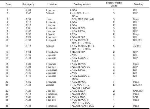
[image:2.612.62.561.303.667.2]The location of the AVMs treated, the arterial supply, the expected risk of surgical morbidity according to the Spetzler-Martin grading system, and the clinical presenta-tion are given in Table 1. According to the Spetzler-Martin grading system (9), one AVM was grade 1, six were grade 2, 11 were grade 3, six were grade 4, and two were grade 5.
TABLE 1: Clinical and neuroradiologic presentation of 26 patients with arteriovenous malformations
Case Sex/Age, y Location Feeding Vessels Spetzler-Martin
Grade Bleeding
1 M/67 R par occ R MCA 1 ICH
2 F/40 Callosal R1L ACA; R1L
PChA
3 ICH*
3 F/57 L par L ACA; MCA (M1 perf) 3 None
4 F/12 R rolandic R MCA 2 ICH
5 F/15 L par occ R MCA 2 ICH
6 F/8 R rolandic R MCA; R ACA 3 ICH
7 M/46 L par occ L MCA; L PCA 3 ICH†
8 F/30 R frontal R ACA 2 None
9 F/27 R par occ R MCA; R PCA 3 ICH
10 M/37 R trigonal R ACA; R MCA; R
PCA; R1L PChA
4 ICH, IVH
11 M/15 Callosal R ACA; R AChA; R1L
PCA; R1L PChA
5 4x ICH
12 F/51 R cerebellar R PICA; R SCA 2 ICH*
13 M/59 L rolandic L ACA 3 ICH
14 M/44 L rolandic L MCA; L ACA; L
AChA
4 ICH*
15 F/23 R rolandic R ACA; R MCA 3 None
16 M/24 R par occ R MCA; R PCA; VA 3 ICH*
17 F/32 L temporal L MCA; L PCA 3 ICH
18 M/48 L rolandic L ACA 3 ICH
19 F/18 L rolandic L MCA; L AChA; L
PChA
4 ICH
20 F/59 R rolandic R ACA; R MCA 4 None
21 M/36 Callosal R1L ACA; R1L
MCA; R1L PCA
5 ICH, IVH
22 M/26 L par occ L MCA; L ECA 2 SAH, ICH
23 M/14 R temporal R MCA; R ECA 2 None
24 F/27 L par occ L ACA; L MCA 4 SAH
25 M/24 R par occ R ACA; R MCA; R
PCA; R1L ECA
4 None
26 M/46 R temporal R MCA; R PCA; R ECA 3 None
Note.—SAH indicates subarachnoid hemorrhage; ICH, intracerebral hemorrhage; IVH, intraventricular hemorrhage; par, parietal; occ, occip-ital; ACA, anterior cerebral artery; MCA, middle cerebral artery; PCA, posterior cerebral artery; AChA, anterior choriodal artery; PChA, posterior choroidal artery; ECA, external carotid artery; VA, vertebral artery; PICA, posterior inferior cerebellar artery; SCA, superior cerebellar artery; and perf, perforating artery.
The embolizing substance used in all cases was a mix-ture of NBCA (Histoacryl Blue; Braun, Melsungen, Ger-many) and a nonionic contrast agent (iodized oil; Lipiodol ultrafluide; Guerbet, Aulnay-sous-Bois, France). In five cases, glacial acetic acid was added to the embolizing agent to decrease the velocity of polymerization without altering the viscosity (10).
Diagnostic angiography was performed when the pa-tients were medically stable unless angiograms were ob-tained at another center before the patient was transferred. Examination of the feeding vessels was performed with superselective digital substraction angiography in con-junction with embolization. The extent of angiographic nidus reduction after embolization was estimated from the final angiograms. The endovascular interventions were usually performed under local anesthesia. General anes-thesia was required only in critically ill or uncooperative patients. In every case, the partially embolized AVM nidus was resected in one session under microsurgical condi-tions. In 25 cases (96%), a total extirpation of the AVM was possible. Total extirpation was confirmed by intraoperative digital subtraction angiography (11) in each case. In one instance of a giant callosal AVM (case 25), a small residual nidus was subsequently treated with gamma knife radio-surgery. Angiographic follow-up was obtained in every patient. The clinical outcome of the patients was stratified according to the Glascow Outcome Scale, in which 1 in-dicates no deficit; 2, mild deficit; 3, severe deficit; 4, veg-etative state; and 5, death.
The resected surgical specimens were routinely fixed in buffered formaldehyde and embedded in paraffin. Serial sections were cut to a thickness of approximately 5 mm and stained with hematoxylin-eosin and Gieson’s Elastica stains. Additionally, a staining technique designed to de-pict the intranidal cast was performed, as proposed by Lundie et al in 1985 (12), which allowed clear differentia-tion between the embolizing agent and the surrounding tissue (Fig 1). Representative sections were examined by means of immunohistochemistry with antibodies against membrane-bound factor VIII to show the endothelium of the capillaries sprouting into the intranidal cast.
Results
Clinical Findings
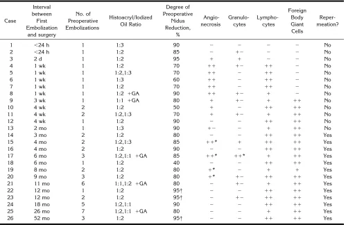
Fifty-four endovascular embolizations were performed in 26 patients. The time of tissue exposure to NBCA ranged from less than 24 hours to 52 months. The extent of angiographic nidus reduction after the final embolization is shown in Table 2. A residual flow through the malformation was present on preoperative an-giograms in every case. In no case was a solid casting of the entire nidus achieved. However, in three cases, a transient angiographic occlu-sion was followed by reperfuocclu-sion of the
malfor-mation via branches of the external carotid ar-tery 12 to 52 months after the first embolization. Total extirpation of the malformation was ac-complished in 25 cases. In one instance of a giant callosal AVM (case 25), a small residual nidus was subsequently treated with gamma knife radiosurgery. Clinical outcomes on the Glascow Outcome Scale were as follows: 17 cases (65%) were scored 1, six cases (23%) were scored 2, and one case (4%) each was scored 3, 4, and 5. Endovascular morbidity and mortality were due to severe intracerebral hem-orrhage in three patients during or after embo-lization. Surgical morbidity (ie, focal neurologic deficits following resection of the AVM) oc-curred in six cases. The higher the surgical dig-nity of the lesion, stratified according to the Spetzler-Martin grading system, the more fre-quently surgical morbidity was encountered. In contradistinction, endovascular morbidity was not related to the Spetzler-Martin grades of the lesions. Additionally, there was no correlation between outcome and degree of preoperative endovascular nidus reduction.
Preoperative embolization with NBCA facili-tated extirpation of the malformation in most cases. However, embolized feeding arteries were sometimes rigid and therefore difficult to cut. In addition, retraction of solidly casted compartments of the AVM during dissection was more difficult. In four cases, in which
[image:3.612.320.558.81.250.2]gery was performed later than 3 months after the first embolization, surgical extirpation was complicated by a diffuse perinidal capillary net-work, providing collateral blood supply to the nidus. However, these setbacks could not out-weigh the overall advantages of significantly re-duced blood loss and rere-duced operating time provided by sufficient preoperative emboliza-tion.
Embolization-Related Tissue Lesions and Repermeation of Intranidal Cast
The time course of mural angionecrosis, flammation, and repermeation of the casted in-tranidal structures is summarized in Table 2. The cases are listed according to the time of exposure to NBCA. In the case of a single em-bolization, exposure was defined as the interval between embolization and surgery. If a patient had staged embolizations, the time between the
first embolization and surgery was recorded. Each specimen was examined histologically for the degree of inflammation, mural angionecro-sis, extravasation of NBCA, and recanalization of embolized structures in relation to the time elapsed between the first embolization and sur-gery.
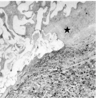
[image:4.612.63.562.362.686.2]Among embolization-related tissue lesions, inflammation (Fig 2) was seen in 25 cases (96%). Acute inflammation, comprising pre-dominantly polymorphonuclear cells, was seen as soon as 24 hours after embolization. Chronic inflammation, marked by the appearance of transmural lymphocytic infiltration and foreign body giant cells within the casted areas, was detected in 23 cases (88%). Lymphocytic infil-tration occurred 1 week after the intervention. Foreign body giant cells were not seen before 1 month after embolization, but they were detect-able for as long as 52 months. Mural angione-crosis (Fig 3) was seen as soon as 2 days after
TABLE 2: Embolization-related tissue lesions and recanalization of cast
Case
Interval between First Embolization
and surgery
No. of Preoperative Embolizations
Histoacryl/Iodized Oil Ratio
Degree of Preoperative
Nidus Reduction,
%
Angio-necrosis
Granulo-cytes
Lympho-cytes
Foreign Body Giant Cells
Reper-meation?
1 ,24 h 1 1:3 90 2 2 2 2 No
2 ,24 h 1 1:2 85 2 12 2 2 No
3 2 d 1 1:2 95 1 1 2 2 No
4 1 wk 1 1:2 70 11 12 11 2 No
5 1 wk 1 1:2,1:3 70 11 2 11 2 No
6 1 wk 1 1:3 60 11 2 11 2 No
7 1 wk 1 1:2 70 11 2 11 2 No
8 1 wk 1 1:2 1GA 90 11 12 1 2 No
9 3 wk 1 1:1 1GA 80 1 12 1 11 No
10 4 wk 2 1:2 50 1 2 11 11 No
11 4 wk 2 1:2,1:3 70 1 12 1 11 No
12 4 wk 1 1:2 90 2 2 11 11 No
13 2 mo 1 1:3 90 12 2 1 11 No
14 3 mo 2 1:2 80 2 2 11 11 Yes
15 4 mo 2 1:2,1:3 85 11* 1 11 11 Yes
16 4 mo 2 1:2 90 2 2 11 11 Yes
17 6 mo 3 1:2,1:1 1GA 85 11* 11* 1 11 Yes
18 6 mo 1 1:2 40 2 2 11 11 Yes
19 8 mo 2 1:2 80 1* 2 1 1 Yes
20 9 mo 3 1:2 80 1* 12 11 11 Yes
21 11 mo 6 1:1,1:2 1GA 80 2 12 1 11 Yes
22 12 mo 1 1:2 95† 2 2 11 11 Yes
23 12 mo 2 1:2 95† 2 12 11 11 Yes
24 18 mo 5 1:2,1:1 90 2 2 11 11 Yes
25 26 mo 7 1:2,1:1 1GA 80 2 2 1 11 Yes
26 52 mo 3 1:2 95† 2 2 11 11 Yes
Note.—GA indicates glacial acetic acid;2, no tissue reaction was observed;12, slight tissue reaction;1, moderate tissue reaction; and11, severe tissue reaction.
embolization, reaching a maximum after 1 week. Owing to fibrosis of the vessel wall, an-gionecrosis was not seen later than 2 months after embolization. In four cases, signs of necro-sis were detectable as long as 9 months after the first embolization. These patients underwent staged embolizations and had surgery within 4 weeks after the final intervention. No clear-cut extravasation of cyanoacrylate was observed in any specimen in our series.
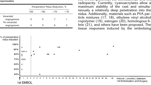
[image:5.612.62.300.82.245.2]Histologic examination of the resected nidus disclosed a repermeation of embolized vessels in 13 cases (50%). Trabeculae of connective tissue were seen bridging the lumen of initially obliterated malformed vessels (Fig 4). Inside these trabeculae, capillaries with endothelial cell lining were identified by immunohistochem-istry with antibodies against membrane-bound factor VIII (Fig 4A). Repermeation did not occur until 3 months after embolization. In contrast, intranidal recapillarization was seen in every case in which surgery was performed later than 3 months after the initial embolization. Recap-illarization of previously embolized malformed vessels was seen as long as 52 months after the first embolization (case 26). The location of the AVM did not influence the onset of reperme-ation, nor were the pattern and time course of intranidal recapillarization influenced by the Histoacryl/iodized oil ratio of the embolizing substance, the addition of glacial acetic acid to the compound (Table 3), the degree of preop-erative nidus reduction (Table 4, Fig 5), or the Spetzler-Martin grade of the malformation. In addition, lacunae without endothelial lining were identified inside the cast, representing ei-ther areas of dissolved intranidal thrombus or
Fig 3. Photomicrograph shows an area of marked mural an-gionecrosis (open star) in proximity to intraluminal cast (solid star) (hematoxylin-eosin, original magnification320).
[image:5.612.100.264.333.502.2]Fig 4. Photomicrographs of an NBCA-occluded malformed vessel 26 months af-ter embolization. Angiogenic repermeation of the casted lumen is clearly demon-strated with immunohistochemistry for factor VIII (A) and Gieson’s Elastica stain (B). Trabeculae of connective tissue and embedded vascular channels (arrows), bridging the original vessel lumen (stars), are identified (original magnification340). Fig 2. Photomicrograph of vascular channel containing
[image:5.612.63.393.550.727.2]artifacts caused by processing of the surgical specimens.
Discussion
Endovascular embolization of cerebral AVMs will lead to definitive cure if complete oblitera-tion of the nidus is achieved and patients remain stable at follow-up angiography. Repermeation of a previously embolized malformation
there-fore represents a major threat to the quality of the treatment. Recanalization after embolization with NBCA was previously demonstrated by Brothers et al (13) in domestic swine. In their series, repermeation of casted rete mirabile was encountered as early as 21 days after emboli-zation. The surgical specimens of 10 human cerebral AVMs were investigated by Klara et al (14), with both light and scanning electron mi-croscopy. In their series, surgery was not per-formed earlier than 3 weeks after embolization with isobutyl 2-cyanoacrylate (IBCA). Endothe-lial proliferation was documented immunohisto-chemically by Schweitzer et al (15) in 18 hu-man cerebral AVMs embolized with either IBCA, polyvinyl alcohol (PVA) particles, or Avitene. Our study provides a long-term histologic fol-low-up of human cerebral AVMs embolized with NBCA.
Embolization-Related Tissue Lesions
The ideal embolizing agent ought to meet the criteria postulated by Clarisse et al in 1976 (16): (a) controllable viscosity, (b) elasticity of the substance after setting, (c) in vivo stability, (d) biocompatibility, (e) sterilizability, and (f) radiopacity. Currently, cyanoacrylates allow a maximum stability of the cast and simulta-neously a relatively deep penetration into the nidus. Additionally, materials such as PVA par-ticle mixtures (17, 18), ethylene vinyl alcohol copolymer (19), estrogen (20), homologous fi-brin (21), and others have been proposed. The tissue responses induced by the embolizing
[image:6.612.63.302.236.366.2]Fig 5. Angiogenic repermeation of intranidal NBCA. Specimens are plotted according to their interval between first embolization and surgery (months) and according to the extent of preoperative endovascular nidus reduction (percent). The graph demonstrates the positivecorrelation between repermeation and interval between first embolization and surgery and the negativecorrelation between repermeation and extent of preoperative endovascular nidus reduction. Repermeation did not occur earlier than 3 months after the first embolization.Asterisksindicate specimens with histologically proved repermeation of NBCA;circles, specimens without repermeation. TABLE 3: Influence of embolic material on angiogenic
reperme-ation
Embolic Material
Histoacryl/ Iodized Oil Ratio of
1:2
Histoacryl/ Iodized Oil Ratio Other
than 1:2
Histoacryl/ Iodized Oil
Plus Glacial
Acetic Acid
Angiogenic
repermeation 8 2 3
No angiogenic
[image:6.612.63.559.398.678.2]repermeation 6 5 2
TABLE 4: Influence of preoperative nidus reduction on angiogenic repermeation
Preoperative Nidus Reduction, %
100 .90 .70 ,70
Intranidal
angiogenesis 0 5 7 1
No intranidal
substance need to be relatively mild, as the cast is intended to remain in vivo for a lifetime. Heat production in the initial exothermic polymeriza-tion and release of formaldehyde during the breakdown process of cyanoacrylates have been described as causative factors for the em-bolization-related tissue lesions encountered (22, 23). Some degree of mural inflammation, however, seems to act in synergy with the em-bolizing substance to render permanent occlu-sion of the nidus through the formation of scars and fibrosis. This study emphasizes that the tis-sue responses to NBCA can be considered rel-atively benign, as angionecrosis was replaced by fibrosis of the vessel wall within 2 months and acute inflammation developed into a chronic lesion within 1 month after emboliza-tion. The observation of a typical pattern and time course of angionecrosis and inflammation after embolization (Table 2) unites our results with those of previous studies (13, 24). In con-trast, such previously reported lesions as intima loss, shrinking and calcification of the media, intramural hemorrhage, extravasation of glue, and inflammation of neuronal tissue after em-bolization with cyanoacrylates other than NBCA (13, 24 –32) were not encountered in our series.
Repermeation of Embolized Malformed Vessels
Collateralization—that is, the dilatation of preexistent collateral vessels, reconstituting the blood flow to the AVM nidus (33)—is an impor-tant mechanism for repermeation of embolized malformations. This phenomenon usually oc-curs in the perilesional tissue, where the number of preexisting vessels relates to the anatomic location of the nidus. Maximum angiographic reperfusion after embolization has been de-scribed for occipital AVMs, even after total oc-clusion (33). Our study indicates that recapil-larization of the embolized nidus is not influenced by the anatomic location of the AVM. In addition, resorption of intranidal cyanoacry-late has been reported (34). Although resis-tance to biodegradation may be altered by a change in the Histoacryl/iodized oil ratio of the compound, our study produced no evidence that either this ratio or the addition of glacial acetic acid influences the onset of angiogenic repermeation (Table 3).
Previous reports (31, 35) emphasize that po-lymerized glue occludes only a part of the
in-jected lumen while the remainder is filled with thrombus. In the absence of solid casting of the entire nidus with glue, repermeation of embo-lized vessels can occur by recapillarization of intraluminal thrombus. The release of angio-genic cytokines, promoting the recapillarization of thrombus, is triggered by ischemia, inflam-mation, and changes in shear stress to the vas-cular endothelium. Because endovasvas-cular em-bolization with permanent materials like NBCA induces intranidal ischemia, the question arises whether ischemia is causally related to the re-capillarization of partially embolized malforma-tions. The same may be true for embolization-related inflammation, as vasoactive factors such as vascular endothelial growth factor (36 – 38), fibroblast growth factor (39, 40), trans-forming growth factor (41, 42), platelet-derived growth factor (43, 44), granulocyte-macroph-age colony-stimulating factor (45, 46), interleu-kin 6 (47), and tumor-necrosis factor (48) were shown to be produced by leukocytes, predom-inantly activated macrophages and T lympho-cytes (49). In addition, vascular endothelial shear stress, the rheological force shearing the luminal surface of the blood vessel when a vis-cous fluid such as blood flows over it, is altered but not abolished in partially embolized malfor-mations.
Clinical Implications
The most important finding of the present study was that angiogenic repermeation of in-tranidal NBCA is possible as soon as 3 months after the first embolization. This finding may have implications for the timing of subsequent surgery. Physicians involved in the treatment of AVMs should be aware of a potential for angio-genic repermeation of the nidus later than 3 months after the first embolization. As histo-logic repermeation had no prominent angio-graphic correlate, the definitive clinical signifi-cance of this finding, however, needs to be clarified.
The question of whether angiogenic reperme-ation occurs only in subtotally embolized le-sions or also in totally casted malformations is still unclear. We had no patients with permanent total nidus obliteration who went on to surgery, as these subjects were considered definitively cured if stable at angiographic follow-up. In contradistinction, the patients reported in the present study all went on to surgery, as solid casting of the entire nidus was not accom-plished. In this group, 23 patients had preoper-ative nidus reductions of 40% to 95% after the last embolization. As pointed out previously, residual hemodynamic stress induced angio-genic repermeation of the cast in 10 of these cases. In the remaining three patients, total an-giographic obliteration of the malformation was obtained immediately after treatment. Reperfu-sion of the nidus via branches of the external carotid artery was, however, seen 12 to 52 months later at follow-up angiography. The density of the intranidal cast was erroneously overestimated in these cases and dilatation of collateral vessels (32) redirected blood through the incompletely casted nidus. There was con-siderable time for recapillarization, as none of these particular three lesions was resected ear-lier than 12 months after the first embolization. This finding is in agreement with reliable angio-graphic follow-up studies (54), demonstrating stable angiographic results only in cases with total and solid casting of the nidus. The clinical significance of our findings could be clarified if human cerebral AVMs, totally casted with NBCA, were examined histologically for signs of angiogenic recapillarization.
Conclusion
Twenty-six surgical specimens of human ce-rebral AVMs resected less than 24 hours to 52 months after embolization were examined his-tologically. Pattern and time course of emboli-zation-related tissue lesions were typical. Recapillarization of previously embolized mal-formed vessels was demonstrated in every case in which surgery was performed later than 3 months after the first embolization. It is sug-gested that nidus repermeation may act in syn-ergy with recruitment of collateral vessels in the reperfusion of embolized malformations.
Acknowledgment
We thank H. Flicker for her excellent technical assis-tance.
References
1. Picard L, Moret J, Lepoire J. Endovascular treatment of intrace-rebral arteriovenous malformations.J Neuroradiol1984;11:9 –28 2. Deruty R, Lapras C, Pierluca P, Patet JD, Pialat J, Garcia C. Embolisation preoperatoire des malforations arterio-veineuses cerebrales par le butyl-cyanoacrylate.Neurochirurgie 1985;31: 21–29
3. Spetzler RF, Martin N, Carter LP, Flom R, Raudzens P, Wilkinson E. Surgical management of large AVMs by staged embolization and operative excision.J Neurosurg1987;67:17–28
4. Vinuela F, Dion J, Duckwiler G, et al. Combined endovascular embolization and surgery in the management of cerebral arterio-venous malformations: experience in 101 cases. J Neurosurg 1991;75:856 – 864
5. Lunsford LD, Kondziolka D, Flickinger J, Bissonette D. Stereotac-tic radiosurgery for arteriovenous malformations of the brain.J Neurosurg1991;75:512–524
6. Hamilton MG, Spetzler RF. The prospective application of a grad-ing system for arteriovenous malformations.Neurosurgery1994; 34:2–7
7. Marks M, Lane B, Steinberg G, Chang P. Hemorrhage in intrace-rebral arteriovenous malformations: angiographic determinants. Radiology1990;176:807– 813
8. Vinuela F, Nombela L, Roach M, Fox A, Pelz D. Stenotic and occlusive disease of the venous drainage system of deep brain AVMs.J Neurosurg1985;63:180 –184
9. Spetzler RF, Martin N. A proposed grading system for arterio-venous malformations.J Neurosurg1986;65:476 – 483
10. Spiegel SM, Vinuela F, Goldwasser JM, Fox AJ. Adjusting the polymerization time of isobutyl-2-cyanoacrylate. AJNR Am J Neuroradiol1986;7:109 –112
11. Hieshima GB, Reicher MA, Higashida RT, Halbach VV. Intraoper-ative digital subtraction neuroangiography: a diagnostic and ther-apeutic tool.AJNR Am J Neuroradiol1987;8:759 –767 12. Lundie MJ, Ellyatt D, Kaufmann JCE, Vinters HV. Staining
13. Brothers MF, Kaufmann JC, Fox AJ, Deveikis JP.n-Butyl 2-cy-anoacrylate: substitute for IBCA in interventional neuroradiology: topographic and polymerization time study.AJNR Am J Neuro-radiol1989;10:777–786
14. Klara PM, Eugene DJ, McDonnell DE, Pevsner PH. Morphological studies of human arteriovenous malformations: effects of isobutyl 2-cyanoacrylate embolization.J Neurosurg1985;63:421– 425 15. Schweitzer JS, Chang BS, Madsen P, Vinuela F, Martin NA,
Vin-ters HV. The pathology of arteriovenous malformations of the brain treated by embolotherapy, II: results of embolization with multiple agents.Neuroradiology1993;35:468 – 474
16. Clarisse J, Gozet G, Cornil J, Jomin P, Delandsheer J, Laines E. Les emboles plastiques fluides: etude experimentale et rapport de deux cas cliniques de fistules arterio-veineuses traintees.J Neu-roradiol1975;2:29 –38
17. Germano LM, Davis RL, Wilson CB, Hieshima GB. Histopatholog-ical follow up study of 66 cerebral arteriovenous malformations after therapeutic embolization with polyvinyl alcohol.J Neurosurg 1992;76:607– 614
18. Lylyk P, Vinuela F, Vinters HV, Dion J, Duckwiler C, Lin T. Use of a new mixture for embolization of intracranial vascular malforma-tions.Neuroradiology1990;32:304 –310
19. Taki W, Kikuchi H, Iwata H, Nishi S. Embolization of arteriovenous malformations using EVAL mixture (a new liquid embolization material).Neuroradiology1991;33(Suppl):195–196
20. Suzuki J, Komatsu S. New embolization method using estrogen for dural arteriovenous malformations and meningiomas.Surg Neurol1981;16:438 – 442
21. Richling B. Homologous controlled viscosity fibrin for endovascu-lar embolization.Acta Neurochir(Wien) 1982;62:159 –170 22. Mickey BE, Samson D. Neurosurgical applications of the
cyano-acrylate adhesives.Clin Neurosurg1981;28:429 – 444
23. Pevsner PH, George ED, Doppmann JL. Interventional radiology polymeric update: acrylic.Neurosurgery1982;10:314 –316 24. Vinters H, Lundie MJ, Kaufmann J. Long term pathological follow
up of cerebral arteriovenous malformations treated by emboliza-tion with bucrylate.N Engl J Med1986;314:477– 483
25. Diaz FG, Mastri AR, Chou SN. Neural and vascular tissue reac-tions to aneurysm coating adhesive (ethyl-2-cyanoacrylate). Neu-rosurgery1978;3:45– 49
26. Green AR, Milling MA. Butyl cyanoacrylate adhesives in micro-vascular surgery: an experimental pilot study.J Reconstr Micro-surg1986;2:103–105
27. Hood T, Mastri A, Chou S. Neural and vascular tissue reactions to cyanoacrylate adhesives: a further report.Neurosurgery1982;11: 363–366
28. Lanman TH, Martin NA, Vinters HV. The pathology of encephalic arteriovenous malformations treated by prior embolotherapy. Neuroradiology1988;30:1–10
29. Vinters H, Galil K, Lundie M, Kaufmann JCE. The histotoxicity of cyanoacrylates, a selective review.Neuroradiology1985;27: 279 –291
30. Vinters H, Debrun G, Kaufmann J, Drake C. Pathology of arterio-venous malformations embolized with isobutyl-2- cyanoacrylate (bucrylate).J Neurosurg1981;55:819 – 825
31. Zanetti P, Sherman F. Experimental evaluation of a tissue adhe-sive as an agent for the treatment of aneurysms and arteriovenous abnormities.J Neurosurg1972;36:72–76
32. Zumpano J, Jacobs L, Hall J, Margolis G, Sachs E Jr. Bioadhesive and histotoxic properties of ethyl-2-cyanoacrylate.Surg Neurol 1982;18:452– 457
33. Fournier D, Terbrugge K, Rodesch G, Lasjaunias P. Revascular-ization of brain arteriovenous malformations after embolRevascular-ization with bucrylate.Neuroradiology1990;32:497–501
34. Rao VRK, Mandalam KR, Gupta AK, Kumar S. Dissolution of isobutyl 2-cyanoacrylate on long-term follow-up. AJNR Am J Neuroradiol1989;10:135–141
35. Vinuela F, Fox AJ, Pelz D, Debrun G. Angiographic follow up of large cerebral AVMs incompletely embolized with isobutyl-2-cy-anoacrylate.AJNR Am J Neuroradiol1986;7:919 –925
36. Neufeld G, Tessler S, Gitay-Goren H, Levi BZ. Vascular endothe-lial growth factor and its receptors.Prog Growth Factor Res1994; 5:89 –97
37. Shweiki D, Itin A, Soffer D, Keshet E. Vascular endothelial growth factor induced by hypoxia may mediate hypoxia induced angio-genesis.Nature1992;29:843– 845
38. Brogi E, Wu T, Namiki A, Isner JM. Indirect angiogenic cytokines upregulate VEGF and bFGF gene expression in vascular smooth muscle cells, whereas hypoxia upregulates VEGF expression only.Circulation1994;90:649 – 652
39. Weich HA, Iberg N, Klagsbrunn M, Folkman J. Transcriptional regulation of basic fibroblast growth factor gene expression in capillary endothelial cells.J Cell Biochem1991;47:158 –164 40. Villaschi S, Nicosia RF. Angiogenic role of endogenous basic
fibroblast growth factor released by rat aorta after injury.Am J Pathol1993;143:181–190
41. RayChaudhury A, D’Amore PA. Endothelial cell regulation by transforming growth factor-beta.J Cell Biochem1991;47:224 – 229
42. Iruela-Arispe ML, Sage EH. Endothelial cells exhibiting angiogen-esis in vitro proliferate in response to TGF beta-1.J Cell Biochem 1993;52:414 – 430
43. Risau W, Drexler H, Mironov V, Heldin CH. Platelet derived growth factor is angiogenic in vivo.Growth Factors1992;7:261–266 44. Sato N, Beitz JG, Kato J, Raymond A, Frackelton AR Jr. Platelet
derived growth factor stimulates angiogenesis in vitro. Am J Pathol1993;142:1119 –1130
45. Sunderkotter C, Steinbrink K, Goebeler M, Bhardwaj R, Sorg C. Macrophages and angiogenesis.J Leukoc Biol1994;55:410 – 422 46. Sunderkotter C, Goebeler M, Schulze-Osthoff K, Bhardwaj R, Sorg C. Macrophage-derived angiogenesis factors. Pharmacol Ther 1991;51:195–216
47. Rosen EM, Liu D, Setter E, Bhargava M, Goldberg ID. Interleu-kin-6 stimulates motility of vascular endothelium.EXS1991;59: 194 –205
48. Fajardo LF, Kwan HH, Kowalski J, Prionas SD, Allison AC. Dual role of tumor necrosis factor-alpha in angiogenesis.Am J Pathol 1992;140:539 –544
49. Ribatti D, Loria MP, Tursi A. Lymphocyte induced angiogenesis: a morphometric study in the chicken embryo chorioallantoic mem-brane.Acta Anat(Basel) 1991;142:334 –338
50. Risau W. Angiogenesis and endothelial cell function. Arzneimit-telforschung1994;44:416 – 417
51. Ando J, Kamiya A. Blood flow and vascular endothelial cell func-tion.Front Med Biol Eng1993;5:245–264
52. Gimbrone MA Jr. Platelet-derived growth factor beta chain pro-moter contains a cis-acting fluid shear-stress-responsive element. Proc Natl Acad Sci U S A1993;90:4591– 4595
53. Ciricillo SF, Edwards MS. Occult malformations with anomalous venous drainage (letter).J Neurosurg1995;82:311–312 54. Wikholm G. Occlusion of cerebral arteriovenous malformations