With 13 text-figures Printed in Great Britain
STATOCYST-INDUCED EYE MOVEMENTS IN THE
CRAB SCYLLA SERRATA
III. THE ANATOMICAL PROJECTIONS
OF SENSORY AND MOTOR NEURONES AND THE RESPONSES OF THE MOTOR NEURONES
BY D. C. SANDEMAN AND A. OKAJIMA*
Department of Neurobiology, Research School of Biological Sciences, Australian National University, Canberra, A.C.T., Australia
(Received 12 December 1972)
INTRODUCTION
The detection of horizontal rotational acceleration by crabs follows the same principle as that of vertebrates in that the crab statocyst is equipped with a horizontal circular canal containing fluid and sets of receptor hairs which detect displacements of the fluid. If a blind but otherwise intact crab is rotated about its vertical axis, its stalked eyes undergo the typical slow forward and quick return phases of nystagmus. We have found that displacement of the canal fluid, even in isolated eye-brain preparations of the crab, is an adequate stimulus for the production of nystagmus (Sandeman & Okajima, 1972a, b). The crab therefore provides an opportunity to study, in a fairly simple way, the neural mechanisms of a basic eye movement reflex which is common to most animals with movable eyes.
The structure of the statocyst canals and the qualitative responses of the different groups of receptor hairs in the statocyst are described in the first paper of this series. The similarity between the optokinetic and statocyst-induced eye movements and the responses of the eye muscles in isolated preparations are dealt with in the second paper (Sandeman & Okajima, 1972a, b). The purpose of this study is (1) to examine the anatomical projections of the statocyst and oculomotor nerves and (2) to explore the physiological responses and relationship between antagonist eye-muscle systems during statocyst-evoked nystagmus.
We have found that the branches of the sensory neurones of the statocyst and the motor neurones of the eye muscles are close to one another in the brain and are confined to the side of the brain ipsilateral to their nerve trunks. Different hair receptors in the statocyst have nerve cells of different sizes and shapes associated with them. Electrical recordings from the motor neurone cell bodies produced evidence of direct inhibition of motor neurones during the fast phase of nystagmus. Direct inhibition of motor neurones during the slow phase could not be convincingly demon-strated but is not excluded. The outputs to the antagonist muscles are balanced against one another and this balance is maintained both during a stimulus and in its absence.
• Present address: Biological Institute, Yokohama City University, Yokohama, Japan.
i 8 D. C. SANDEMAN AND A. OKAJIMA Microelectrode
Eye clamp
Fig. i. The isolated eye-brain preparation showing the placement of the stimulating pipettes in the horizontal canals of the statocyst, the microelectrode for motor neurone cell somata recordings and the extracellular electrodes supporting branches of the oculomotor nerve. The oculomotor nerve branches have been dissected away from the individual muscles in the eye.
MATERIAL AND METHODS
The Australian mud crab, Scylla serrata, was used, and the details of the preparation are the same as those reported previously. The statocyst stimulator which allows a small controlled displacement of the fluid within the statocyst canals in either direction is also described in a previous paper (Sandeman & Okajima, 19726).
Electrical recordings were taken intracellularly (by micropipettes filled with 3M-KCI) from the somata of the oculomotor neurones and extracellularly from specific branches of the oculomotor nerves at a point near where they run into the respective muscles. The extracellular recordings were made by supporting the nerve branches on pairs of wire hooks surrounded by a wire ring containing a drop of mineral oil (Fig. 1).
Recordings of eye-muscle potentials were also made on some occasions by inserting fine wires, insulated except at the tips, into the muscles.
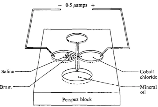
— 0-5 /(amps +
Saline
Brain
Cobalt chloride Mineral
[image:3.451.94.352.63.238.2]oil
Fig. 2. The cobalt chloride electrophoresis bath. Mineral oil in the upper and lower chambers flows along the groove linking them, electrically insulating the two lateral chambers from each other. The whole brain is placed in saline in the left-hand chamber and the nerve branch is drawn across the mineral oil gap and immersed in a 1:1 mixture of saline and 10% aqueous solution of cobalt chloride. Platinum wires dipped into the lateral chambers carry a current which is regulated to be 0-5 /AA or less.
maintained for 3-4 h produced the best results, but the range and size of axon dia-meters in the nerve appears to be an important factor in the success of the method. For example, in a nerve bundle where the axon diameters are all similar the distribu-tion of cobalt chloride is fairly uniform, but in a nerve bundle containing a few large axons and many fine axons only the large axons will take up the cobalt chloride. We were unable to get cobalt chloride to go any distance down very fine nerves (<^ 2 fim) such as those in the olfactory bundle of the antennular nerve. Application of electro-phoretic currents stronger than 0-5 /jamps produced an abrupt stoppage of the cobalt half way along the axons. Considerable diffusion of the cobalt chloride along the axons occurs without any forcing current but we found that without a small current the finer branches of neurones were not as satisfactorily filled. The cobalt chloride will sometimes contaminate neighbouring nerve branches, and to obtain the clean prepara-tions such as we have figured in this paper may require 10-30 trials.
B
Free hook hair receptor
cells Thread
hair bases
Free hook" /Statolith\
hair nerve ftreceptors- III B ar cells
Fig. 3. Bipolar cells associated with the hair bases of the receptor hairs. 3 A, 47 nerve cells have been filled with cobalt sulphide and these lie opposite 30 thread hair bases. 3 B, Bipolar cells of the free hook hairs are larger and rounder than those of the thread hairs and similar to most of the cells in the statolith area (3 C), with the exception of the very large bipolar cells which are probably associated with an outer circle of statolith hook hairs. The central projections of the numbered receptor nerves are given in Table 1.
RESULTS
During the fast and slow phases of nystagmus all but two of the eleven eye muscles of the crab change their pattern of activity in some way (see Burrows & Horridge, 1968, for details). The activity of three muscle blocks, numbers 20a, 21 and 19b (numbering according to Cochrane, 1935) is particularly relevant to the study of nystagmus because their activity is closely correlated with the position of the eye in the horizontal plane. Muscle 20 a moves the eye away from the midline and is active during both slow and fast movements in that direction but is silent when the eye is moved in the opposite direction by muscles 21 and 19b. Similarly, muscles 21 and 19b are silent when 20 a is activated. None of the other eye muscles exhibit the same clear association between their activity and the horizontal movements of the eyes.
What follows is a description of the anatomy and physiology of the eye-movement system with particular reference to these muscles and the sense organ which activates them.
Anatomy
1. Sensory nerves
There are three types of sensory hairs - thread hairs, free hook hairs and statolith hairs - arranged in definite areas in the lumen of the statocyst. Cobalt perfusion has shown that the different hairs are associated with sensory cells of different sizes.
II
in
Fig. 4. The lower part of the vertical canal of a statocyst showing the origins of the various statocyst nerve bundles. Cross-sections of all the bundles of the antennular nerve are shown on the right of the figure, in which the relative sizes and numbers of the axons contained in the bundles are indicated. The numbers and characteristics of the nerve bundles are almost exactly the same in different animals. Details of the origins and destinations of all the nerve bundles are given in Table 1.
Table 1. The subdivisions of the antennular nerve and their central projections
Category
I
II
I I I
Sub-division A B A B C D A B C D
Type of nerves
Mechanosensory and motor Olfactory
Thread hair sensory Free hook hair sensory Unknown sensory Motor
Inner hook hair sensory
Outer hook hair sensory and pre-sumably mechanosensory Sensory from lateral part of basal
joint of antennule Motor
Terminal region of central pro-jection of sensory nerves
— —
D i , D2, E2, F2, G2, G3 E2, F 2 . F 3
— —
D I . E I
F i , F 2
F i , F2
The free hook hair receptor cells are more globular in shape and are larger than the thread hair receptors. The bases of the free hook hairs are not arranged in orderly rows so that an accurate count of hair bases and associated cells was not possible (Fig. 3 B). However, the distal processes of some bipolar cells were seen to fuse and may terminate beneath a single hair.
22
Anterior 3 4 5
Anterior 3 4 5
Oc^ophagcul connective
I _ | I
Posterior
Ooophiigcal connective
Tcgumcnl
2J0/im
Posterior
much larger bipolar cells associated with them. In ten separate preparations we observed only one large cell to each outer hair but there are more small cells than inner hair bases (Fig. 3 C). We could not find any sensory cells belonging to the very fine statolith hairs which lie directly beneath the statocyst.
The bundles of axons from the thread hairs, free hook hairs and statolith area main-tain their integrity in the antennular nerve (Fig. 4 and Table 1), and although they fuse when they reach the brain we can trace the peripheral and central projections of known cell groups by electrophoretically forcing cobalt chloride along the axons of selected bundles of the antennular nerve. The central projections of these nerves are mapped in relation to a grid superimposed on a diagram of the brain where the spaces between the grid lines represent 250 /im (Fig. 5).
The axon arborizations of the thread hair receptors have the widest distribution in the brain when compared with the other statocyst receptors and cover almost the whole of the dendritic field of the oculomotor neurones (see below). The arborizations are confined to the dorsal part of the brain and no branches were traced to the contra-lateral side. A slender bundle of axons approaches to within 20-30 jim of the midline and fine unfilled branches from this bundle may spread to the other side, although we never observed this (Fig. 5 A).
The free hook hair nerve bundle contains more large-diameter sensory axons than any of the sensory nerves and these have a fairly limited projection, with most axons ending in squares E2 and F2 of the grid and a subsidiary branch in square F3 (Fig. 5B). Like the thread hairs, the free hook hair projections are confined to the ipsilateral part of the brain.
Two main bundles of the nerve from the statolith area can be distinguished. The first (A in category III in Fig. 4) contains axons from the inner circlet of statolith hook hairs, and like the free hook hairs and thread hairs these project to the dorsal ipsilateral part of the brain but here they spread more medially and anteriorly than the fibres of the free hook hairs and thread hairs (D1 and E1, Fig. 5 C). The second bundle of the statolith nerve (B in category III in Fig. 4) contains large-diameter axons from the large bipolar receptors associated with the outer circlet of statolith hook hairs. The bundle also carries a number of smaller axons which probably come from mechano-receptors on the outer surface of the basal joint of the antennule. The central projec-tions of these axons is significantly different from other statocyst receptors in that they end in the ventral and medial part of the brain with the projections of mechano-receptors in the oculomotor and tegumentary nerve branches (Fig. 5 C).
Axons of mechanoreceptors in the distal joints of the antennule (category I in Fig. 4) project to the ventral part of the brain and the fine olfactory fibres enter the outer layers of the olfactory lobe.
Op and ( : Anterior ic tra xulo lerve HA
Ct j . «
motor
Ml
Dorsal Sensory neurones / ~ & ^ M o t o r new
/— s UIB/C | ! ones
i—
2tJr5
250/an H VentralFig. 6. Lateral view of the central projections of the sensory and motor oxons of the eye and the statocyst. Most sensory axons from the statocyst project dorsally and end in close proximity to the dendrites of the oculomotor neurones. A group of axons which come from the large bipolar cells in the statocyst region project ventrally and share this area with the sensory neurones in the oculomotor nerve. The grid system in this figure corresponds with that of Figs. 5 and 7 and the projections shown are an accurate representation of their real locations in the brain.
2. Motor nerves
Motor neurones in the nerve bundles are identified by the location of their cell bodies in the brain, and a number of motor neurones in the antennular nerve were identified in this way. All of these have their cell somata located on the ventral side of the brain and their dendritic fields distributed dorsally, ipsilaterally and contra-laterally.
H
[image:9.451.28.417.75.583.2]Posterior
26 D. C. SANDEMAN AND A. OKAJIMA
^*J^*«m%
m^*>>>A«i>«MJ^
! j
i i
^A«~*~~^^^
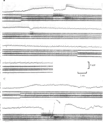
5mV
1 sec
Fig. 8. Intracellular responses of an oculomotor neurone which did not show any marked direc-tional sensitivity to statocyst stimulation but which was strongly inhibited when nerves con-taining sensory inputs to the eye-withdrawal system were electrically stimulated. The asterisk marks the stimulus to the oculomotor nerve which carried sensory axons from mechanoreceptors on the eye. An antidromic action potential appears and is followed by a hyperpolarization and increased synaptic activity. This inhibition is fairly long-lasting in spite of being initiated by a single shock. In 8 B it can be seen that the inhibition will temporarily over-ride spikes pro-duced by direct polarization of the cell soma. The arrows show the point of application and removal of the depolarization, and the asterisk marks the point at which the oculomotor nerve is stimulated.
Physiology
Recordings were always taken from the right-hand side and the motor neurones referred to here all belong to the right eye.
Penetration of an oculomotor neurone is accompanied by the registration of a resting potential of 48-54 mV and the appearance of small synaptic potentials. Many oculomotor neurones were found to be spontaneously active, and attenuated axon spikes are recorded in the cell bodies. Coincidence observed between the action potentials in the cell body with those recorded from a peripheral branch of the oculomotor nerve going to a known muscle allows positive identification of the impaled cell body.
1. Generation of slow and fast phases of nystagmus
Very few motor neurones give no response to statocyst stimulation and most can be clearly divided into those which increase their firing frequency irrespective of the direction of fluid displacement in the statocyst and those which increase their rate of firing only when the statocyst fluid is displaced in one particular direction. A pro-portion of the first group of neurones are strongly inhibited during stimulation of the tegumentary or oculomotor nerve of the same side. This inhibition must be directly applied to the motor neurone because the membrane potential becomes hyper-polarized and repeated IPSPs appear following the stimulus (Fig. 8).
The directdonally sensitive motor neurones are not inhibited by tegumentary or oculomotor nerve stimulation. They can be further identified as 20 a or 21 type neurones depending upon the particular direction of statocyst fluid displacement which produces excitation. Clockwise rotation of statocyst fluid for example is known to excite no. 20 a motor neurones of the right eye while anticlockwise rotation of the fluid excites no. 21 motor neurones. In addition these neurones show a characteristic break in the excitation which is interpreted to be the fast phase of nystagmus (Sande-man & Okajima, 19726).
The responses of two directionally sensitive motor neurones are shown in Fig. 9. Taking the tonic no. 20 a type unit in Fig. 9 A as an example it can be seen that after the stimulus is applied the unit increases its frequency from 7 impulses/sec to 40 impulses/sec. The increase is accompanied by a general depolarization of the mem-brane. Three seconds after the onset of the stimulus there is a sudden strong repolari-zation of the membrane which reaches its original level in about 100 msec and spike activity is abolished. The repolarization coincides with a burst of activity in a motor unit in the nerve going to muscle 21 and a complete cessation of at least five motor units (identified by spike amplitude) in the nerve to muscle 20 a. In this preparation all the branches of the oculomotor nerve were cut peripherally and there was no possibility of any sensory feedback from the eye.
The cessation of spike activity in the 20 a motor neurones lasts for about 200 msec after which there is a renewed excitation which reaches a peak of 80 impulses/sec. The re-excitation of the 20 a motor neurone coincides with a temporary cessation of the activity in the 21 motor neurones, but 1-5-2 sec later no. 21 begins to discharge again, this time at a significantly higher rate than before the stimulus had been applied.
Reversal of the stimulus slows down the rate of the 20 a neurones and increases that of the 21 motor neurones. The membrane potential of the 20 a motor neurone is repolarized with the change in frequency.
i ; ! i ! ! i : f
5mV
lsec
Fig. 9 B). The fast phase is accompanied by a repolarization of the motor neurone and followed by a resumption of excitation. Reversal of the stimulus repolarizes the membrane gradually to its original level and there is a clear decrease in the number of EPSPs. There is no significant difference in the response of 21 type neurones when compared with the 20 a type neurones apart from the obvious one of directional sensitivity.
The rate of a motor neurone's discharge can be altered by hyperpolarizing or depolarizing the neurone through the electrode in its cell body. Depolarization pro-duces the repetitive firing common to crustacean motor neurones but never led to a fast phase nor did it have any effect on the rate of firing of the antagonist motor neurones. Statocyst stimulation applied to depolarized motor neurones produces the same slow and fast responses. The only observable difference is that the peak discharge frequency of about 80 impulses/sec was achieved earlier and maintained for a longer period before the fast phase occurred (Fig. 10 A).
Hyperpolarization slows down or stops the firing of a tonic unit but if applied in conjunction with a stimulus to the statocyst it will not prevent the occurrence of a fast phase. A tonic unit, hyperpolarized to the extent that its resting discharge is just blocked and then subjected to an input from the statocyst, gives the characteristic slow and fast response except that the fast phase occurs when the unit has reached only 20 impulses/sec instead of its normal 40 impulses/sec (Fig. 10B). A further hyperpolarization of this neurone results in an almost complete block during slow phase excitation, but a fast phase is still produced (Fig. 10C). The longer delay between the onset of the stimulus and the appearance of the fast phase is more likely to be caused by adaptation than by the hyperpolarization of the cell because the same delays can be produced by repeated application of the statocyst stimulus to preparations in which a motor neurone is depolarized.
Further evidence of there being no direct correlation between the frequency of any one motor neurone and the occurrence of the fast phase is provided by stimulation of only one statocyst. Only the ipsilateral statocyst was stimulated in one preparation and this produced a motor neurone discharge of 40 impulses/sec but without a fast phase occurring. The same motor neurone, subject to bilateral statocyst stimulation, underwent a fast phase after the firing rate had reached only 8-10 impulses/sec (Fig. 11).
Finally, antidromic stimulation of one set of motor neurones was never seen to have any effect on the discharge frequency of the antagonists.
Direct polarization of a single neurone through its cell body will affect its own spike frequency but not that of its synergists. Hyperpolarization of the cell eventually blocks the action potentials but does not prevent the appearance of EPSPs which are synchronous with the bursts of the synergist cells (Fig. 10).
2. Interaction between antagonist motor systems
Sfi
4 riii
5 ZSXS&
I • •
m i M m i
i m n j i i i ! n i
5 ,"!
5 mV
[image:14.451.47.401.54.376.2]1 see
Fig. ioA. For legend sec facing page.
fast phase occurs (as in Fig. 12 A) the final frequencies of both sides may be higher than what they were originally. In some cases the fast phase is followed by fluctuations in the motor neurone discharges and when this happens the outputs maintain their complementary relationship to each other. The neurones in the first histogram in Fig. 12 A for example are responding to clockwise rotation of the fluid in the statocysts. This produces the initial increase in 20 a motor neurones of the right eye and a decrease in 21 motor neurones of the same eye. A fast phase occurs with the vigorous excitation of 21 and decrease of activity in 20 a. The second histogram in Fig. 12 A shows that the reverse occurs when the fluid in the statocysts is moved in an anti-clockwise direction. In this case, however, the response of the 21 motor neurone rises to a second peak after the fast phase and 20 a decreases. The same interdepen-dence of the antagonist motor neurones is seen when no stimulus is applied and a slow drift in the frequency of one set of motor neurones is matched by the other (Fig. 12 C).
uJiiiili*>Uuu^^
3 4 5
I 2 3 4 5
I
4 * 5
-5mV
I I 1 I I 1 I H I I I H I I l l II 1 HI
[image:15.451.43.392.54.476.2]1 sec
• • • • 1
5mV
1 sec
Fig. 11. A directional oculomotor neurone (20 a upper trace) and the myogram from muscle 21 (lower trace). A fast phase appears a short time after the stimulation of both statocysts (11 A) but does not occur during stimulation of only the ipsilateral statocyst (11 B) despite the high frequency of discharge of the motor neurone. The onset and reversal of the statocyst stimulus are marked by the large artefacts on the lower trace.
33
10
Seconds
7 0 6 0
50
4 0
3 0
2 0
1 0
-4
1
1
[image:17.451.103.336.75.591.2]5 10 15 20 25 Seconds
Fig. 12. For legend see facing page. 30
by a decrease in the other. The same oscillatory phenomenon described above then follows with frequency increases of one side matched by decreases in the other (Fig. 12 B).
DISCUSSION
i. Anatomical considerations
The cobalt chloride perfusion technique which we have developed from two previous neurone-marking methods (Pitman, Tweedle & Cohen, 1972; lies & Mulloney, 1971) provides a convenient way to identify the central cell bodies of motor neurones with axons in specific nerve bundles and to relate sensory nerve bundles to specific groups of receptors. It also allows maps to be made of the central dendritic fields of known motor and sensory nerves and has provided new information about the neural components of the eye-movement system of the crab.
In the statocyst we can identify three sets of receptors, the thread hairs, free hook hairs and statolith hook hairs, and we know now that these receptors send their axons to discrete areas in the brain. It is of interest to find that the axons of the very large bipolar cells innervating the outer circlet of statolith hook hairs terminate in the same part of the brain as the axons from mechanoreceptors on the carapace and eye, and that the terminals of the large eye-withdrawal axons are also found there. The inference which can be drawn is that excessive displacement of the statolith will activate withdrawal of the eyes. All the other statocyst hairs project directly to an area occupied by the branches of the oculomotor neurones.
One of the most striking anatomical features revealed by the cobalt chloride mapping is the characteristically open and wide dendritic branching of the motor neurones and the more densely structured sensory field. There is always the problem that the entire nerve cell has not been filled by the cobalt; but presumably this restric-tion applies equally to both the sensory and motor neurones, and more complete filling would not remove the distinction already conferred by the low-order branches. Also, the results from different individual preparations are consistent as to the extent, shape and position of the dendritic fields.
The individual branch patterns of all the motor neurones are anatomically similar and can be grouped as a class. Branch profiles of other motor neurones, for example the eye-withdrawal neurones, are a different shape (Sandeman, 1971, and unpublished observations) and belong to a different anatomical class. In the Crustacea as in insects (Burrows, in preparation) groups of neurones with similar function apparently share the same approximate shape.
The branches of both the motor neurones and sensory neurones associated with the statocyst eye-movement system are clearly ipsilateral with respect to their nerve branches. Transference of sensory information across the brain must be performed by interneurones.
2. Physiological considerations
35
Left statocyst Right statocyst
Right eye
21 20a
Left statocyst Right statocyst
Slow phase to right
Left statocyst Right statocyst
[image:19.451.48.406.35.595.2]Fast phase to left
Fig. 13. A model of the crab eye-movement system. The dotted lines enclose a central area where the elements and their interactions are surmised. The part of the system which is known is shown above and below the dotted lines. Solid lines in the model represent dominant (thick) and subdominant (thin) excitatory pathways. The broken lines show the fast-phase inhibitory pathways. Fig. 13 B shows the pathways active during a slow-phase movement of the eyes to the right and 13 C shows the subsequent fast-phase eye movement to the left.
one muscle system is inhibited while the antagonist is excited. There is general agree-ment that the slow-phase system is simpler than the fast-phase system but the site of action of the inhibition proposed in the models has been open to question. It has been supposed to be entirely pre-synaptic to the motor neurone (Horcholle-Bossavit & Tyc-Dumont, 1969; Lorente de N6, 1938) but there is also good evidence of direct post-synaptic inhibition of motor neurones at least during the fast phase (Maeda, Shimazu & Shinoda, 1971).
The motor output of the eye-movement system in the crab is the same as in the vertebrate but there is a difference in the nature of the sensory input which produces the typical nystagmus movements. In the vertebrate the vestibular nerve transmits a steady tonic discharge from the receptors in the horizontal canal. This is increased when the animal is turned in one direction and decreased when it is turned in the opposite direction. Section of the nerve on one side leads to a tonic imbalance between the two sides, and spontaneous nystagmus movements occur which can be counter-acted and even reversed by appropriate stimulation of the cut nerve (Baker & Berthoz, 1971). In the crab the level of tonic activity in the thread hairs of one statocyst is relatively low and is increased during rotation in both directions. Section of the nerve of one statocyst does not produce nystagmus and direct stimulation of the nerve raises the level of excitation in all the eye motor neurones. In spite of this difference the principle upon which the two systems operate may still be similar because in both vertebrate and crab the transient change in velocity resulting from an imposed rotation of the animal is transmitted in the sensory nerve as a change in the discharge frequency of much longer duration. This is brought about by the properties of the transducing mechanism which is very similar in vertebrates and in crabs (Sandeman & Okajima, 1972 a).
The diagram in Fig. 13 summarizes what we know about the crab system and what we infer to be taking place within the neuronal circuits of the central nervous system. The boxes representing the central components can easily be modelled as real inter-neurones (two excitatory and one inhibitory neurone would be sufficient) but we do not at this stage have any evidence that the system is so simple.
Kery rapid repolarization of the cells at this time. It is impracticable to measure changes in conductance of the cell with the electrode placed in the cell soma and remote from the site of synaptic activity.
The relationship between the output to the antagonist muscles is flexible to a point. An increase in the frequency of both can be produced by stimulating the statocysts in opposite directions, but if the frequency of one side increases significantly its antagonist decreases. This interdependence is not governed by the sensory input because spontaneous shifts of the frequency of one motor neurone are matched by equivalent but opposite changes in the frequency of the antagonists. It is also clear that the interaction is pre-synaptic to the motor neurone because direct electrical stimulation of one set of motor neurones has no effect on the antagonists and so a reciprocal inhibitory path is shown to be between interneurones in the model. It is not known whether the motor neurones themselves are directly inhibited during the slow phase but the EPSPs impinging upon the motor neurones certainly decrease in number when the antagonist system is activated, so that pre-synaptic inhibition is more likely. Also, inhibition of non-directional units is accompanied by an easily measurable hyper-polarization and the appearance of presumed IPSPs. This does not occur in the directional units during a slow phase of nystagmus opposite to their preferred direction.
The extracellular recordings from the motor nerves to eye muscles reveal that they discharge in bursts, and simultaneous intracellular recordings show the appearance of phase-related action potentials (Fig. 10). Polarization of the one motor neurone, however, fails to affect the frequency of its partners and so a low-resistance electrical link between similar motor neurones does not seem to be present. The EPSPs in a hyperpolarized motor neurone, however, still maintain the phase relationship with the synergists, suggesting that a common interneuronal driving system is responsible for the observed synchrony of the action potentials.
The arrangement of the motor neurones, ipsilateral to their muscles, is established anatomically as is the coincidence of their dendritic fields with the relevant receptor neurones. Clearly, contralateral excitation of the motor neurones and the synchrony of the fast phases relies on interneurones, and further knowledge of the whole system now depends on the location and identification of these.
SUMMARY
1. The sensory axons of the thread hair receptors, free hook hair receptors and most receptors of the statolith area of the crab statocyst all project to the same dorso-lateral part of the brain. Large sensory receptors which innervate some hairs sur-rounding the statolith project to a more ventral site, and send some branches across to the contralateral side of the brain.
2. The central projections of oculomotor neurones have a characteristically open branch pattern and their dendritic field corresponds closely with that of the thread hairs. There are no branches extending to the contralateral side of the brain.
slow-phase eye movement opposite to their preferred direction the input to the motor neurones is diminished pre-synapticalry.
4. Sets of antagonist motor neurones maintain a fairly rigid relationship to one another so that an increase in activity of one set leads to a decrease in the antagonists. Neither this, nor the onset of the fast phase of nystagmus, is governed by proprio-ceptive input or by the frequency of discharge of the motor neurones themselves.
REFERENCES
BAKER, R. G. & BERTHOZ, A. (1971). Spontaneous nystagmus recorded in trochlear motoneurons follow-ing labyrinthine lesion. Brain Res. 33, 239-45.
BETHE, A. (1897). Dan Nervensystem von Carcinus maenas; ein anatomische-physiologischer Versuch.
Arch, mikrosk. Anat. EntwMech. (I. Thai. I Mitheil.) 50, 460-546.
BURROWS, M. The morphology of an elevator and a depressor motoneuron of the hindwing of the locust (in preparation).
BURROWS, M. & HORRIDGE, G. A. (1968). The action of the eyecup muscles of the crab, Carcinus, during optokinetic movements. J. exp. Biol. 49, 223-50.
COCHRANE, D. M. (1935). The skeletal musculature of the blue crab, Callinectes sapidus, Rathbun.
Smithson. misc. CoUns 93, 1-76.
HoRCHOLLE-BossAViT, G. & TYC-DUMONT, S. (1969). Phenomenes synaptiques au nystagmus. ExplBram
Res. 8, 201-18.
ILES, J. F. & MULLONEY, B. (1971). Procion yellow staining of cockroach motor neurones without the use of micro-electrodes. Brain Res. 30, 397-401.
LORKNTK DE N6, R. (1938). Analysis of the activity of the chains of internuncial neurons. J.
neuro-physiol. 1, 207-44.
MABDA, M., SHIMAZU, M. & SHINODA, Y. (1971). Nature of synaptic events in cat abducens motoneurons at slow and quick phase of vestibular nystagmus. J. neuropkysiol. 35, 279-96.
PANTIN, C. F. A. (1949). Notes on Microscopical Technique for Zoologists. Cambridge University Press. PHASE, D. C. (1962). Buffered formaldehyde as a killing agent and a primary fixative for electron
microscopy. Anat. Record 143, 342.
PITMAN, R. M., TWEHDLE, C. D. & COHEN, M. J. (1972). Branching of central neurons: intracellular cobalt iryection for light and electron microscopy. Science 176, 412-15.
ROBINSON, D. A. (1971). Models of oculomotor neural organization. In The Control of Eye Movements (ed. P. Bachy-Rita, C. C. Collins and J. E. Hyde). New York and London: Academical Press. SANDBMAN, D. C. (1971). The excitation and electrical coupling of four identified motoneurons in the
brain of the Australian mud crab, Scylla serrata. Z. vergl. Physiol. 73, 111-30.
SANDEMAN, D. C. & OKAJIMA, A. (1972a). Statocyst-induced eye movements in the crab Scylla serrata: I. The sensory input from the statocyrt. J. exp. Biol. 57, 187-204,