REVIEW ARTICLE
Interventional Neuroradiology of the Head and
Neck
D. Gandhi J.J. Gemmete S.A. Ansari S.K. Gujar S.K. Mukherji
SUMMARY:Interventional neuroradiology procedures are a valuable asset in the diagnosis, treatment, and surgical management of various disorders affecting the extracranial head and neck. A detailed understanding of cross-sectional and vascular anatomy and an awareness of potential collateral pathways between extracranial and intracranial vessels are essential for ensuring safe and successful procedures. With the use of high-quality imaging and a meticulous technique, the incidence of major complications is extremely low.
I
nterventional neuroradiology has undergone a rapid evolu-tion in the last 2 decades. Continuing advances in imaging technology and the introduction of newer tools, including more trackable microcatheters and safer embolic materials, have resulted in expanding applications of this discipline in the treatment of intracranial as well as head and neck lesions. Compared with open surgical methods, interventional neuro-radiology techniques offer minimally invasive alternatives for many head and neck disorders.Interventions in the head and neck can be performed via percutaneous, endovascular, or a combination of these ap-proaches. Procedures that predominantly require percutane-ous access include biopsies and aspirations, sclerotherapy, and newer techniques like radio-frequency ablation and cryoabla-tion. On the other hand, a transarterial (or endovascular) ap-proach forms the mainstay of treatment for head and neck bleeding as well as for transarterial chemotherapy for head and neck neoplasms. A combination of percutaneous and transar-terial approaches may be needed in the embolization of high-flow craniofacial vascular malformations (VMs) and hypervascu-lar tumors. This article provides a review of the current clinical applications of a variety of percutaneous and endovascular inter-ventional procedures of the extracranial head and neck.
Image-Guided Biopsies
Percutaneous image-guided biopsies play an important part in the diagnosis and management of deep-seated head and neck lesions. Superficial and palpable masses are often accessible to clinical examination and can be safely biopsied in the office by an otolaryngologist or cytopathologist without image guid-ance. On the other hand, the facial skeleton, airway, and major vessels may prevent a direct access to the deep-seated lesions. CT is the technique of choice for image-guided biopsies of these deep-seated lesions,1,2though sonography and
interven-tional MR imaging systems can also be used.
With knowledge of cross-sectional anatomy and careful at-tention to the anatomic structures that could potentially be in the needle path, safe needle trajectories can be planned to al-most all the major spaces of the head and neck. Although con-trast injection is often not needed, it may be helpful when the mass to be biopsied is in close relationship to the vascular
structures (Fig 1C, -D). Most of biopsies can be performed with local anesthesia and conscious sedation, but transoral biopsies of prevertebral and upper cervical vertebral lesions need general anesthesia.1
A coaxial needle technique is very useful, with placement of an 18- to 19-gauge guiding (introducer) needle close to the target mass and subsequent advancement of the biopsy needle into the mass. We use thin 20- to 22-gauge Chiba or Franseen needles for obtaining aspirates for the cytologic analysis and, if required, 20-gauge cutting needles for obtaining core speci-mens. Injury to vascular structures and nerves can be avoided by using the Hawkins-Akins needle (Meditech, Westwood, Mass) with a blunt trocar.1,3,4Modifications of the head
posi-tion and opening the jaw with a bite block can be very helpful in permitting and facilitating access to many sites in the head and neck (Fig 1A). For example, mild hyperextension of the head can provide easier access to the skull base lesions via a paramaxillary or transbuccal approach. We often prefer to turn the patient’s head contralateral to the side of the lesion. Suprahyoid neck, skull base, and upper cervical vertebral le-sions can be biopsied via subzygomatic, retromandibular, paramaxillary, submastoid, transoral, and posterior approaches (Fig 1). Infrahyoid neck masses and lower cervical vertebrae re-quire anterolateral, posterolateral, or posterior approaches. A de-tailed discussion of these approaches is beyond the scope of this article. However, the interested reader is referred to a recent arti-cle by Gupta et al,1which discusses different approaches along
with their particular anatomic considerations.
Percutaneous aspiration and biopsy techniques have a very high yield of diagnostic samples approaching 90%, with very few reported complications.2,5Minor complications include pain, limited infection, or bleeding and vasovagal reactions.1
Injury to blood vessels and nerves is a theoretic concern but is very rarely encountered if careful attention is paid to needle-trajectory planning and small-caliber needles are used.
Radio-frequency Ablation and Cryoablation for Tumors Radio-frequency and cryoablation represent focal tumor ab-lation strategies, most often used for the palliative treatment of unresectable malignancies or resectable lesions in patients with poor surgical risk. Radio-frequency ablation uses an al-ternating electric current to create ionic agitation, which pro-duces frictional heat and subsequent tissue necrosis6and is
performed by using small percutaneous probes that produce heat, resulting in electrocauterization. This technique is gain-ing wide acceptance in the treatment of solid tumors of the liver, lung, kidney, and bone. The complication and postabla-From the Divisions of Interventional Neuroradiology (D.G., J.J.G., S.A.A.) and Diagnostic
Neuroradiology (S.K.G., S.K.M.), Department of Radiology and Neurosurgery (D.G.), Uni-versity of Michigan Health System, Ann Arbor, Mich.
tion bleeding rates are fairly low.7On the other hand,
cryoab-lation produces rapid freezing of tissue, with resultant cell death due to ice crystallization, desiccation, and ischemic in-jury during thawing.8Percutaneous cryoablation has been
re-ported in the treatment of primary as well as metastatic tu-mors, most notably in the liver and the prostate.9
The advantages of radio-frequency ablation and cryoabla-tive techniques include absence of a surgical scar, reduced re-covery time, and the ability to visualize the treated tumor both during and after the procedure to determine treatment suc-cess. With cryoablation, direct visualization of the ice ball can be performed with sonography, CT, or MR imaging, and the treatment margins can be monitored in real time. Experience with radio-frequency ablation and cryoablation techniques in
the head and neck is still in the very early stages, and only a handful of case descriptions exist, for example in the treatment of circumscribed solitary fibrous tumor,9adenoid cystic car-cinoma,10and recurrent thyroid cancer.11
Recently Brook et al6have reported the use of this
tech-nique in 14 patients with advanced unresectable head and neck cancers, in which radio-frequency ablation was per-formed with the intent of palliative therapy (Fig 2). The tech-nical success of the procedure was reported to be 100%. The University of Washington quality-of-life surveys completed by 6 of the 14 patients (43%) showed an index increase by a median of 3.1 percentage points, with 4 of 6 patients (67%) demonstrating improvement. There were 3 major complica-tions, all relating to proximity of the electrodes to the carotid Fig 1.A, Paramaxillary approach to the left parapharyngeal space mass, proven to be an oncocytoma. Slight turning of the head to the opposite side simplifies the approach to this parapharyngeal space lesion.B, Subzygomatic approach to the masticator space mass via the intercondylar notch. The core specimens in this patient with previously treated squamous cell carcinoma revealed scar tissue with no evidence of malignant cells.C, CT image in a patient with a mass at the C2 level reveals a subtle left-sided epidural soft-tissue (arrow) and cortical irregularity of the vertebral body (arrowhead). This image was acquired with contrast to map the location of the adjacent vertebral artery.D, A posterolateral approach to the epidural mass was planned. A 22-gauge Franseen needle is advanced through a guiding needle, and aspiration biopsy is performed. Aspiration biopsy was consistent with a diagnosis of chordoma.
Fig 2.A 59-year-old man with severe dyspnea and dysphagia secondary to a large squamous cell carcinoma treated with radio-frequency ablation.A, Axial contrast-enhanced CT scan demonstrates a large necrotic tumor (arrows) in the floor of the mouth and hypopharynx.B, 3D volume-rendered reconstruction demonstrates the radio-frequency probe (Starburst XLi; RITA Medical Systems, Mountainview, Calif) and electrode deployment within the tumor by means of a submental approach. Note that the tumor anterior and posterior to the hyoid bone could be ablated simultaneously. Reproduced with permission from theJournal of Vascular and Interventional Radiology.6
Copyright 2008, Elsevier.
REVIEW
[image:2.594.134.453.42.267.2] [image:2.594.133.452.330.509.2]artery. These included stroke, carotid blowout leading to death, and threatened carotid blowout with subsequent stroke. The authors cautioned that tumors surrounding or immediately adjacent to the carotid artery should not be treated at the energy levels used in their study. Additionally, a larger distance between the tumor and the electrodes may be helpful in preventing these complications.
Percutaneous Sclerotherapy
Percutaneous sclerotherapy (ST) is a technique that can be used to treat a variety of abnormalities in the head and neck, including low-flow VMs, plunging ranulas, sialoceles, and other benign cysts of the head and neck. Percutaneous cathe-terization of vascular channels or cysts is generally performed under direct visualization or sonography by using a needle or Teflon (Dupont, Wilmington, Del)–sheathed needle cannula. The procedure is typically performed in the interventional suite with conscious sedation or general anesthesia. We prefer to use general anesthesia in children or when the lesion is large and may involve the airway. Contrast material is first injected under fluoroscopic guidance to assess the lesion and its venous drainage, exclude arterial cannulation, and calculate the vol-ume of the sclerosant that can be injected before filling the draining vein (Fig 3). Injected contrast material is then aspi-rated out of the malformation and replaced with a slightly lower amount of the sclerosing agent. Commonly used sclero-sants include the following: absolute alcohol, sodium tetrade-cyl sulfate (STS), polidocanol, sodium morrhuate, and doxy-cycline. Foaming the sclerosant agent can be performed by mixing it with air. Theoretically, foaming allows more pro-longed displacement of the blood within the lesion and better contact of the sclerosant with the venous wall.
Low-Flow VMs
Low-flow VMs (LFVMs) of the head and neck include venous, lymphatic, and capillary malformations.12 They represent congenital defects in vascular morphogenesis. A discussion of capillary malformations is beyond the scope of this article and will be deferred because treatment is rarely performed with endovascular or percutaneous techniques. Venous and lym-phatic malformations constitute the most common indication for ST of the head and neck.
Symptoms can range from pain, swelling, infection, or
bleeding; or the problem can be a cosmetic.12,13Functional
impairment may be present, depending on the size, location, hemodynamic effects, and type of vessel involved. Large le-sions may compress the aerodigestive tract and can sometimes present with compromise of the airway. Venous malforma-tions consist of dilated venous channels that enlarge in a de-pendent position or with the Valsalva maneuver.14The
over-lying skin may have a bluish-purple discoloration, and the lesions are compressible on palpation. Lymphatic malforma-tions (LMs) are similar, except that the overlying skin does not show discoloration.
Evaluation is best performed with MR imaging. Sequences should include T1-weighted with fat-saturation before and after the infusion of gadolinium, inversion-recovery or T2-weighted with fat-saturation, and gradient-echo. We addi-tionally prefer to obtain dynamic MR imaging with gado-linium to study the dynamics of contrast uptake within these lesions.
Venous malformations demonstrate high signal intensity with septations on T2-weighted images.15Phleboliths may be evident as areas of signal-intensity void and are best seen on the gradient-echo images or on CT. Contrast administration results in a variable enhancement pattern, ranging from atten-uated enhancement similar to that in adjacent veins to non-homogeneous or delayed enhancement. LMs are generally classified as macrocystic, microcystic, or mixed. On MR imag-ing, lesions are generally of high signal intensity on T2-weighted and isointense to fluid on T1-T2-weighted sequences.16 Unlike venous malformations, LMs generally do not enhance. Treatment of LFVMs is challenging and often multidisci-plinary, with involvement of the plastic surgeon, pediatric sur-geon, otolaryngologist, and radiologist. Therapeutic options include compression, laser photocoagulation, resection, radio-frequency ablation, and obliteration of the lesion by percutane-ous injection of a sclerosant agent.17,18The most commonly used
sclerosant agents are ethanol, STS, and polidocanol for VMs. LMs may be treated with OK-432 or doxycycline.19At our center, we
prefer to use ethanol and STS for venous malformations and doxycycline for LMs. For larger lesions, the sclerotherapy sessions are usually spaced 6 –12 weeks apart, until there is no further recanalization or swelling.
[image:3.594.56.536.43.179.2]tionnaires as the assessment of treatment outcome. Lee et al20 advocated the use of ethanol and demonstrated a 95% fair-to-good clinical response after treatment. Cabrera et al21performed
the largest study of polidocanol foam in the treatment of VMs and showed a 92% beneficial response rate. Seventy-five percent of patients with facial lesions report good or excellent results with ethanol or STS.17Poor outcomes have been reported with diffuse
VMs. Complete cure is unusual with diffuse lesions, and multiple treatment sessions are often necessary.17,20
Other Applications for Sclerosants
The mechanism of action of sclerosant agents involves pro-ducing an inflammatory reaction of the adjacent tissue as well as endothelial necrosis. Because of these properties, sclerosant agents have been used with variable success in many other cystic lesions of the head and neck, including plunging ranu-las, sialoceles, benign lymphoepithelial cysts of the parotid gland, branchial cysts, and other benign cysts of the head and neck.22-24Complications are generally minor, including fever
and tender swelling for few days. Extracystic extravasation of sclerosants should be avoided, especially in the oral cavity and oropharynx because it can cause mucosal swelling, discom-fort, and swallowing difficulties. The sclerotherapy should be withheld for the acutely infected cysts, and the infection should be controlled first.22
Preoperative Tumor Embolization
The tumors that require embolization in the head and neck most commonly include glomus tumors, angiofibromas, and meningiomas. Many other types of tumors that may also re-quire preoperative embolization include the following: hyper-vascular metastases, esthesioneuroblastomas, schwannomas, rhabdomyosarcomas, plasmacytomas, chordomas, and hemangiopericytomas.
The goal of tumor embolization is to occlude selectively the external carotid artery (ECA) feeders through intratumoral deposition of embolic material. The embolic agents in com-mon use are polyvinyl alcohol (PVA), Embospheres (Bio-Sphere Medical, Rockland, Mass), liquid embolic agents (glue, ethylvinyl alcohol copolymer [EVOH], or Onyx [ev3, Irvine, Calif]), gelatin sponge (Gelfoam; Phadia, Uppsala, Sweden), and coils. The embolization is ideally performed 24 –72 hours before surgical resection to allow maximal thrombosis of the occluded vessels and prevent recanalization of the occluded arteries or formation of collateral arterial channels. Pretive embolization is cost-effecPretive and tends to shorten opera-tive time by reducing blood loss and the period of recovery.25,26
Treatment consists of performing a detailed cerebral an-giography including selective injections of the internal carotid artery (ICA) and the ECA. A microcatheter is advanced into the artery supplying the tumor and angiography is performed checking for dangerous anastomoses between the ECA and ICA and or vertebral branches. The appropriate embolic agent is then injected under constant fluoroscopic monitoring, making sure to avoid reflux of embolic material and opening up of any dangerous anastomoses. If critical anastomoses are present, the anastomotic connection is first occluded with coils and then the particulate embolization is performed. Ide-ally, the embolic material is deposited at the
arteriolar/capil-lary level. If there is arteriovenous shunting, particle size may need to be increased to prevent passage into the venous side. Proximal occlusion is inadequate because it allows arterial collat-eralization and may make surgical removal more difficult.
Embospheres of 100 –300m are preferred by the authors because these particles allow more distal penetration into the tumor bed and better devascularization.27 However, one
should always be aware of the possible risk of devascularizing the cranial nerves (the vasa nervorum are usually smaller than 150m) and the skin. Smaller particles may also increase the risk of tumoral hemorrhage and swelling.28Therefore, when
embolizing the arterial pedicles that might also supply the cra-nial nerves (for example the stylomastoid branch of the occip-ital artery or neuromeningeal trunk of the ascending pharyn-geal artery), we upsize the particle size to 300 –500m. We generally do not use liquid embolic agents via a transarterial approach because they can potentially occlude the arterial supply to the cranial nerves and may pass through tiny anas-tomoses into the intracranial circulation.
Direct percutaneous puncture under fluoroscopic guid-ance, CT, or sonography has also been described to embolize a number of different types of tumors. This method was initially reported for use in tumors in which conventional transarterial embolization was technically impossible due to the small size of the arterial feeders (Fig 4) or the posed risks that were con-sidered too high.29Examples include large tumors with supply
from the ICA or ophthalmic artery, where devascularization from an intra-arterial approach with a microcatheter may not be pos-sible or there may be significant risk of reflux of particles into the intracranial circulation or the retina. Excellent results obtained by this technique have extended its application to smaller and less complex tumors.30Direct and easy access to the vascular tumor
bed that is not hampered by arterial tortuosity, the small size of the feeders, atherosclerotic disease, or induced vasospasm is the main advantage of this technique (Fig 4).
Complete devascularization of the tumor can be obtained with decreased risk to the patient by direct tumoral injection of n-butyl cyanoacrylate (n-BCA, Trufill; Cordis, Miami Lakes, Fla) or Onyx (ev3).29,30Onyx is a recent US Food and
Drug Administration–approved nonadhesive embolic agent, which is supplied in ready-to-use vials with a mixture of EVOH, dimethyl sulfoxide solvent (DMSO), and tantalum. Currently 6% (Onyx 18) and 8% (Onyx 34) EVOH concen-trations (dissolved in DMSO) are available in the United States for presurgical cerebral arteriovenous malformation (AVM) embolization. Onyx is mechanically occlusive but nonadher-ent to the vessel wall. Its nonadhernonadher-ent properties allow a slow single injection of the embolic agent over a long period of time. During direct injection, if unfavorable filling of the normal vascular structures occurs, the injection can be stopped and resumed after 30 seconds to 2 minutes. Solidification will oc-cur in the embolized portion of the tumor. The injection can then be restarted with Onyx taking the path of least resistance and filling another portion of the tumor. Due to its properties, Onyx may potentially allow a more controlled injection with better penetration into the tumor bed compared withn-BCA. Another benefit is that it advances in a single column, thus reducing the risk of involuntary venous migration.
Serious complications occur in⬍2% of patients.29,31These
non-visualization of dangerous anastomoses resulting in blindness or irreversible neurologic deficits.
Embolization of Cervicofacial High-Flow Malformations AVMs of the head and neck are rare in contrast to low-flow vascular anomalies. These can involve the soft-tissue struc-tures of the face and neck and/or the maxillofacial skeleton. The clinical presentation can be cosmetic defects, pain, bleed-ing, ischemic ulceration, and congestive heart failure.
The management of high-flow vascular anomalies is chal-lenging because of their unpredictable biologic behavior and a relatively high incidence of recurrence if not managed correct-ly.32 A multidisciplinary team approach is required for the
assessment and optimal treatment of high-flow VMs. Al-though smaller lesions can be managed by endovascular em-bolization alone, larger and complex lesions may require ad-ditional surgical resection.
Angiographically, the craniofacial high-flow VMs may consist of a nidus (AVMs) or direct communications between arteries and veins (arteriovenus fistulas),33though the precise
distinction between these 2 types may not always be possible. A variety of embolic agents have been used for the treatment of high-flow lesions, including ethanol,n-BCA, Onyx (Contour PVA particles; Boston Scientific, Natick, Mass), and coils.33,34
Transarterial endovascular embolization of high-flow lesions with particles is generally not effective because of frequently incomplete closure as well as recurrence of the AVM. Transar-terialn-BCA is more effective and permanent,35but staged
procedures may be necessary and distal microcatheter access may be difficult when there is excessive vascular tortuosity.
Direct-puncture (percutaneous) embolization is used for direct access into the vascular nidus or the adjacent vein.33,35,36
[image:5.594.53.532.38.454.2]cation techniques are frequently required to limit the venous egress of the embolic material and to facilitate complete filling of the nidus. This can be accomplished by manual compres-sion of the vein if there is a single large outflow channel. How-ever, when an AVM has multiple venous drainage channels, a compression device for circumferential flow reduction can be more effective than manual digital compression.37 Alterna-tively, a temporary balloon may be inflated in the feeding artery33to decrease the rate of shunt surgery and limit the
backward reflux of embolic material in the feeding vessel. Di-rect-puncture embolization of an AVM is technically easier compared with transarterial embolization, reduces the proce-dure time, and has the advantage of reducing the risk of emic complications (skin necrosis and brain or retinal isch-emia).37If Onyx is used for the percutaneous injection, care should be taken to avoid subcutaneous extravasation because it can result in black skin discoloration.
Because of the rarity of high-flow VMs, there is a lack of consensus on the optimal method of treatment. However, em-bolization forms an integral part of treatment. The fistulous lesions are cured more often by embolization compared with the AVMs.37
Management of Bleeding from the Head and Neck
Transarterial Embolization for Epistaxis
Epistaxis is a common clinical problem, with 60% of the nor-mal population experiencing an episode of varying severity in
their lifetime.38Only 6% of patients require medical or
surgi-cal attention, and intractable epistaxis is relatively uncom-mon.39Causes of epistaxis include hereditary hemorrhagic tel-angiectasia (HHT), craniofacial trauma, infections, tumors, bleeding disorders, vascular abnormalities, and anticoagula-tion therapy.39,40In some patients, no definite cause can be
identified, and it is labeled as idiopathic.
Uncontrolled epistaxis can be a life-threatening condition. The treatment is variable and often depends on institutional preference and available expertise. If the bleeding source is identified, it can be treated with electrocautery. Nasal packing can be useful if the bleeding source is occult. Nasal packing, with or without balloon tamponade, is however associated with risks of necrosis of the nasal cartilage, infections, aspira-tion, hypoxia, cardiac arrhythmia, and sepsis. Although transantral ligation of the internal maxillary arteries can be performed, a failure rate of up to 24% has been described with this technique.39
[image:6.594.52.534.41.335.2]the ECA and ICA as well as to delineate the predominant supply to the ophthalmic artery. Moreover, ICA injection also helps to delineate the ethmoidal artery supply to the nasal cavity. It is gen-erally unsafe to embolize the anterior ethmoidal arteries with par-ticulate agents because of the associated risk of blindness.
The embolization is generally performed under systemic anticoagulation and with superselective catheterization of tar-get vessels. We generally prefer microcatheters with larger bores (0.019 – 0.021 inches) and use 100 –300 or 300 –500m particles (Embospheres) for embolization. The microcatheter is positioned just proximal to the branches supplying the nasal mucosa, and care is taken to avoid nontargeted vessel emboli-zation. If smaller particles (100 –300m) are chosen, they are typically used in small quantities because aggressive emboliza-tion with small particles is associated with a risk of necrosis of the embolized territory. Gelfoam pledgets may be placed in the vessel lumen after completing embolization with particulate agents. Permanent occlusion of vessels is avoided in patients with epistaxis unless the bleeding is related to trauma, pseu-doaneurysm, and nasal VMs.
Embolization is highly effective for treatment of intractable epistaxis with reported success rates ranging from 71% to 100%.39,41The most common cause of the failure of this tech-nique is bleeding from the anterior ethmoidal artery.39,42
Re-current bleeding is not uncommon in patients with HHT; however, embolization often decreases the severity of hemor-rhage and improves the quality of life in these patients.40
Complications resulting from the treatment are usually minor, including groin hematoma, facial numbness, mucosal necrosis, and sinusitis. A cerebrovascular accident or blind-ness can occasionally occur as a complication of the treatment, though the incidence of this complication is very low.41,43
Bleeding from Carcinoma of the Head and Neck
Traditional management of hemorrhage in a patient with head and neck carcinoma consists of open surgical exploration and ligation of the involved vessel. The hemorrhage can be difficult to control because of associated problems such as recurrent tumor, postsurgical anatomic changes, fistulas, infection, and radiation necrosis. Endovascular embolization may be a better alternative due to decreased morbidity, a shorter hospital stay, and greater efficacy.44,45
Patients who present with severe bleeding should be managed as if they were hemodynamically unstable (blowout precautions). A large-bore intravenous catheter line should be established, and hemodynamic parameters should be closely monitored. The blood should be typed and cross-matched, and complete blood count and coagulation studies should be obtained. A head and neck examination, includ-ing careful fiberoptic examination of the nasal cavity, nasopharynx, oropharynx, larynx, and hypopharynx, fre-quently can help in determining the site and/or the side of hemorrhage.
CT angiography (CTA) of the neck is a very useful tech-nique and often the first imaging study performed at our in-stitution. CTA can help identify the location of the hemor-rhage and provides valuable information regarding the integrity of the carotid artery (Fig 6). Initial CTA examination may demonstrate a pseudoaneurysm or an area of active hem-orrhage.46CTA can also determine the location and the extent
of the tumor and its relation to the major neck vessels. Treatment consists of obtaining a detailed cerebral angio-gram, including an aortic arch and selective injections of the common carotid artery, ICA, and ECA, as well as the bilateral thyrocervical trunks. An active site of hemorrhage is uncom-monly seen on the angiogram. However, focal arterial narrow-ing and irregularity from tumor erosion or encasement of the artery can point toward the possible site of arterial hemor-rhage. If a definite area of hemorrhage is not identified, on the basis of the location of the tumor on CT, a microcatheter is advanced into the artery supplying the tumor. Particles form the mainstay of tumor embolization. Embolization is initially performed with the particles in the 150- to 250-m range. This can be followed with larger particles and/or Gelfoam pledgets until the main arterial branches to the tumor are occluded. The goal of embolization is tumor devascularization through intratumoral deposition of embolic material at the arteriolar/ capillary level.
To our knowledge, data from large series comparing differ-ent techniques and results for endovascular treatmdiffer-ent are lacking. In our institutional experience (unpublished data; D. Gandhi, University of Michigan, June 2008) and in a few re-ported small series, this technique has good efficacy with min-imal risk of complications.47,48
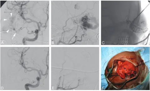
[image:7.594.51.539.41.203.2]Carotid Blowout Syndrome
Carotid blowout syndrome (CBS) refers to rupture of the ca-rotid artery and its branches.49,50It is a life-threatening de-layed complication of extensive or recurrent squamous cell carcinoma of the head and neck. The cause may be related to tumor involvement of the vessel wall, postoperative radiation, or prior surgical exposure. The reported morbidity and mor-tality rates associated with this complication are 40% and 60% respectively.51,52Survival is usually⬍2 years.
Surgical ligation of the carotid artery with or without a bypass is the traditional form of treatment. This is technically demanding because exploration and repair of a previous sur-gical and irradiated field can be difficult. Endovascular therapy is an excellent alternative to surgery and the method of choice.
The use of a covered stent has been reported in this condition, and a high rate of technical success is reported in achieving immediate hemostasis.53,54 However, several recently pub-lished series have shown unfavorable long-term outcomes due to rebleeding, delayed thrombosis, and abscess formation from contamination of the stent with the skin or oral flora.54-56
The poor long-term patency of the stent-grafts and the delayed complications like infection and rebleeding suggest that stent-graft placement may be beneficial for temporary rather than permanent management for patients with CBS (Fig 7). Appro-priate candidates for the use of stent-grafts may include those at high risk of neurologic morbidity from carotid occlusion, patients with acute massive bleed that prevents temporary bal-loon test occlusion, or those with a short life expectancy.54
[image:8.594.113.473.46.494.2]Permanent occlusion of the carotid artery is usually per-formed with a detachable balloon or coils. Approximately 15%–20% of patients treated with permanent vessel occlusion may have an immediate or delayed cerebral ischemia.52,53A
temporary balloon occlusion test can be performed before sac-rificing the carotid artery and may help in identifying patients at risk for immediate or delayed ischemic complications from occlusion of the carotid artery.
Intra-Arterial Chemotherapy for Head and Neck Carcinoma
The prognosis for patients with advanced head and neck can-cer treated conventionally with surgery, radiation, or both is extremely poor, with a long-term survival of 15%– 40%.57,58
Patients who are fortunate to survive their cancer often face a lifetime of significant morbidity, predominantly because of cosmetic and functional disabilities from extensive resection of the aerodigestive tract. Radiation therapy alone has the ad-vantage of organ preservation; however, its use in advanced disease only shows benefit with surgical resection.59
Intrave-nous chemotherapy for advanced disease has shown little benefit as a single-technique treatment, and the use of concur-rent chemoradiation is limited by severe toxicity with higher doses.60A large prospective study of 332 patients with stage III
or IV laryngeal cancer showed no difference in survival when induction intravenous chemotherapy followed by radiation therapy was compared with surgical resection and radiation therapy (P⫽.98).61
The radiation and platinum (RADPLAT) protocol was de-veloped at the University of California, San Diego and the University of Tennessee, Memphis, to address these limita-tions.62The basis of this approach is to deliver an extremely
high dose of the anticancer drug directly into the artery sup-plying the tumor, while circulating the antagonist of the drug in the venous system. This approach increases the amount of cisplatin delivered to the tumor, thus increasing its cytotoxic effects. In addition, the cisplatin also acts as a radio-sensitiza-tion agent, increasing the effectiveness of the concurrent radi-ation. The drug’s antagonist thiosulfate neutralizes the effects of cisplatin, thus minimizing the systemic toxicity.
Transfemoral carotid arteriography is carried out to assess the vascular anatomy before superselective catheterization of the tumor’s dominant vascular supply. A microcatheter is then placed coaxially into the ECA at the level of the orifice of the dominant artery supplying the tumor. This procedure allows local intra-arterial infusion of cisplatin (150 mg/m2for
3–5 minutes) in conjunction with an intravenous infusion of sodium thiosulfate (9 g/m2 for 3–5 minutes, followed by
12 g/m2for 6 hours). Bilateral catheterizations and infusions are performed in patients with disease extending across the midline. The goal of intra-arterial infusion is to target the por-tion of the tumor considered bulky or infiltrative and likely to fail treatment with radiation therapy alone.63
Four cycles of intra-arterial chemotherapy are adminis-tered on days 1, 8, 15, and 22. Radiation therapy is started on day 1 before chemotherapy and continued once daily 5 days a week. Opposed lateral fields are used to encompass the pri-mary and overt nodal disease at 2.0 Gy per fraction once daily 5 days a week to a planned total dose of 70.0 Gy. Uninvolved lower neck is treated with a single anteroposterior
supraclavic-ular field at 50.0 Gy at 2.0 Gy per fraction once daily. Several studies have evaluated the response rate and survival of pa-tients using this technique.62,64-66Although this form of
ther-apy has been studied for many years, the results have been variable, with heterogeneous use of this technique depending on institution or practitioner preference. However, several tri-als, particularly those using cisplatin-based regimens, indicate that a high response rate can be achieved. Additionally, there has been 1 phase II trial evaluating the feasibility and effective-ness of this form of treatment in a multi-institutional setting.67
The results have been encouraging, with reported complete response rates ranging from 55% to 90%. Locoregional con-trol rates of 57%–96% have been quoted at 2–5 years. Survival has ranged from 32%– 63% between 2 and 5 years.
References
1. Gupta S, Henningsen JA, Wallace MJ, et al.Percutaneous biopsy of head and neck lesions with CT guidance: various approaches and relevant anatomic and technical considerations.Radiographics2007;272:371–90
2. Sherman PM, Yousem DM, Loevner LA.CT-guided aspirations in the head and neck: assessment of the first 216 cases. AJNR Am J Neuroradiol
2004;25:1603– 07
3. Akins EW, Hawkins IF Jr, Mladinich C, et al.The blunt needle: a new percuta-neous access device.AJR Am J Roentgenol1989;152:181– 82
4. Mukherji SK, Turetsky D, Tart RP, et al.A technique for core biopsies of head and neck masses.AJNR Am J Neuroradiol1994;15:518 –20
5. DelGaudio JM, Dillard DG, Albritton FD, et al.Computed tomography– guided needle biopsy of head and neck lesions.Arch Otolaryngol Head Neck Surg2000;126:366 –70
6. Brook A, Gold MM, Miller TS.CT-guided radiofrequency ablation in the pal-liative treatment of recurrent advanced head and neck malignancies.J Vasc Interv Radiol2008;19:725–35. Epub 2008 Mar 17
7. Rhim H, Dodd GD 3rd.Radiofrequency thermal ablation of liver tumors.
J Clin Ultrasound1999;27:221–29
8. Shafir M, Shapiro R, Sung M, et al.Cryoablation of unresectable malignant liver tumors.Am J Surg1996;171:27–31
9. Schirmang TC, Davis LM, Nigri PT, et al.Solitary fibrous tumor of the buccal space: treatment with percutaneous cryoablation.AJNR Am J Neuroradiol
2007;28:1728 –30
10. Bui Q, Dupuy DE.Percutaneous CT-guided radiofrequency ablation of an adenoid cystic carcinoma of the head and neck.AJR Am J Roentgenol
2002;179:1333–35
11. Monchik JM, Donatini G, Iannuccilli J, et al.Radiofrequency ablation and percutaneous ethanol injection treatment for recurrent local and distant well-differentiated thyroid carcinoma.Ann Surg2006;244:296 –304
12. Mulliken JB, Glowacki J.Hemangiomas and vascular malformations in infants children: a classification based on endothelial characteristics.Plast Reconstr Surg1982;69:412–20
13. Enjolras O, Mulliken JB.Clinical and laboratory investigations: the current management of vascular birthmarks.Pediatr Dermatol1993;10:311–33 14. Yakes WF.Extremity venous malformations: diagnosis and management.
Semin Intervention Radiol1994;11:332–39
15. Claudon M, Upton J, Burrows PE.Diffuse venous malformations of the upper limb: morphologic characterization by MRI and venography.Pediatr Radiol
2001;31:507–14
16. Burrows PE, Laor T, Paltiel H, et al.Diagnostic imaging in the evaluation of vascular birthmarks.Dermatol Clin1998;16:455– 88
17. Berenguer B, Burrows PE, Zurakowski D, et al.Sclerotherapy of craniofacial venous malformations: complications and results.Plast Reconstr Surg1999; 104:1–11, discussion 12–15
18. Choi YH, Han MH, O-Ki K, et al.Craniofacial cavernous venous malfor-mations: percutaneous sclerotherapy with use of ethanolamine oleate.J Vasc Interv Radiol2002;13:475– 82
19. Cordes BM, Seidel FG, Sulek M, et al.Doxycycline sclerotherapy as the pri-mary treatment for head and neck lymphatic malformations.Otolaryngol Head Neck Surg2007;137:962– 64
20. Lee BB, Do YS, Byun HS, et al.Advanced management of venous malformation with ethanol sclerotherapy: mid-term results.J Vasc Surg2003;37:533–38 21. Cabrera J, Cabrera J Jr, Garcia-Olmedo A, et al.Treatment of venous
malfor-mations with sclerosant in microfoam form.Arch Dermatol2003;139:1409 –16 22. Rho MH, Kim DW, Kwon JS, et al.OK-432 sclerotherapy of plunging ranula in 21 patients: it can be a substitute for surgery.AJNR Am J Neuroradiol
2006;27:1090 –95
24. Chang HS, Yoon JH, Chung WY, et al.Sclerotherapy with OK-432 for recur-rent cystic thyroid nodule.Yonsei Med J1998;39:367–71
25. Macpherson P.The value of pre-operative embolization of meningiomas esti-mated subjectively and objectively.Neuroradiology1991;33:334 –37 26. Dean BL, Flom RA, Wallace RC, et al.Efficacy of endovascular treatment of
meningiomas: evaluation with matched samples.AJNR Am J Neuroradiol
1994;15:1675– 80
27. Wakhloo AK, Juengling FD, Delthoven VV.Extended preoperative polyvinyl alcohol microembolization of intracranial meningiomas: assessment of two embolization techniques.AJNR Am J Neuroradiol1993;14:571– 82 28. Kallmes DF, Evans AJ, Kaptain GJ, et al.Hemorrhagic complications in
embo-lization of a meningioma: case report and review of the literature. Neuroradi-ology1997;39:877– 80
29. Quadros RS, Gallas S, Delcourt C, et al.Preoperative embolization of a cervi-codorsal paraganglioma by direct percutaneous injection of Onyx and endo-vascular delivery of particles.AJNR Am J Neuroradiol2006;27:1907– 09 30. Abud DG, Mounayer C, Benndorf G, et al.Intratumoral injection of
cyano-acrylate glue in head and neck paragangliomas.AJNR Am J Neuroradiol
2004;25:1457– 62
31. Gruber A, Bavinzski G, Killer M, et al.Preoperative embolization of hyper-vascular skull base tumors.Minim Invasive Neurosurg2000;43:62–71 32. Erdmann MW, Jackson JE, Davies DM, et al.Multidisciplinary approach to the
management of head and neck arteriovenous malformations.Ann R Coll Surg Engl1995;77:53–59
33. Arat A, Cil BE, Vargel I, et al.Embolization of high-flow craniofacial vascular malformations with Onyx.AJNR Am J Neuroradiol2007;28:1409 –14 34. Lee BB, Do YS, Yakes W, et al.Management of arteriovenous malformations: a
multidisciplinary approach.J Vasc Surg2004;39:590 – 600
35. Rodesch G, Soupre V, Vazquez MP, et al.Arteriovenous malformations of the dental arcades: the place of endovascular therapy—results in 12 cases are pre-sented.J Craniomaxillofac Surg1998;26:306 –13
36. Resnick SA, Russell EJ, Hanson DH, et al.Embolization of a life-threatening mandibular vascular malformation by direct percutaneous transmandibular puncture.Head Neck1992;14:372–79
37. Ryu CW, Whang SM, Suh DC, et al.Percutaneous direct puncture glue embo-lization of high-flow craniofacial arteriovenous lesions: a new circular ring compression device with a beveled edge. AJNR Am J Neuroradiol
2007;28:528 –30
38. Turowski B, Zanella FE.Interventional neuroradiology of the head and neck.
Neuroimaging Clin N Am2003;13:619 – 45
39. Andersen PJ, Kjeldsen AD, Nepper-Rasmussen J.Selective embolization in the treatment of intractable epistaxis.Acta Otolaryngol2005;125:293–97 40. Layton KF, Kallmes DF, Gray LA, et al.Endovascular treatment of epistaxis in
patients with hereditary hemorrhagic telangiectasia.AJNR Am J Neuroradiol
2007;28:885– 88
41. Strutz J, Schumacher M.Uncontrollable epistaxis, angiographic localization and embolization.Arch Otolaryngol Head Neck Surg1990;116:697–99 42. Tseng EY, Narducci CA, Willing SJ, et al.Angiographic embolization for
epistaxis: a review of 114 cases.Laryngoscope1998;108:615–19
43. Christensen NP, Smith DS, Barnwell SL, et al.Arterial embolization in the management of posterior epistaxis. Otolaryngol Head Neck Surg
2005;133:748 –53
44. Remonda L, Schroth G, Caversaccio M, et al.Endovascular treatment of acute and subacute hemorrhage in the head and neck.Arch Otolaryngol Head Neck Surg2000;126:1255– 62
45. Morrissey DD, Anderson PE, Nesbit GM, et al.Endovascular management of hemorrhage in patients with head and neck cancer.Arch Otolaryngol Head Neck Surg1997;123:15–19
46. Goodman DN, Hoh BL, Rabinov JD, et al.CT angiography before emboliza-tion for hemorrhage in head and neck cancer.AJNR Am J Neuroradiol
2003;24:140 – 42
47. Sittel C, Jungelhulsing M, Gossmann A, et al.Superselective embolization as palliative treatment of recurrent hemorrhage in advanced carcinoma of the head and neck.Ann Otol Rhinol Laryngol2001;110:1126 –28
48. Wilner HI, Lazo A, Metes JJ, et al.Embolization in cataclysmal hemorrhage caused by squamous cell carcinoma of the head and neck. Radiology
1987;163:759 – 62
49. Chaloupka JC, Roth TC, Putman CM, et al.Recurrent carotid blowout syndrome: diagnosis and therapeutic challenges in a newly recognized sub-group of patients.AJNR Am J Neuroradiol1999;30:1069 –77
50. Mcdonald S, Gan J, Mckay AJ, et al.Endovascular treatment of acute carotid blowout syndrome.J Vasc Interv Radiol2000;11:1184 – 88
51. Citardi MJ, Chaloupka JC, Son YH, et al.Management of carotid artery rupture by monitored endovascular therapeutic occlusion (1988 –1994).Laryngoscope
1995;105:1086 –92
52. Chaloupka JC, Putnam CM, Citardi MJ, et al.Endovascular therapy for the carotid blowout syndrome in head and neck surgical patients: diagnostic and managerial considerations.AJNR Am J Neuroradiol1996;17:843–52 53. Lesley WS, Chaloupka JC, Weigele JB, et al.Preliminary experience with
endo-vascular reconstruction for the management of carotid blowout syndrome.
AJNR Am J Neuroradiol2003;24:975– 81
54. Chang FC, Lirng JF, Luo CB, et al.Carotid blowout syndrome in patients with head-and-neck cancers: reconstructive management by self-expandable stent-grafts.AJNR Am J Neuroradiol2007;28:181– 88
55. Chang FC, Lirng JF, Tai SK, et al.Brain abscess formation: a delayed compli-cation of carotid blowout syndrome treated by self-expandable stent-graft.
AJNR Am J Neuroradiol2006;27:1543– 45
56. Pyun HW, Lee DH, Yoo HM, et al.Placement of covered stents for carotid blowout in patients with head and neck cancer: follow-up results after rescue treatments.AJNR Am J Neuroradiol2007;28:1594 –98
57. Marcial VA, Pajak TF.Radiation therapy alone or in combination with surgery in head and neck cancer.Cancer1985;55:2259 – 65
58. Laramore GE, Scott CB, Al-Sarraf M, et al.Adjuvant chemotherapy for resect-able squamous cell carcinoma of the head and neck: report on Intergroup Study 0034.Int J Radiat Oncol Biol Phys1992;23:705–13
59. Robertson AG, Soutar DS, Paul J, et al.Early closure of a randomized trial: surgery and postoperative radiotherapy versus radiotherapy in the manage-ment of intra-oral tumours.Clin Oncol (R Coll Radiol)1998;10:155– 60 60. Vokes EE, Weichselbaum RR.Concomitant chemotherapy: rationale and
clinical experience in patients with solid tumors.J Clin Oncol1990;8:911–34 61.Induction chemotherapy plus radiation compared with surgery plus
radia-tion in patients with advanced laryngeal cancer: The Department of Veterans Affairs Laryngeal Cancer Study Group.N Engl J Med1991;324:1685–90 62. Robbins KT, Kumar P, Wong FS, et al.Targeted chemoradiation for advanced
head and neck cancer: analysis of 213 patients.Head Neck2000;22:687–93 63. Kumar P, Robbins KT.Treatment of advanced head and neck cancer with
intra-arterial cisplatin and concurrent radiation therapy: the “RADPLAT” protocol.Curr Oncol Rep2001;3:59 – 65
64. Robbins KT, Kumar P, Regine WF, et al.Efficacy of targeted supradose cispla-tin and concomitant radiation therapy for advanced head and neck cancer: the Memphis experience.Int J Radiat Oncol Biol Phys1997;38:263–71 65. Robbins KT, Wong FS, Kumar P, et al.Efficacy of targeted chemoradiation and
planned selective neck dissection to control bulky nodal disease in advanced head and neck cancer.Arch Otolaryngol Head Neck Surg1999;125:670 –75 66. Balm AJ, Rasch CR, Schornagel JH, et al.High dose superselective intra-arterial
cisplatin and concomitant radiation (RADPLAT) for advanced head and neck cancer.Head Neck2004;26:485–93
67. Robbins KT, Kumar P, Harris J, et al.Supradose intra-arterial cisplatin and concurrent radiation therapy for the treatment of stage IV head and neck squamous cell carcinoma is feasible and efficacious in a multi-institutional setting: results of Radiation Therapy Oncology Group Trial 9615.J Clin Oncol