ISSN Online: 2163-9469 ISSN Print: 2163-9450
DOI: 10.4236/ojgas.2017.710028 Oct. 31, 2017 271 Open Journal of Gastroenterology
Analysis of Recent Cases of Postbulbar
Duodenal Ulcer in Japan
Tateki Yamane
1*, Akira Umeda
2, Hitoshi Shimao
31Division of Gastroenterology, Department of Internal Medicine, The International University of Health and Welfare, Shioya
Hospital,Tochigi, Japan
2Division of Pulmonology, Department of Internal Medicine, The International University of Health and Welfare, Shioya
Hospital,Tochigi, Japan
3Department of Surgery, The International University of Health and Welfare, Shioya Hospital,Tochigi, Japan
Abstract
Although the incidence of postbulbar duodenal ulcers is low, many complica-tions occur, such as hemorrhage, stenosis and perforation, and there are many points to be paid attention to with regard to its pathology. A high acid level has been pointed out as a cause, but this is not conclusive, and its association with Helicobacter pylori (Hp) infection is also unclear. We investigated chronic cases excluding acute cases such as acute duodenal mucosal lesion we encountered between 2001 and 2015. Five cases corresponded, accounting for 1.4% of all chronic duodenal ulcer cases, and there were more male patients (four patients were male), as previously reported. All cases were solitary punched-out ulcer on the medial side near the superior duodenal flexure, and many complications were observed such as melena in all patients, narrowing in 2, and penetration in 1. No specific pathology, such as Crohn’s disease, vasculitis, or Zollinger-Ellison syndrome, was noted, no patient was previous-ly medicated with NSAIDs or low dose aspirin, and 3 patients were infected with Hp. All cases were healed by proton pump inhibitor administration or gastric acid secretion-suppressing treatment with selective proximal vagoto-my. Recurrence after Hp eradication occurred in all of the 3 Hp-infected pa-tients, suggesting that involvement of Hp in the cause of disease is unlikely.
Keywords
Postbulbar Duodenal Ulcer, Hp Infection, Peptic Ulcer
1. Introduction
Postbulbar duodenal ulcer (postbulbar ulcer) is defined as an ulcer developing in
How to cite this paper: Yamane, T., Ume-da, A. and Shimao, H. (2017) Analysis of Recent Cases of Postbulbar Duodenal Ulcer in Japan. Open Journal of Gastroenterology, 7, 271-278.
https://doi.org/10.4236/ojgas.2017.710028
Received: August 3, 2017 Accepted: October 28, 2017 Published: October 31, 2017
Copyright © 2017 by authors and Scientific Research Publishing Inc. This work is licensed under the Creative Commons Attribution International License (CC BY 4.0).
DOI: 10.4236/ojgas.2017.710028 272 Open Journal of Gastroenterology the duodenum deeper than Kerckring’s fold on contrast imaging and distal to the superior duodenal flexure on endoscopy [1]. Reportedly, many complica-tions occur, such as hemorrhage, stenosis, and perforation, although the inci-dence of the ulcer is low [2][3][4][5]. A high acid level has been suggested as a cause [1], but many points remain unclear, and the association with Helicobac-ter pylori (Hp) infection has not been clarified. We investigated cases we en-countered in a recent.
2. Case Reports
The following items were investigated in chronic open cases excluding acute cases such as acute duodenal mucosal lesion encountered at our department and related institutions between 2001 and 2015 (15 years): The incidence, sex differ-ence, age, symptoms, underlying disease, Hp infection, NSAID-low dose aspirin (LDA) medication history, ulcer properties, accompanying lesions, development site, endoscopic gastric mucosal atrophy (by Kimura-Takemoto classification), serum gastrin level, complications, treatment, and course.
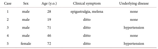
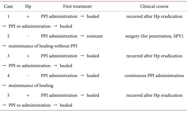
[image:2.595.208.542.617.721.2]Five cases corresponded, accounting for 1.4% of all chronic open duodenal ulcer cases (involving bulbar ulcer cases). Four and one patient were male and female, respectively, and 1, 1, 1, and 2 patients were in their 10 s, 20 s, 40 s, and 70 s, respectively. The symptoms were epigastralgia and melena in all patients, and none of them had a specific underlying disease (Table 1). Three patients were infected with Hp, and none of them were medicated with NSAIDs-LDA. The lesion was a solitary oval punched-out ulcer located on the medial side near the superior duodenal flexure in all cases, and no accompanying lesion was noted on endoscopy in any case (Table 2). Atrophy of the gastric mucosa was endoscopically mild even in the cases with Hp infection. The serum gastrin level was within the normal range in all patients. Regarding complications, hemorr-hage was noted in all 5 cases, and penetration into the gallbladder and narrowing of the lumen were noted in 1 and 2 cases, respectively (Table 3). Four of the 5 cases were healed by proton pump inhibitor (PPI) administration. Penetration into the gallbladder was confirmed in the remaining one case. This case was Hp-negative and resistant to PPI, and therefore treated with cholecystectomy, closure of the ulcer region, and selective proximal vagotomy (SPV), and the healing state after surgery was maintained without PPI administration. In the 3
Table 1. Clinical profile of the patients.
Case Sex Age (y.o.) Clinical symptom Underlying disease
1 male 28 epigastralgia, melena none
2 male 19 ditto none
3 male 71 ditto hypertension
4 male 46 ditto none
DOI: 10.4236/ojgas.2017.710028 273 Open Journal of Gastroenterology patients with Hp infection, the bacteria were eradicated after healing the ulcer by PPI administration, but the ulcer recurred in all of them and the healing state was maintained by re-administration of PPI (Table 4).
3. Case Presentation
[image:3.595.207.540.226.350.2]Case 1: A 28-year-old male. Hp-positive. A large punched-out ulcer was present on the medial side of the descending part of the duodenum near the superior duodenal flexure on upper GI series (Figure 1(a)) and endoscopy (Figure 1(b)).
Table 2. Characters of the ulcers.
Case Hp NSAIDs ~LDA Properties of the ulcers/ Accompanying lesions Development site of the ulcers
1 + − solitary oval puched-out/none medial side near the superior
duodenal flexure
2 − − ditto/ditto ditto
3 + − ditto/ditto ditto
4 − − ditto/ditto ditto
[image:3.595.208.540.386.497.2]5 + − ditto/ditto ditto
Table 3. Background and complication of the ulcers.
Case Endoscopic gastric mucosal atrophy (Kimura-Takemoto classification) Serum gastrin level (pg/ml) Complication
1 C-1 112 hemorrhage
2 C-0 47 hemorrhage, penetration
3 C-3 98 hemorrhage, narrowing
4 C-0 58 hemorrhage, narrowing
5 C-3 121 hemorrhage
Table 4. Treatment and clinical course of the ulcers.
Case Hp First treatment Clinical course
1 + PPI administration → healed recurred after Hp eradication
→ PPI re-administration → healed
2 − PPI administration → resistant surgery (for penetration, SPV)
→ maintenance of healing without PPI
3 + PPI administration → healed recurred after Hp eradication
→ PPI re-administration → healed
4 − PPI administration → healed continuous PPI administration
→ maintenance of healing
5 + PPI administration → healed recurred after Hp eradication
[image:3.595.210.531.528.726.2]DOI: 10.4236/ojgas.2017.710028 274 Open Journal of Gastroenterology
Figure 1. (a) Upper GI series finding. A niche was found in the duodenal post bulbar re-gion (arrow). (b)-(e): Gastrointestinal endoscopic findings. (b) A large punched-out ulcer was observed. (c) The ulcer scarred after PPI administration. (d) The ulcer recurred after Hp eradication. (e) The ulcer scarred by PPI re-administration.
On histopathological examination of a biopsy specimen collected from the ulcer margin, only erosive duodenal mucosa was noted without significant findings. After PPI administration, the ulcer scarred (Figure 1(c)) and then Hp eradication was performed. The ulcer recurred (Figure 1(d)), and PPI was re-administered to cicatrize it (Figure 1(e)).
Case 2: A 19-year-old male. Hp-negative. A large punched-out ulcer was ob-served on the medial side of descending part of the duodenum near the superior duodenal flexure on endoscopy. The ulcer was resistant to PPI administration. Penetration into the gallbladder was recognized. Surgery (cholecystectomy, clo-sure of the ulcer region and SPV) was performed. After surgery healing state of the ulcer was maintained without PPI administration.
Case 3: A 71-year-old male. Hp-positive. A large punched-out ulcer was present on the medial side of the descending part of the duodenum near the su-perior duodenal flexure on endoscopy (Figure 2(a)). The ulcer scarred by PPI administration (Figure 2(b)). The ulcer recurred after Hp eradication (Figure 2(c)). It scarred again by re-administration of PPI (Figure 2(d)).
Case 4: A 46-year-old male. Hp-negative. A large punched-out ulcer was ob-served on the medial side of descending part of the duodenum near the superior duodenal flexure on endoscopy. After PPI administration the ulcer scarred. Healing state of the ulcer was maintained with PPI administration.
DOI: 10.4236/ojgas.2017.710028 275 Open Journal of Gastroenterology
Figure 2. Gastrointestinal endoscopic findings. A large punched-out ulcer was observed. (b) The ulcer scarred after PPI administration. (c) The ulcer recurred after Hp eradica-tion. (d) The ulcer scarred by PPI re-administraeradica-tion.
[image:5.595.219.527.418.685.2]DOI: 10.4236/ojgas.2017.710028 276 Open Journal of Gastroenterology ulcer recurred (Figure 3(c)). It was cicatrized again by re-administration of PPI
(Figure 3(d)).
4. Discussion
No studies on postbulbar ulcers have been reported recently. According to old reports, the incidence was 5% - 1% [2][3] in autopsied cases in other countries, being relatively high, but it was only 0.73% [6] on endoscopy in Japan. In our study, the frequency was also low and it accounted for only 1.4% of all duodenal ulcer cases. The male/female ratio was 23.1 [7] in other countries and 26.1 [8] in Japan, showing a high incidence in males. Four patients were male, accounting for 80%, in the present study. The mean age with a high incidence is 45.5 - 54.9 years old [1][2] in Western countries and 59.8 years old [9] in Japan, and these were higher than that of the bulbar duodenal ulcer (bulbar ulcer) patients. The ages ranged widely in our study and there was no specific age with a high inci-dence. Reportedly, epigastralgia is the most frequent symptom of postbulbar ulcer, followed by right hypochondralgia, and some cases are asymptomatic [1] [3][6][9]. In our study, the main symptoms were epigastralgia and melena in all patients. The reported most frequent development site is the medial region near the superior duodenal flexure, followed by the medial side of this region over the main duodenal papilla [10], and all cases developed in the former in our study. Regarding the ulcer properties, many lesions are reportedly oval and solitary, the shape is large and deep in the acute phase in many cases [11]. The property of our cases was similar. Postbulbar ulcers are frequently complicated by hemorr-hage, stenosis, and perforation, and this is considered to be due to the anatomy of this region: abundant blood vessels and thin wall [1] [3] [5]. The reported frequency of hemorrhage is 37% - 86%, being higher than that for bulbar ulcers [1][2][3][4][5], complete stenosis is rare, narrowing is frequently observed [4] [12], and the frequency of free perforation is low compared with that for bulbar ulcers, but the frequency of penetration is high because of the positional rela-tionship with the surrounding organs [3][4][7][12]. In our study, hemorrhage was noted in all patients (100%) narrowing was noted in 2 (40%), and penetra-tion into the gallbladder was noted in 1 (20%).
A high acid level is considered to be the cause of postbulbar ulcers, and the causes other than acid include NSAIDs and ischemia due to serious underlying disease [2][4]. Crohn’s disease, Behçet’s disease, vasculitis, viral infection, and Zollinger-Ellison syndrome (ZES)which is a state with a morbidly high acid lev-el, have been pointed out as rare pathologies to be differentiated from [2][4]. In the present study, no specific underlying disease, such as Crohn’s disease, was noted. ZES was also negative based on the serum gastrin level, and no patient had been treated with oral NSAIDs-LDA.
DOI: 10.4236/ojgas.2017.710028 277 Open Journal of Gastroenterology It has been reported that the maximum acid output level on gastric juice testing was 26.6 ± 6.4 mEq/h in postbulbar ulcer cases and 18.4 ± 6.4 mEq/h in bulbar ulcer cases [13], demonstrating that the acid level for postbulbar ulcers was higher than that for bulbar ulcers [1][8][13]. Regarding treatment, it was in-tractable before the appearance of potent acid secretion inhibitors, such as H2 receptor antagonist and PPI, and surgery was mainly performed because com-plications frequently occur [1] [2] [3] [9] [14]. However, responses to these drugs are favorable, excluding special pathologies, such as Crohn’s disease. In the present study, PPI was effective for 4 of the 5 cases. The PPI-resistant case was considered intractable due to penetration into the gallbladder, and the heal-ing state was maintained without PPI after SPV, suggestheal-ing the effectiveness of SPV [15]. Therefore, treatment by inhibition of gastric-acid secretion was effec-tive in all cases.
Bulbar ulcers are closely associated with Hp infection and rarely recur after eradication of Hp. On the other hand, the association of postbulbar ulcers with Hp has not been clarified. The mechanism of bulbar ulcers is assumed as follows: Hp colonizes the antrum of the stomach and stimulates gastrin secretion, which promotes gastric acid secretion. Hp flows into and colonizes the bulbar region in which gastric metaplasia is induced by stimulation with a high acid level, and induces production of mucosa-damaging factors, such as ammonia and inflam-matory cytokines, leading to ulceration. Eradication of Hp contributes to pre-vention of recurrence by removal of damaging factors and inhibition of gas-tric-acid secretion. In the present study, three patients (60%) were infected with Hp, but recurrence occurred after eradication of Hp, suggesting that the pathol-ogy was different from that of bulbar ulcers.
Gastric acid may have been the main cause of the disease in all 5 patients, but the possibility of involvement of gastric acid retention in the descending part of the duodenum and impaired bicarbonate secretion in the duodenal mucosa was also considered.
5. Conclusion
The amount of gastric acid secretion in Japanese people is likely to increase due to reduction of the Hp infection rate and an increase in fat intake. It will be in-teresting to see how the incidence of postbulbar ulcer changes in Japan.
References
[1] Hines, J.R., Geurkink, R.E. and Gordon, R.T. (1976) Postbulbar Peptic Ulcerlation of the Duodenum. SurgGynecolObstect, 142, 13-15.
[2] Lonergan, W.M. and Kahn, A. (1951) Post-Bulbar Duodenal Ulcerlation. Gastroen-terology, 17, 494-503.
[3] Bergner, L.H. and Gold, E. (1964) Postbulbar Peptic Ulcerlation of the Duodenum.
The American Journal of Gastroenterology, 42, 285-291.
DOI: 10.4236/ojgas.2017.710028 278 Open Journal of Gastroenterology https://doi.org/10.1016/S0002-9610(66)80005-3
[5] Ramsdell, J.A., Bartholomew, L.G., Gain, J.C. and Davis GD. (1957) Postbulbar Duodenal Ulcer. Annals of Internal Medicine, 47, 700-710.
https://doi.org/10.7326/0003-4819-47-4-700
[6] Akasaka, Y., Koori, Y. and Nakajima, M. (1974) Clinical Aspects of Postbulbar Ulc-er of the Duodenum—Including Its Endoscopic Follow-Up Study. Stomach and In-testine, 9, 225-232.
[7] Cooke, L. and Htton, C.F. (1958) Postbulbar Duodenal Ulcerlation. Lancet, 1, 754-757. https://doi.org/10.1016/S0140-6736(58)91572-1
[8] Masuda, H., Miyamori, A. and Fukuhawa, N. (1967) Postbulbar Duodenal Ulcer.
Stomach and Intestine, 2, 51-60.
[9] Yanai, H., Tada, M. and Fuji, T. (1990) Clinical Features of Postbulbar Duodenal Ulcer. Endoscopia Digestiva, 2, 81-87.
[10] Nagai, N., Hayakawa, K., Yamada, K., Tsuchiya, F., Hara, T., Suzuki, H., Ooi, I., Kobayashi, S. and Takemoto, T. (1974) Endoscopic Diagnosis of Postbulbar Duo-denal Ulcer. Gastrointestinal Endoscopy, 16, 120-125.
[11] Rao, S.S.C. and Murthy, K.V.R. (1993) Post-Bulbar and Coexisting Ulcerlation; Unique Features of Peptic Ulcer in Hyderabat. Gut, 34, 1327-1330.
https://doi.org/10.1136/gut.34.10.1327
[12] Ritchie, J.D. (1976) Postbulbar Ulcer. ANZ Journal of Surgery, 46, 116-120. [13] Kaminishi, M., Shimazu, R., Kuramoto, S., Kuramoto, S., Sadatsuki, H., Johjima, Y.
and Oohara, T. (1987) A Clinicopathological Study of Post-Bulbar Duodenal Ulcer.
The Japanese Journal of Surgery, 17, 140-145. https://doi.org/10.1007/BF02470655
[14] Konno, Y., Kameyama, J., Miyakawa, H., Naito, H., Tsuchiya, H., Narui, H. and Sa-to, T. (1983) Surgical Management of Postbulbar Duodenal Ulcer. The Japanese Journal of Surgery, 16, 1324-1327.