LEABHARLANN CHOLAISTE NA TRIONOIDE, BAILE ATHA CLIATH TRINITY COLLEGE LIBRARY DUBLIN OUscoil Atha Cliath The University of Dublin
Terms and Conditions of Use of Digitised Theses from Trinity College Library Dublin
Copyright statement
All material supplied by Trinity College Library is protected by copyright (under the Copyright and Related Rights Act, 2000 as amended) and other relevant Intellectual Property Rights. By accessing and using a Digitised Thesis from Trinity College Library you acknowledge that all Intellectual Property Rights in any Works supplied are the sole and exclusive property of the copyright and/or other I PR holder. Specific copyright holders may not be explicitly identified. Use of materials from other sources within a thesis should not be construed as a claim over them.
A non-exclusive, non-transferable licence is hereby granted to those using or reproducing, in whole or in part, the material for valid purposes, providing the copyright owners are acknowledged using the normal conventions. Where specific permission to use material is required, this is identified and such permission must be sought from the copyright holder or agency cited.
Liability statement
By using a Digitised Thesis, I accept that Trinity College Dublin bears no legal responsibility for the accuracy, legality or comprehensiveness of materials contained within the thesis, and that Trinity College Dublin accepts no liability for indirect, consequential, or incidental, damages or losses arising from use of the thesis for whatever reason. Information located in a thesis may be subject to specific use constraints, details of which may not be explicitly described. It is the responsibility of potential and actual users to be aware of such constraints and to abide by them. By making use of material from a digitised thesis, you accept these copyright and disclaimer provisions. Where it is brought to the attention of Trinity College Library that there may be a breach of copyright or other restraint, it is the policy to withdraw or take down access to a thesis while the issue is being resolved.
Access Agreement
By using a Digitised Thesis from Trinity College Library you are bound by the following Terms & Conditions. Please read them carefully.
L E A B H A R L A N N C H O L A IS T E N A T R 1 0 N 6 ID E , BAILE A TH A C L IA T H Ollscoil Atha Cliath
f K ' l -
lU
T R IN IT Y COLLEGE LIB RA RY D U B L IN The University of Dublin
THIS THESIS M AY BE READ ONLY IN THE LIBRARY
Reader's Declaration
I undertake not to reproduce any portion of, or use any inform ation derived from this thesis w ith o u t first obtaining the permission, in w ritin g , o f the Librarian, T rinity College. If permission is granted, I shall give appropriate acknowledgem ent fo r any portion o f the
thesis used or reproduced.
Date consulted
Name and address in block letters
University or institution
Title- A comparison of mandibular
incisor proclination when using
clear aligners and fixed labial
orthodontic brackets
Subm itted in accordance w ith th e requ irem ents for the degree of Clinical D octorate in Dental Surgery (O rthodontics)
Trinity College Dublin, Dublin Dental University Hospital
20 1 5
Student- Dr Joe H ennessy
Supervisors- Dr Therese Garvey
- Dr Ebrahim Al-Awadhi
Declaration
I declare that this thesis has not been submitted as an exercise fo r a
degree a t this or any other university and it is entirely my own work. I
agree to deposit this thesis in the University's open access institutional
repository or allow the library to do so on my behalf, subject to Irish
Copyright Legislation and Trinity College Library conditions o f use and
acknowledgement
Joe Hennessy
0 6 OCT ?015 ^LIBR ARYDU BLIN ^
Summary
Title-
A random ised clinical trial com paring m andibular incisor
proclination produced by fixed labial appliances and clear aligners.
Introduction-
The objective of this ‘2-arm parallel' clinical trial w as to
com pare the m andibular incisor proclination produced by fixed labial
appliances and 3‘‘‘* generation clear aligners.
M ethods-
Patients underw ent a course of orthodontic treatm en t using
fixed lalDial appliances or clear aligners (Invisalign®). M andibular
incisor proclination was m easured by com paring p re-treatm en t and
n ear end treatm en t lateral cephalograms. Eligibility criteria included
adult patients w ith mild m andibular incisor crowding (<4 mm) and
Class I skeletal bases (ANB 1-4°). The main outcome was the
cephalom etric change in m andibular incisor inclination to the
m andibular plane at the end of treatm ent. Eligible patients picking a
sealed opaque envelope, which indicated th eir group allocation, was
used to achieve random ization. Data w ere analysed using a Welch two
sam ple t-test.
Results-
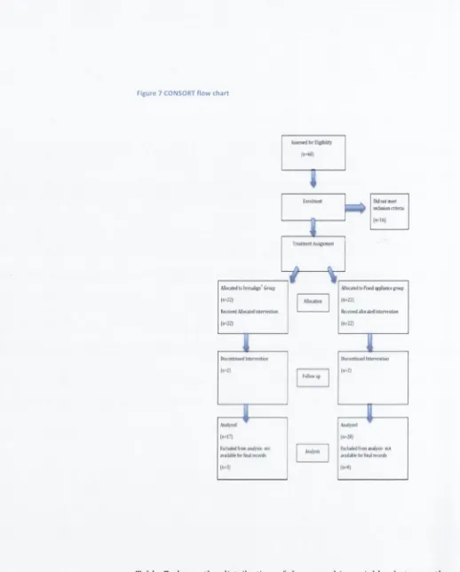
Forty-four patients (m ean age 26.4 ± 7.7 years) w ere
random ised in a 1:1 ratio to either the fixed labial apphance or the clear
aligner group. Baseline characteristics w ere sim ilar for both groups,
fixed appliance m ean crowding- 2.1 ± 1.3 mm vs clear aligner m ean
crow ding 2.5 ± 1.3 mm, p re-treatm en t m ean m andibular incisor
inclination for the fixed appliance group 90.8 ± 5.4° vs 91.6 ± 6.4° for
the clear aligner group. Seven p atients [5 clear aligner and 2 fixed
appliance) w e re excluded from the study due to compliance issues. Fixed appliances produced 5.3 ± 4.3° of m andibular incisor proclination. Clear aligners proclined th e m andibular incisors by 3.4 ± 3.2°. The difference betw ee n the tw o groups w as n ot statistically significant [p >0.05).
C onclusion- There w as no significant difference in the am o u n t of m andibular incisor proclination produced by clear aligners and fixed labial appliances in mild crow ding cases.
Acknowledgements
1 w o uld like to thank the staff at the D ublin Dental School and Hospital fo r th e ir assistance w ith this project.
1 w ould like to thank m y supervisors, Dr Al-A w adhi and Dr Garvey, for all th e ir tu to rin g and advice over the last ten years. They have been in fluentia l in my career progression and th e ir door is always open when 1 need help.
To my family, and in pa rticu la r m y parents, w ho granted me every o p p o rtu n ity possible. I w ill never repay my debt to them and m y gratitude is indescribable.
Finally to Laura, who has been by m y side every step o f the way. She halved every problem and her supp ort was inexhaustible.
Table of Contents
D e c la ra tio n ... 2
S um m ary...3
A cknow ledgem ents... 5
Table of C o n te n ts ... 6
Table of T a b le s ...8
Table of F ig u re s ...9
1 In tro d u c tio n ... 10
2 L iterature R ev iew ... 12
2.1 Fixed appliances and to o th m o v e m e n t...12
2.2 H istory of Clear A lig n e rs... 15
2.2.1 Classification of Clear A ligners... 18
2.3 The Clear A ligner P ro c e ss... 20
2.4 A dvantages of Clear A ligners...23
2.4.1 A e sth e tic s... 23
2.4.2 P ain... 23
2.4.3 Oral H ygiene...25
2.4.4 S p eech ...26
2.4.5 Root R eso rp tio n ... 26
2.4.6 O ther suggested a d v a n ta g e s ... 27
2.4.7 S um m ary... 28
2.5 Lim itations of Clear Aligner T re a tm e n t...28
2.5.1 T rea tm e n t tim e ... 29
2.5.2 A ccuracy of to o th m o v e m e n t...29
2.5.3 C o m pliance... 35
2.5.4 Practical L im itations... 36
2.5.5 S um m ary...36
2.6 C ephalom etric m e a su re m e n ts... 38
2.7 M andibular incisor p ro c lin a tio n ... 40
3 M aterials and M eth o d s... 47
3.1 Study O u tlin e... 47
3.2 Sam ple Size... 47
3.3 Inclusion C rite ria ... 47
3.4 Exclusion C rite ria ... 48
3.5 R e c ru itm e n t... 48
3.6 P re -tre a tm e n t R ecords... 48
3.7 Crow ding A sse ssm e n t... 49
3.8 Group A llocation... 51
3.9 Fixed A ppliance g ro u p ... 51
3.10 Invisalign g ro u p ... 52
3.11 R adiographic M easu rem e n ts...55
3.12 E rro r of th e m e th o d ... 56
3.13 Statistical A nalysis...58
4 R esults...59
5 D iscussion... 65
6 C onclusion... 73
7 R eferences... 74
8 A ppendices... 81
i) Ethical ap prov al application fo rm ...81
ii) Ethical A p p ro v a l...108
iii) Fixed appliance inform ed co n sen t and ag reem en t for tre a tm e n t ... 109
iv) Invisalign appliance inform ed co n sen t and a g re e m e n t for tr e a tm e n t... 113
v) P atien t invitation le tte r...121
vi) P atient inform ation le a fle t...122
Table of Tables
Table 1- Clear aligner m a n u fa ctu re... 17 Table 2 Comparing accuracy o f tooth movements produced by Clear
aligners and Fixed appliances...30 Table 3 Comparing Clear aligner and Fixed appliance results...37 Table 4 Studies com paring low er Incisor proclination produced by
different orth odon tic appliances...42 Table 5 Paired t-tests to assess intra and inter-examiner errors for
cephalometric measurements... 56 Table 6 Paired t-tests to assess intra and inter-examiner errors for crowding
measurements... 57 Table 7 Descriptive statistics for the demographic and clinical
characteristics o f pre-treatm ent study groups [n = 4 4 ]...61 Table 8 Invisalign® group m andibular incisor inclination pre-treatm ent, post-treatm ent and inclination change... 62 Table 9 Fixed labial appliance group m andibular incisor inclination p re treatm ent, post-treatm e nt and in clination change...63 Table 10 Welch tw o sample t-test comparing m andibular incisor
proclination produced by Invisalign® and Fixed labial appliances.. 64
Table of Figures
Figure 1 Downs' method to measure lo w e r incisor inclination, Lateral
cephalogram o f patie nt 12 Invisalign g ro u p ... 38
Figure 2 Nance Brass W ire technique... 50
Figure 3 Fixed appliance tre a tm e n t...52
Figure 4 Clincheck softw are...54
Figure 5 Pre alig n e rs...55
Figure 6 Aligners in s itu ... 55
Figure 7 CONSORT flo w c h a rt... 60
1 Introduction
Clear aligners have been introduced as an aesthetic alternative to fixed labial appliances. Over the years these appliances have evolved in an attempt to achieve improved tooth alignment and occlusion. There have been three generations of aligners. The first generation aligners were solely reliant on the removable appliance to move teeth. Following this, a second generation of aligner depended more on attachments being bonded to teeth- The most recent third generation, has been modified to place different shaped attachments automatically where difficult tooth movements are required. Although fixed appliances have been the backbone of orthodontics for years, patient demands have been the driving force for the development of clear aligners. In addition to improved aesthetics it has been reported that clear aligners are less painful, allow for improved oral hygiene and cause less root resorption than conventional fixed appliances(Miethke and Brauner, 2007; Miller
etal., 2007; Barbagallo etal., 2008; Jeremiah etal., 2011).
Despite over 15 years of commercial availability and many millions of cases treated worldwide, very little research has assessed how clear aligners achieve their results. From the available studies, clear aligners would appear to have poorer clinical results when compared to fixed appliances. The aligners ability to extrude, derotate and torque teeth has been questioned (Djeu
et al, 2005; Kravitz
et al, 2009; Krieger
et
al, 2011; Krieger et al. 2012). It has, however, been suggested that they
can accurately retrocline teeth (Kravitz etal., 2009).Mandibular incisor proclination is often an unwanted side effect of
orthodontic treatment. Excessive proclination can cause poor
aesthetics, gingival recession and unstable results. Many studies have
compared the amount of lower incisor proclination produced by
different orthodontic appliances (Toth and McNamara, 1999; Gill and
Lee, 2005; Pandis et al, 2007; Scott et al, 2008). The effect of clear aligners on incisor proclination, however, has yet to be determined.
Case reports account for the majority of the literature available with
regards to clear aligner treatment. A limited num ber of poorly designed
clinical trials have appraised the ability of aligners to move teeth (Djeu
et ai, 2005; Kravitz et al., 2009; Krieger et al., 2011; Krieger et al., 2012]. This research compares the mandibular incisor proclination
produced by fixed labial brackets and 3rd generation clear aligners
[Invisalign®, Align Technology Inc, Santa Clara, CA, USA) when treating
patients with mild mandibular crowding.
2 Literature Review
2.1 Fixed appliances and tooth m ovem ent
Fixed o rth o d o n tic appU ances have evolved since Angle described the
edgew ise b rack e t in 1928 [Angle, 1928). Initially th e slot w as placed
vertically in th e bracket, until he realised m ore control of too th
m ovem ents could be ob tain ed by placing th e slots horizontally. Three-
dim ensional control of a to o th w as achieved by using a rectan g u lar w ire
w ithin a rectan g u lar slot. The stan d ard edgew ise system had a n u m b er
of disadvantages, n o t least th e fact th a t a passive b rack et resu lted in a
re q u ire m e n t for com plex w ire bending du ring finishing p rocedu res.
T hese b ends also en su red space closure needed to be u n d erta k en using
closing loops.
Begg w as th e next to develop a b rack e t system , basing it on th e concept
of differential force (Begg, 1956). His appliance w o rked by using
elastics to tip crow ns to th e ir co rrect p osition s and th en uprig htin g
th e ir ro o ts w ith auxiliary springs. It relied on ro u n d arch w ires w hich
m ade finishing p ro ced u res com plex and tim e consum ing. Kesling, in th e
1980s, atte m p te d to resolve som e of th e se prob lem s by creatin g th e
Tip-Edge appliance® [TP O rthodontics, Inc, 100 C enter Plaza, La Porte,
Indiana 4 63 50 -9 67 2, USA)[Kesling, 1988). A lthough this system again
tips te e th in th e early stages of tre a tm e n t it allow s for th e use of
rectan g u lar arch w ires, w hich a re essential w hen th ree-d im en sio n al
control is req uired.
Lawrence Andrews was the next to produce a new fixed orthodontic
apphance (Andrews, 1979). His system was based on the idea of having
prescribed brackets, for each tooth, that moved the teeth into their
respective ideal positions. This removed the need for the majority of the
wire bends that were required when using the original edgewise
appliance. A number of different prescriptions are available for these
brackets and this is now the most widely used system in modern
orthodontics.
More recently, the use of self-ligating brackets has become popular.
Stolzenberg created the original self-ligating bracket in the 1930s
[Stolzenberg, 1946). A number of proposed advantages, such as quicker
treatm ent times and more secure ligation of the archwire, have been
suggested with the use of these appliances but very few have been
scientifically verified (Chen
et al,
2010). There are two types of self-
ligating brackets, active and passive. Active brackets have a spring-
loaded ligation mechanism which presses against the wire when closed.
The passive clip does not encroach on the bracket slot lumen and
therefore the clip doesn’t place a force on the wire.
The theory of how fixed appliances move teeth has created a degree of
controversy over the years. There are two types of tooth movements
that can be achieved with a fixed appliance. Tipping movements, where
the crown moves while the tip of the root stays stationary, and bodily
movement where the crown and root move in unison (Profit
et al,
2013). It was assumed that the type of movement produced depended
on the relation of the applied force to the centre of resistance and the
location of the centre of rotation of the tooth (Isaacson et ai, 1993). The centre of resistance is defined as th a t point on the tooth w here, if a single force passed through, p u re translation would resu lt [Burstone and Pryputniewicz, 1980). It has been determ ined for a single rooted tooth the centre of resistance is one third the root length apical to the alveolar crest along the long axis of the ro o t (Burstone and Pryputniewicz, 1980). The centre of rotation is the p oint about which a body app ea rs to have rotated, as d eterm ined from its initial and final positions (Smith and Burstone, 1984). This point will change depending on the torce being applied. To achieve pure bodily movem ent, the applied force m ust pass directly through the centre of resistance, how ever forces are usually applied to attachm ents on the buccal surfaces of te eth making the force coronal and peripheral to the centre of resistance (Iwasaki etal., 2000).
Smith and Burstone described tw o possible ways force can be applied to a tooth (Smith and Burstone, 1984). A single force can be applied, acting aw ay from th e centre of resistance, which is called th e 'm o m en t of force’ (Isaacson et al, 1993). This force can cause the tooth to tip around the centre of rotation. The second m ethod by which force can be applied is by a pair of equal forces which are parallel and in opposite directions, te rm e d a ‘force couple' (Isaacson et al, 1993). With this force application the centre of rotation equals the centre of resistance (Isaacson e t al, 1993). As fixed appliances are not able to apply a force directly thro u g h the centre of resistance, bodily m o v e m e n t could only be achieved by applying a force a t the attachm ent in the direction of the requ ire d m o v e m en t while a t the sam e time having a c o u n te r m o v e m en t
to p re v e n t tipping (Isaacson et ah, 1993). It vi^as Isaacson e t al w ho first suggested th a t p ure bodily m ov e m en t w as impossible to achieve and th a t bodily m ovem ents actually occur through a series of tipping and uprighting movements, w h ere crow n m ov e m en t (m o m en t of force) exceeds root m ovem ent (m o m en t of couple) and th e n root m ove m en t exceeds crown m ovem ent (Isaacson e t a l , 1993).
2.2 H is to ry o f C lear A ligners
The th e o ry of using an aligner to straighten te eth w as first postulated in the 1940s. In 1945, Kesling produced a tooth positioning appliance to refine the final stages of orthodontic tre a tm e n t (Kesling, 1946). This positioner was a piece of pliable ru b b e r m anufactured from a laboratory wax up of the te eth in class 1 occlusion (Phan and Ling, 2007). This appliance allowed for m inor tooth m ovem ents to be achieved while maintaining the alignm ent of the rem aining teeth in the arch. Kesling foresaw th a t m ore am bitious tooth m ovem ents could be realised with a series of aligners while recognising the limitations of the technology available to him a t the time:
Thirty years later, Ponitz introduced an "Invisible Retainer” which used Kesling's idea of prepositioning teeth on a master study model [Ponitz, 1971). Again this appliance could only produce minor tooth movements.
In the early nineties Sheridan described a technique of using clear aligners in conjunction with interproximal tooth reduction (Sheridan
et
al.,
1993). The principle of producing minor tooth movements withindividual aligners had not changed. A new ‘Kesling set up’ was required for every tooth movement and therefore a new impression was taken at almost every visit. This process demanded a large amount of clinical and laboratory time.
In 1999, Align technology released their Invisalign® system. It was the first orthodontic appliance to use computer-aided design (CAD) and computer-aided manufacturing (CAM). Instead of requiring a new impression for each tooth movement, this technology allows for multiple tooth set-ups to be created from a single impression (Hajeer
et
al,
2004). The advent of this digital process removed the impracticalityof previous aligner systems and made Kesling's concept a reality. Other aligner systems use similar principles to achieve their results (Jones
et
al,
2009) (Table 1). These systems have evolved over time.T ab le 1- Cl ear a l i g n e r m a n u f a c t u r e
N am e of appliance C ountry of origin
3D technology used
W ebsite Attach m en ts
How m any aligners
G enerati on
Average Price
Clear aligner™ UK Laser w w w .clearali B ner.co.uk /
No Unlimited 1» £114
Clear path USA Laser w w w .clearp at h d en tal.m m
No U nlimited 1» £890
C le a rs te p '" now Smilelign
UK Laser w w w .sm ilelif ILCOm/
Yes Unlimited 2 n d £750
Simplifive -Red, W hite and Blue Aligner
USA M anual w w w .orm co.c om
No Seven
aligners
1« £500
MTM C lear- A lig n e r'"
USA Laser w w w .m tm cle a r a l i B n e r . c o m I
No Unlim ited 1“ £450
N im rodental Clear aligner™
UK Laser w w w .nim rod en tal.co m /
No Unlim ited !'■ £400
Clear Image Aligners™
USA M anual w w w .snecialt vaoDliances.c om
No Unlim ited 1" £35
ClearAligner™ USA M anual w w w .clear-alipner.com
No Unlimited 1« £125
ClearCorrect™ USA Laser w w w .clearcor rect.com
Yes Unlim ited 2 n d £760
Invisalign™ USA Laser w w w .invisalig n.com
Yes Unlimited £1690
[image:19.543.13.527.41.751.2]2 .2 .1 C lassification o f Clear Aligners
Generation Aligners
The earh est forms of these system s w ere solely reliant on the aligners to achieve their results. No auxiliary elem ents w ere incorporated. These aligners can be fabricated using CAD/CAM technology or manually following Kesling’s method.
G eneration Aligners
As the aligner system s developed, m anufacturers encouraged the use of attachm ents to improve tooth movements. Clinicians could request composite buttons to be placed on teeth and also could attach inter maxillary elastics.
3 '‘‘ G eneration Aligners
In an attem p t to improve the results, m anufacturers again attem pted to alter the way aligners delivered force. A ttachm ents w ere automatically placed by the m anufacturer's softw are w here
Extrusion of Incisors a n d /o r canines are required D erotations are required
Root m ovem ents are required
attem pted. They a re 3m m high, 2mm w ide and 0 .7 5 -lm m thick and are
available for incisors, canines and prem olars. W hen they are used
singly, sim ilar to th e d evelopm ent of w id e r brackets in fixed appliances,
ellipsoid a ttach m en ts should allow for g re a te r rotational control. Using
them in pairs should allow for p roduction of m om ents of couple to
upright roots. They may also allow the appliance to achieve bodily
movement, like labial brackets, through the use of m om ents of couple
and m om ents of force. Bevelled attach m en ts are used m ost often w h en
trying to extrude a tooth. They can be 3,4 or 5 mm wide, 2 mm high and
from 0.25 to 1.25 mm thick. They have an active border, just like fixed
brackets, th a t should limit the slipping (or loss of tracking) th a t can
occur b e tw ee n the aligner and the tooth. Rectangular attachm ents are
used w hen large mesiodistal m ovem ents are requested. These are 3,4
or 5 mm high, 2 mm w ide and 0.5 to 1 mm thick. It is proposed th a t
these a ttach m en ts will allow teeth to be m oved bodily by allowing for a
longer span for force application. All th re e types of attachm ents are not
fully engaged initially w h en they are b o n d ed to the tooth. As the patien t
graduates through the different aligners, the attach m en ts becom e m ore
active until th e y finally fill the aligner slot. This principle is again
similar to w orking through archw ires w h en using the pread ju sted
edgewise appliance. Indentations are placed in the aligners w h e re
lingual ro o t to rq u e is requ ire d for maxillary or m andibular incisors.
These in d entations in the p olyurethane a tte m p t to place increased
p ressu re on specified points on the crow n to produce m om ents of
couple and to rq u e th e root. These suggested tooth m ovem ents have y et
to be verified scientifically. No research has accurately measured the
tooth movements produced by clear aligners.
2.3 The Clear Aligner Process
The first step in clear aligner treatm ent is the production of clinical
records (joffe, 2003). These include maxillary and mandibular
impressions, a silicone record of maximum intercuspation, intraoral
and extraoral photographs, a panoramic and cephalometric radiograph.
Digital or paper copies of these records are sent to the manufacturers.
An accurate set of impressions is very important. Materials that can be
used include polyvinyl silicone or polyether.
The majority of clear aligner manufacturers use CAD/CAM technology
to produce their clear aligners [Table 1). Some companies manufacture
the aligners manually which can be more time consuming and limit the
amount of appliances that can be requested. Each company has a
slightly different manufacturing process with the Invisalign® aligner
fabrication the most widely described digital manufacturing procedure.
This will now be discussed in greater detail.
After receiving the clinical records Align scan the impressions with a
FlashCT® (HYTEC Inc, Los Alamos, New Mexico) to produce a 3
dimensional digital model (Vardimon e ta l, 2010). This initial scan does not capture all the dental anatomy accurately, particularly in the
interproximal region [Beers et al, 2003). Align uses ClinCheck® software to approximate these missing surfaces. Krieger et al compared
p retreatm en t study models and virtual scans produced by ClinCheck® for 35 Invisalign® patients (Krieger et al, 2011). They used Toothmeasure®, a softw are application developed by Align, to m easure the virtual models. This application has not been independently verified bu t was reported as being accurate by an internal Align study of 10 cases (Miller et al, 2003). Krieger e t al found a minimal m ean difference in m easurem ents of overjet, overbite and dental midline shift (Krieger e t al, 2011). They concluded th a t ClinCheck® provides an accurate virtual p retreatm en t model. An internal review of 2000 Invisalign® cases found similar results (Beers e t al., 2003).
Following production of the virtual p retreatm en t models the maxilla and m andible are placed in centric occlusion using ToothShaper® Autobite softw are (Ali and Miethke, 2012). Another softw are package. Treat®, aligns individual teeth according to the orthodontist’s prescription (Ali and Miethke, 2012). The paths the teeth take to move from the initial to final positions are then specified. The treatm en t plan is then retu rn ed to the clinician for verification. At this stage the clinician can assess the tooth m ovem ents th at are being proposed and alter the treatm en t plan w here it is necessary.
W hen the clinician has approved the treatm en t plan the com puter softw are converts com puter images to physical models using a process know n as sterolithography (Wong, 2002).
"Sterolithography technology is a solid imaging process th a t uses a laser beam to expose and solidify successive layers of a photosensitive
liquid until the desired mold is formed in acrylic resin" [Vardimon et ah, 2 0 1 0).
In the case of Invisalign, these models are used to manufacture polyurethane aligners (polyurethane from methylene diphenyl diisocyanate and 1,6-hexanedial, additivies) by using a pressure molding machine (Great lakes Orthodontic Products, Tonawanda, NY) (Vardimon et al., 2010). Each aligner is 0.75mm thick and is designed to move teeth in small increments of no more than 0.2mm (Ali and Miethke, 2012).
The complete series of aligners is sent to the clinician for delivery to the patient. The patient is asked to w^ear the appliance full time apart from eating, drinking and during oral hygiene procedures. Each aligner should be replaced every 2 weeks (Boyd, 2007). In the more recent generations of aligners small composite buttons, or attachments, can be placed onto specific teeth to aid retention and allow auxiliaries to be used.
2.4 A dvantages o f Clear Aligners
2.4.1 Aesthetics
Although fixed appliances have been the backbone of orthodontics for
years, patients’ reluctance to wear labial brackets has been the driving force
for the development o f alternatives. Clear aligners have a num ber of
reported advantages w hen com pared to conventional fixed
orthodontics. They remove the appearance of fixed labial braces and
are therefore m ore aesthetic. Jeremiah
et al.
com pared people’sperception tow ards a young female wearing 5 different orthodontic
appliances (Jeremiah
et ai,
2011). A participant was shown a colouredphotograph of the patient with either no appliance, a stainless steel
fixed orthodontic appliance, a ceramic fixed orthodontic appliance, a
gold fixed orthodontic appliance or a clear colourless aligner. The
participant was then asked a series of questions related to the p atient’s
social competence, intellectual ability, psychological adjustm ent and
attractiveness. The no appliance patient was perceived as being more
intelligent followed by the gold and clear appliance. A trend was noted
with regards to the no appliance and clear appliance patient being
perceived as m ore attractive. No other differences w ere found. This
highlights p atien ts’ preconceived ideas about conventional
orthodontics and the aligner's m ajor aesthetic advantage.
2.4.2 Pain
Most orthodontic procedures resu lt in some degree of discomfort for
the patient (Scheurer
et al,
1996). A num ber of studies haveinvestigated the pain produced by different orthodontic appliances
(Nedwed and Miethke, 2005; Miller
et al,
2007; Shalishet al,
2012; Cooper-Kazazet al,
2013). Nedwed and Miethke asked 54 consecutive Invisalign® patients to complete a pain related questionnaire between 3and 6 months after the start of Invisalign® treatm ent (Nedwed and
Miethke, 2005). Thirty five percent of patients reported never having
any pain during appliance wear. Miller
et al.
compared the pain experienced by Invisalign® and fixed appliance patients during the firstweek of treatm ent [Miller
et al,
2007). Using a visual analogue scale the fixed appliance patients reported that their pain started after 4 hrs ofappliance wear, it peaked at 24 hrs and had not returned to baseline
measurements by the end of the 7‘*’ day. The Invisalign® patient’s pain
levels had returned to baseline levels by the 5‘^ day of treatment. The
overall pain experience of both groups was similar (Miller
et al,
2007). Shalishet al
compared labial, lingual and Invisalign® treatments (Shalishet al,
2012). Patients were asked to complete a Health-Related Quality of Life Questionnaire for the first 7 days of treatm ent and thenagain on the 14th day of treatment. Lingual appliances caused the most
discomfort and resulted in the most analgesic use. Invisalign® patients
reported the highest pain scores in the first 3 days after insertion but
had similar levels of general activity disturbances when compared to
the labial appliances. A study of the psychological traits of patients
receiving orthodontic treatm ent concluded that anxious patients
preferentially chose to be treated with lingual or clear aligner
appliances (Cooper-Kazaz
et al,
2013). Overall clear aligner therapy may cause increased pain at the start of treatm ent when compared tofixed appliances but the symptoms may resolve quicker.
2.4.3 Oral Hygiene
A significant risk of fixed orthodontic treatm ent is enamel
decalcification (Lucchese and Gherlone, 2013). This can lead to
unaesthetic white spot lesions and an increased risk of cavitation of
enamel. Difficulty in maintaining a high standard of oral hygiene can
predispose fixed appliance patients to enamel decalcification. Clear
aligner treatm ent has less impact on the patient's oral hygiene
procedures than other forms of orthodontic therapy. Miethke's two
papers compared Invisalign® treatm ent with labial fixed appliance
therapy in 2005 and with lingual fixed appliance therapy in 2007
(Miethke and Vogt, 2005; Miethke and Brauner, 2007). Patients
receiving lingual fixed appliance treatm ent had plaque scores that were
twice as high as Invisalign patients. Gingival inflammation was also
higher in lingual appliance patients. Plaque scores w ere significantly
lower in Invisalign® patients when compared to labial fixed appliance
patients. However the authors do mention thoroughly cleaning some
patient’s teeth, during the research, when time permitted. This may
make the results questionable. Many authors have suggested clear
aligners could be used preferentially in patients who had previous
periodontal disease due to the aligners limited effect on oral hygiene
(Ali and Miethke, 2012). No clinical trials are available to verify this but
Turatti
et al described a case report of a periodontally compromised
patient with extruded, protrusive and labially inclined upper incisorswith generalised anterior spacing, which was treated successfully with
Invisalign® [Turatti
et al, 2006). More research is required to
conclusively show w h eth er clear aligners can reduce the risks th at coincide w ith poor oral hygiene procedures during orthodontics.
2.4.4 Speech
Most rem ovable orthodontic appliances can have a tem porary effect on the patien t’s enunciation of certain words. This can be a particular concern to adult patients. Conflicting reports exist as to w h eth er clear aligner treatm en t affects patients’ speech. During Nedwed and Miethke’s questionnaire based study, 50% of patients said they had no change to th eir speech (Nedwed and Miethke, 2005). Schaefer and Braumann described a cohort of 31 patients undergoing Invisalign® therapy (Schaefer and Braumann, 2010). The majority of patients reported some effect on their speech for the first three m onths of treatm ent. It seem s likely that patients will notice some alteration in their pronunciation but that this should only be tem porary.
2.4.5 Root Resorption
A nother im portant risk of orthodontic treatm ent is external root resorption (Levander and Malmgren, 1988). Lund
et al,
using cone beam com puter tom ography (CBCT), concluded that 94 % of patients th at had received fixed appliance therapy had at least one tooth with 1mm of root resorption, with 6.6% having a tooth with >4 mm root resorption (Lundet al,
2012). Some authors have postulated th at clear aligners would have a reduced rate of root resorption w hen compared to fixed appliance therapy due to the discontinuous force applied (Boyd, 2008; Ali and Miethke, 2012). In a single case rep o rt Brezniak and W asserstein showed Invisalign treatm ent can produce severe rootresorption (Brezniak and Wasserstein, 2008). The value of this
observation is limited as certain individuals can be more susceptible to
orthodontic induced inflammatory root resorption regardless of what
appliance is used (Ngan
et al,
2004). Barbagalloet al.
did investigate the incidence of root resorption in patients receiving ClearSmile®appliances (ClearSmile, Woollongong, Australia) (Barbagallo
et al,
2008). Barbagalloet al.,
again using CBCT, found teeth receiving no orthodontic therapy had limited resorption lesions. Teeth receivingfixed appliance treatment, with light forces being applied, had
approximately 5 times more resorption lesions than the no treatm ent
group, while ClearSmile® patients had six times more lesions when
compared to patients who received no treatment. However patients
who underw ent fixed appliance therapy, where heavy forces were
applied, resulted in nine times more resorption lesions. These results
were statistically significant. They concluded clear aligners have a
similar incidence of root resorption as fixed appliance therapy when
light forces are applied (Barbagallo
et al,
2008).2.4.6 Other suggested advantages
Multiple case reports have described clear aligners being used to treat
many different malocclusions including premolar extraction cases
(Honn and Goz, 2006), lower incisor extraction cases [Miller
et al,
2002), class II molar correction cases (Fischer, 2010; Schuppet al,
2010a), openbite correction (Schuppet al,
2010c), deep bite cases (Giancottiet al,
2008), joint orthognathtic (Boyd, 2005; Womack and Day, 2008; Marcuzziet al.
2010) and joint restorative cases (Giancottiand Ronchin, 2006) and in conjunction with temporomandibular joint
treatments (Miller, 2009; Schupp et al, 2010b). In addition Invisalign® treatm ent reportedly requires fewer routine/emergency appointments
and less orthodontic equipment when compared to fixed appliance
therapy (Ali and Miethke, 2012). Clear aligners may also be easier to
use in patients with multiple heavily restored teeth because they do not
require extensive bonding (Boyd, 2008). These treatm ent claims have
yet to be scientifically verified.
2.4.7 Summary
Clear aligners would appear to provide the patient with improved
aesthetics and oral hygiene advantages. These appliances appear to
have a transient effect on speech although the impact on the patient
seems to be minor. Whether clear aligners cause less discomfort to the
patient or induce less root resorption requires further investigation.
2.5 Limitations o f Clear Aligner T r e a tm e n t
As with any orthodontic appliance clear aligners have a number of
limitations. Many authors have suggested that clear aligners are most
successful at treating mildly malaligned occlusions (Joffe, 2003; Crosby
skeletal discrepancies greater than 2mm, centric-relation and centric occlusion discrepancies, severely rotated teeth, severe openbltes, teeth requiring extrusion, severely tipped teeth and teeth with short clinical crowns can be difficult to treat with aligner therapy (Phan and Ling, 2007). This may be as result of the clear aligners poorer control of tooth movements when compared to fixed appliances.
2.5.1 Treatment time
Orthodontists are particularly concerned with the time taken to achieve alignment and the quality of the alignment following treatment. This process is dependent on a number of factors including many biological factors beyond the practitioner's control (Sandy
et ai,
1993). The orthodontist has a direct influence over the appliance used during treatment. When comparing treatm ent times, Pavoniet al.
found no difference between Invisalign® and self-ligating brackets in patients with Class 1 occlusion and mild crowding (mean 4.4 +/- 0.8mm) at the start of treatm ent (Pavoniet al.,
2011). The average length of treatm ent was 1.8 years for both groups. The authors suggest that if the roots of teeth are well aligned prior to the commencement of treatment, Invisalign® and fixed appliances will have similar treatment times (Pavoni et a/., 2011).2.5.2 Accuracy of tooth movement
Table 2 C om paring accuracy o f to o th m o v e m e n ts p ro d u c ed by Clear aligners an d Fixed ap p lian ces
A uthors Comparison Results
system
Djeu e t 48 Invisalign 1st
al. 2 0 0 5 cases vs 48
fixed
appliance
cases
K ravitz Alignment 2nd
e t al. with and 2008 without
attachments K ravitz Predicted 2nd
et al. movements
2009 vs actual movements
Drake e t New aligners 2nd al.2 0 1 2 Weekly {15
pts) vs Biweekly (37 pts) American Board of orthodontics objective grading
Similar results for
marginal ridge/root
alignment Fixed better for
buccolingual occlusal
relationship and
overjet reduction No difference
41% of predicted movements were Assess derotation of 51 canines Post treatm ent
study models achieved
vs Virtual
prediction
software
Cone beam CT No difference in scans pre and amount of tooth
post movement achieved
treatm ent
Generation Aligners
From its infancy, it was presumed that clear aligners achieved their
tooth movements by tipping teeth. The earliest forms of these systems
were solely reliant on the aligners to achieve their results. No auxiliary
elements were incorporated.
There is limited research available assessing the tooth movements
produced by 1st generation aligners. Clements et al. found poor post treatm ent peer assessment ratings (PAR) when they used hard or soft
polyurethane clear aligners (Clements et al, 2003). Neither of these aligners became commercially available
In 2005, Djeu et al. compared their first 48 Invisalign® patients with a cohort of fixed appliance patients (Djeu et al., 2005). Using the American Board of Orthodontics objective grading system they
evaluated the results produced by the different treatm en t systems. In
two categories, marginal ridge alignment and root angulation,
Invisalign® and fixed appliances had similar results. However with
regards to buccolingual inclination, occlusal contacts, occlusal
relationship and overjet reduction, fixed appliances had significantly
better scores. These inexperienced authors were using an early version
of the Invisalign® system and unsurprisingly found fixed appliance
therapy to be superior w hen treating patients with moderate to severe
malocclusions.
Using the same patient cohort as Djeu et al, Kuncio et al. assessed the postretention dental changes after Invisalign® and fixed appliance
therapy (Kuncio et al., 2007). They found Invisalign® patients had more
relapse, particularly in the maxillary anterior region. The authors
postulated that, because Invisalign® is reactivated every 2 v»/eeks, it
leads to poorer bone form ation and therefore makes it m ore prone to
relapse. This has yet to be researched.
2"*^ G e n e ra tio n A lig n ers
In two separate studies, Kravitz et al. assessed the accuracy of tooth movements produced by these new er systems [Kravitz et al, 2008; Kravitz et al., 2009). In a prospective clinical study, they com pared the virtual tooth m ovem ents predicted by ClinCheck® softw are with the
clinical results achieved by aligners alone, aligners w ith attachm ents or
aligners with interproxim al reduction (Kravitz et al., 2008). Fifty-one rotated canines w ere treated w ith anterior Invisalign®. The mean
accuracy of derotation achieved when com pared to the predicted
results was 35.8% [Kravitz et al., 2008). The group th at received interproxim al reduction achieved slightly more accurate movements
[43.1%) than the attachm ent only [33.3%) and independent aligner
groups (30.8%), although the differences betw een the th ree groups
w ere not significant (Kravitz et al, 2008). Kravitz e t al. then investigated the accuracy of multiple different tooth movements
predicted in a cohort of 37 patients (Kravitz et al, 2009). The overall accuracy of the tooth m ovem ents was 41%. Only 29.6% of extrusive
movements w ere achieved. The authors found low er incisor
retroclination to be the m ost predictable tooth m ovem ent [Kravitz et al., 2009). Both these clinical trials suggest th ere is a large difference
between the proposed virtual results and the actual clinical movements.
In conclusion, the attachments introduced in the second generation did
not improve the overall accuracy.
Drake et al. investigated w hether placing a new aligner with the same prescription after one week improved the accuracy of the tooth
movements [Drake et al, 2012). Only minor crowding in the maxillary incisor region was assessed. Their results were similar, with 55% of
predicted tooth movements occurring. By taking a polyvinyl silicone
impression, every week for the 8-week period of the trial, the
researchers showed that the majority of the tooth movement occurs in
the first week of aligner wear. They also suggest that each aligner does
not fully express its prescribed tooth movements (Drake etal., 2012).
Krieger et al. found that most tooth movements that were predicted were achieved in their study of 50 clear aligner patients [Krieger et al, 2012). However, they found that the predicted overbite reduction was
not accomplished in the majority of cases when using Invisalign®
[Krieger e ta l, 2012).
Although these studies highlighted the inability of aligners to achieve
the tooth movements predicted, sparse explanation was given for the
discrepancies that were found. It can be hypothesised that earlier
versions of aligners showed poor control of crown and root movements
and that it was necessary to produce a system with more accurate
3 '“^ generation
As m entioned previously the third generation of ahgners is more heavily reliant on composite fixtures being placed on the teeth. These
composite buttons are called 'Precision A ttachm ents' and are
automatically placed by the com puter softw are w here bodily tooth
movements are required. It is proposed th at these attachm ents will
allow moments of couple to be created as the aligner pushes against the
tooth surface. No research is available to verify this suggestion. It
rem ains to be seen w hether more accurate tooth movements can be
expected with this new system.
The composition of the aligners being used has also changed with each
new generation, in an effort to improve the tooth movements produced.
A num ber of studies have investigated the m aterial used to fabricate the
Invisalign® aligners (Bollen et al, 2003; Clements et ai, 2003; Schuster e t ai, 2004; Eliades et al., 2009; Vardimon et al., 2010; Low et ai, 2011). Schuster e t al. investigated the structural changes th at can occur in the
m aterial during aligner w ear (Schuster et al, 2004). After retrieving 10
aligners th at had been w orn for two weeks, they found th at no by
products w ere released by the polyurethane. However they did show an
increase in the Vickers hardness test, which may be caused by the
m asticatory action on the appliance. The authors suggest this may affect
the force being delivered by the aligner (Schuster et al, 2004). An in-
2 .5 .3 C om pliance
With every removable orthodontic appliance, there is a risk that the
patient w^ill not w ear the appliance. Lindauer and Shoff found that 1 in 6
patients wearing a removable retainer lost their appliance within the
first week of use (Lindauer and Shoff, 1998). A major limitation of clear
aligner treatm ent is its dependence on patient compliance. Boyd has
suggested no m oderate/severe malocclusions can be treated without
full time w ear of the appliance (Boyd, 2008). Using the same cohort of
patients as Clements e t al, Bollen et al. evaluated how many patients
completed treatm ent when they used hard polyurethane or soft
polyurethane (Bollen e t al, 2003). There was little difference between
the very low completion rates, 32% for hard versus 27% for soft
respectively. Attempts have been made to improve patient compliance.
Align have manufactured a compliance indicator for use in young
patients in particular. A food dye, Erioglaucine disodium salt, is placed
in the vestibular part of the molar segment of the clear aligner (Schott
and Goz, 2011). When the dye is exposed to oral fluids it begins to fade.
An allowance is made for different saliva compositions with fast and
slow indicators being present. The clinician evaluates the w ear time by
comparing the colour change to 5 potential colours ranging from dark
blue/dark blue to clear/clear (Schott and Goz, 2011). An in vitro study
of these compliance indicators showed the colour change to be very
unreliable and easily manipulated (Schott and Goz, 2011). Ensuring
patients w ear their appliances remains a difficult hurdle for
practitioners to overcome and highlights the importance of treating
extremely motivated patients.
2.5.4 Practical Limitations
Clear aligners have some practical lim ita tions also. When a series o f
aligners is fabricated it can be d iffic u lt to make changes to the o riginal
tre a tm e n t plan. Even though some m anufacturers offer a m id -tre a tm e n t
and an end o f trea tm ent correction, this can s till be cumbersome and
annoying to the patient (Phan and Ling, 2007). Some clinicians find the
additional digital pap e rw o rk to be tim e consuming (Phan and Ling,
2007].
2.5.5 Summary
A num ber o f studies have compared the effects produced by clear
aligners and fixed appliances (Table 3). An im p o rta n t tre a tm e n t result
that has not been investigated is the difference between the am ount o f
lo w er incisor proclinatio n produced by each appliance.
Table 3 Comparing Clear aligner and Fixed appliance results
Effect
Aesthetics
Pain
Improved oral
hygiene
Speech
Root Resorption
Treatment Time
Accuracy of tooth
movement
Comparison Study Result
Jeremiah et al., Clear aligners more aesthetic 2011
Shalish et al., 2012 Clear aligners increased pain at start. Miller et al., 2007 Overall similar pain levels
Nedwed and Miethke, 2005 Cooper-Kazaz et al.,2013
Miethke and Vogt, Results are inconclusive 2005
Miethke and Brauner, 2007
Nedwed and Similar effects Miethke, 2005
Schaefer and Braumann, 2010
Barbagallo et al., Clear aligners and light fixed
2008 appliance forces have similar rates of root resorption
Pavoni et. al., 2011 Similar treatment time
Djeu et. al„ 2005 Poorer results from clear aligners
[image:39.543.29.524.39.772.2]2.6 Cephalometric measurements
Cephalometry has been used to aid diagnosis and monitor treatm ent
and growth since Broadbent introduced the technique in 1931
(Broadbent, 1931). Traditionally, cephalometric analysis was
completed by tracing radiographic landmarks on acetate overlays and
using these landmarks to measure linear and angular values (Chen et al, 2004). A common method of cephalometrically comparing lower incisor proclination is measuring the angle the mandibular central
incisor makes with the mandibular plane. Downs first described this
technique in 1956 (Downs, 1956). Since then many authors have used
this measurement to assess the lower incisor proclination produced by
different orthodontic appliances (Toth and McNamara, 1999; Gill and
Lee, 2005; Pandis et al., 2007; Pandis et al., 2010a; Pandis e t al., 2010b; Aziz et al., 2012; Upadhyay et al, 2012) (Figure 1).
F ig u re 1 D o w n s ' m e th o d t o m e a s u r e lo w e r in c iso r in c lin a tio n . L a te ra l c e p h a lo g r a m o f p a tie n t 12
[image:40.543.8.526.11.831.2]Another method of comparing the labiohngual position of the
mandibular incisors was described Pancherz [Pancherz, 1984). A line
tangent is drawn on a lateral cephalogram, between the distobuccal
cusp of the maxillary first molar and the incisal vertical overlap. A
perpendicular line is then drawn from this line to the sella turcica. A
change in lower incisor position can then be detected by measuring the
horizontal distance from this perpendicular line to the lower incisor
edge (Pancherz, 1984} . More recently, lower incisor to occlusal plane
angulation has been used for comparison[Cattaneo e t a l , 2013].
When comparing mandibular incisor proclination in a non-growing
subject, it would appear Down’s method is more satisfactory. The other
two measurements depend on dental structures which can be unstable
and can bring the accuracy of results into question. No study has
compared the accuracy of these three methods.
Baumrind and Frantz suggested that two types of errors are associated
with headfilm measurements (Baumrind and Frantz, 1971a)
1) Errors of projection- as a result of a three dimensional image being
displayed in two dimensions
2) Errors of identification- as a result of inaccurate identification of
landmarks (Baumrind and Frantz, 1971a)
The authors found that certain landmarks, such as the lower incisor
apex, were more difficult to identify accurately (Baumrind and Frantz,
1971a). The authors also found a higher percentage of errors occurred
when measuring angular values (Baumrind and Frantz, 1971b). They
suggested computer-assisted digital cephalometric analysis could limit
the errors in identification, as it would allow measurements to be
replicated quickly (Baumrind and Frantz, 1971a).
Computer-assisted cephalometry has a number of other advantages
over conventional film-based analysis including speed, easier storage,
transmission and processing (Chen
et al.,
2004). A number of authors have compared both forms of cephalometric analysis (Chenetal,
2004; Santoroetal,
2006; Roden-Johnsonetal,
2008). Santoroetal.
used the 'sandwich technique' to expose the phosphor plate and conventionalradiographic film simultaneously (Santoro
et al,
2006). This ensured radiographs were identically matched. The same operator then traced47 cephalograms digitally and manually. No statistically significant
differences were found with respect to measurement of angles (Santoro
et al,
2006). Roden-Johnsonet al
compared Quick Ceph (Quick Ceph Systems Inc, San Diego, USA) digital tracings with the manual tracingsof 30 cephalograms (Roden-Johnson
et al,
2008). For landmark identification and angular measurements, both methods obtained thesame results (Roden-Johnson
et al,
2008). Chenet al
found digital analysis to be significantly quicker when used by relativelyinexperienced operators (Chen
etal,
2004).2.7 M a n d ib u la r incisor p roclination
Lower incisor position is paramount during orthodontic treatm ent
planning because of the narrow zone of equilibrium around these teeth.
Any unplanned movement of the lower incisors can result in loss of
stability and relapse of the result. Furtherm ore, because a thin gingival
tissue covers these teeth, excessive m ovem ents can resu lt in dam age to
periodontal tissues. This has lead to extensive research on the position
of low er incisors during orthodontic treatm ent. Several rep o rts on the
stability of incisor m ovem ents and the effect of different appliances on
incisor proclination have been published.
To increase the stability of the result, the low er incisors should be
m aintained in their p re tre a tm e n t position w h ere they are in
equilibrium w ith the surro u n d in g soft tissues (Mills, 1966). In certain
circumstances, m andibular incisor proclination can be beneficial, for
example w hen treating patients w ho have retroclined incisors as a
result of a digit sucking habit or w h e re the low er incisors have becom e
tra p p e d by the low er lip or by the palate in deep overbite cases (Mills,
1966). However, m ore often it is an u n w an ted side effect of orthodontic
therapy. Excessive m andibular incisor proclination can have a
detrim ental effect on the result of orthodontic trea tm e n t. It can
predispose the low er labial segm ent to instability, gingival recession
and po o r aesthetics. A recent system atic review has suggested a
reduced thickness of free gingival margin, a n a rro w m a ndibular
symphysis, inadequate plaque control as well as excessive tooth
b rushing may be m ore p e rtin en t factors with regards to gingival
recession (Aziz and Flores-Mir, 2011).
Many orthodontic appliances can p roduce labial m o v e m e n t of
m a ndibular incisors. N um erous investigators have com pared the
incisor proclination produced by different appliances (Table 4)
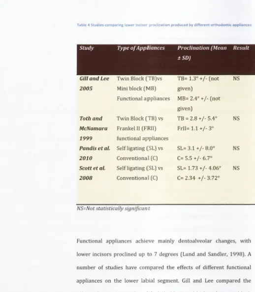
Table 4 Studies comparing lower Incisor procllnatlon produced by different orthodontic appliances
Study Type of Appliances Proclination (Mean Result
±SD)
GUI and Lee
2005
Toth and
McNamara
1999
Pandis et a l
2010
Scott et al.
2008
T w in Block (TB)vs
M in i block [M B )
Functional appliances
T w in Block [T B ) vs
Frankel 11 (F R ll]
functional appliances
Self ligating ([SL) vs
Conventional (C)
Self ligating ([SL) vs
Conventional (C)
TB= 1.3° + /- [n o t NS
given)
M B=2.4° + /- (not
given)
TB = 2.8 + /- 5.4° NS
F r I I = l . l + / - 3 °
S L = 3 .1 + /-8 .0 ° NS
C= 5.5 + /- 6.7°
SL= 1 .7 3 + /-4 .0 6 ° NS
C= 2.34 + /- 3.72°
NS=Not statistically significan t
Functional appliances achieve m ainly dentoalveolar changes, w ith
lo w e r incisors proclined up to 7 degrees [Lund and Sandler, 1998). A
num ber o f studies have compared the effects o f d iffe re n t functional
appliances on the lo w e r labial segment. Gill and Lee compared the
effects o f a conventional m odified Clark’s T w in-block w ith a m in i-b lo ck
appliance [G ill and Lee, 2005). The authors assessed w h eth er
increm ental advancement w ith the m in i-b lo ck affected the
dentoalveolar changes that were produced. T h irty -fiv e age and sex
matched patients were placed in each group. No crow ding
measurements were described. Cephalom etric tracing revealed a
sim ilar a m o u n t of lo w er incisor proclination for both groups. In tra
o p e r a to r e r r o r was assessed by repeating cephalom etric m e a su re m en ts
for 20 random ly selected radiographs, how ev er in te ro p e ra to r e r ro r
w as not calculated. Toth and McNamara retrospectively com pared the
Frankel 11 appliance to a modified Clark's Twin-block appliance and a
control group (Toth and McNamara, 1999). Forty patients w ere trea ted
in each group. The low er incisor to m andibular plane cephalom etric
angulation changed by 0.2° in the control group, 1.1° in the Frankel II
group and 2.8° in the Twin Block group with all groups displaying some
proclination. The a u th o rs did n ot describe the p re -tre a tm e n t crow ding
o r e r r o r of the method. Fixed functional appliances achieve overjet
correction through similar proclination of the low er labial segment.
H ansen etal. described a m ean 10.8° of low er incisor proclination w hen
using a H erbst appliance during a follow up study of 24 patients with
mild low er incisor crow ding [Hansen e t a i , 1997).
Fixed appliance th e ra p y can also result in m andibular incisor
proclination. Minimal differences exist betw ee n the am o u n t produced
by different b racket prescriptions or b e tw ee n conventionally ligated
and self ligated brackets (Fleming and Johal, 2010). Pandis et al, in a
prospective study, com pared m andibular incisor proclination w hen
using a Roth prescription conventional b racket w ith a Damon self
ligating b rack e t (Pandis et ai, 2010b). T w enty-seven patients w ere
tre a te d in each group. All patients had g re a te r th a n 2mm low er incisor
crow ding using Little’s irregularity index w ith similar am ounts of
crow ding in each group. Lower incisor proclination w as m easured
using conventional lateral cephalogram s taken before and after
treatment. No significant difference was found between the groups.
Scott et a l reported on a similar study, again using Damon self-ligating
brackets (32 patients) to compare to Roth prescription conventional
brackets (28 patients] (Scott et al, 2008). Participants had crowding of 5-12mm in the lower arch and were treated with bilateral lower first
premolar extractions. Lateral cephalograms were taken pretreatm ent
and when a 0.019x0.025" stainless steel archwire was placed. These
radiographs were then traced to compare lower incisor proclination.
Again no significant difference was found between the groups. Chen et al. pooled the available research and carried out a systematic review comparing self-ligating and conventional brackets under a number of
different headings (Chen e t al., 2010). They performed a meta-analysis with regards to lower incisor proclination and found conventional
brackets produce slightly more proclination (1.5°)(Chen et al., 2010). Recently Cattaneo et al. compared the proclination produced by active and passive self-ligating brackets (Cattaneo et al., 2013). They used cone beam computer tomography before and after treatment to assess
the lower incisor inclination. They found no difference with regards to
lower incisor proclination. The use of the occlusal plane as a stable
referencing point, however, may question the accuracy of these results.
In the majority of these comparison studies the appliances produced
similar amounts of lower incisor proclination, however no study has
compared the mandibular incisor proclination produced by clear
2.8 In terp ro x im al Enam el R eduction
Many approaches have been used in orthodontics to try and prevent
lower incisor proclination including extraction of teeth, lingual crown
torque, delay in bonding the mandibular incisors and interproximal
reduction. Interproximal reduction decreases the mesiodistal width of
teeth and has become common practice in orthodontic therapy
(Chudasama and Sheridan, 2007). Many authors have described the
benefits of this treatm ent modality where space is required [Peck and
Peck, 1972; Tuverson. 1980; Sheridan, 1985: Sheridan, 1987;
Zachrisson, 2004). The general consensus is that stripping of
mandibular incisors should not exceed 0.75mm at each contact point
with slightly larger amounts possible on posterior teeth (Chudasama
and Sheridan, 2007). Enamel reduction can be performed manually or
mechanically and all surfaces should be polished after completion of the
procedure (Livas
et al,
2013). The available research suggests it doesnot increase the risk of dental caries, sensitivity or periodontal disease
(Zachrisson
et ai,
2007; Zachrissonet al,
2011). Clear alignermanufactures have encouraged the use of interproximal reduction due
to the concerns with closing extraction spaces (Phan and Ling, 2007).
Kravitz
et al.
found that interproximal reduction slightly improved theaccuracy of the tooth movements achieved during aligner therapy
(43.1