Case Report
Pediatric splenic angiosarcoma
Taek Chung, Yoon Ah Cho, Jin Woo Joo, Seok Joo Lee, Cheol Keun Park, Eun Kyung Kim, Sang Kyum Kim
Department of Pathology, College of Medicine, Yonsei University, Seoul, Korea
Received October 24, 2016; Accepted February 13, 2017; Epub July 15, 2019; Published July 30, 2019
Abstract: Angiosarcomas are malignant vascular neoplasms that usually occur in deep soft tissue. Patients di-agnosed with angiosarcoma are usually elderly and are given poor prognoses. Pediatric splenic angiosarcoma is extremely rare, and its pathogenesis is not as well understood as that of older patients. We describe a case of 4-year-old male who had undergone splenectomy for splenic mass. Histopathological examination revealed primary splenic angiosarcoma. We then reviewed published literatures to characterize its clinical characteristics.
Keywords: Angiosarcoma, spleen, pediatric, distant metastasis, prognosis, clinicopathological features
Introduction
Angiosarcomas are malignant tumors of the inner lining of blood vessels that recapitulate the histological features of normal vasculature and endothelium. Most tumors are in the deep muscles of the lower extremities or the abdomi-nal cavity. Patients with angiosarcoma are usu-ally in their 60 s. The prognosis for this cancer is poor.
Primary splenic angiosarcoma arises from the vascular endothelium of the spleen. The mean age at presentation is between 50 and 60 years of age [1, 2]. This malignancy is extremely rare, and pediatric primary splenic angiosarco-ma is rarer still: Only approxiangiosarco-mately 200 cases of primary splenic angiosarcoma have been reported worldwide, and a very small portion of these were in patients aged 18 years or younger.
In this study, we present a case of primary splenic angiosarcoma of a 4-year-old male who had undergone a total splenectomy. We then summarize the information on pediatric splenic angiosarcoma that is available in the literature in the English language to date, and place the new information gained from this case in the context of what is known.
Materials and methods Case
Pediatric splenic angiosarcoma was diagno- sed at Severance Hospital, Yonsei University
College of Medicine, Seoul, Republic of Korea. Representative sections were excised, stained with hematoxylin and eosin (H&E), and reviewed by three pathologists (S. K. Kim, T. Chung, and C. K. Park).
Immunohistochemistry
Tumors were fixed in formalin and embedded in paraffin. Briefly, 5-μm thick sections were cut using a microtome, transferred onto adhesive slides, and dried at 62°C for 30 min. Immuno- histochemistry with antibodies against CD31, CD34, Ki67 (Catalog#s M0823, M7165, and M7240, respectively; DAKO, Glostrup, Denma- rk), Fli-1 (Catalog# 254M-15, Cell Marque, Rocklin, CA, USA) was performed with an auto-mated immunohistochemical staining instru-ment (Ventana Discovery® XT, Ventana Medical System, Inc., Oro Valley, AZ, USA).
Results Case
finding in laboratory tests, although the patient’s hemoglobin level was 9.1 g dL-1 on admission, which was slightly low.
[image:2.612.89.523.72.193.2]The patient underwent a total splenectomy and a wedge-resection of one of the hepatic nod-ules. The spleen measured 16 cm along the
Figure 1. Results from radiological and gross examination. A and B. Computed tomography of abdomen. Note sple -nomegaly with heterogeneous hypervascular and necrotic portions within the spleen and the multiple enhancing lesions within the liver, indicated by the arrow. C. Gross image of the specimen. Note heterogeneous cut surface with
multiple ill-defined grayish soft solid areas, dark red hemorrhagic or sponge-like areas, and yellowish necrotic areas.
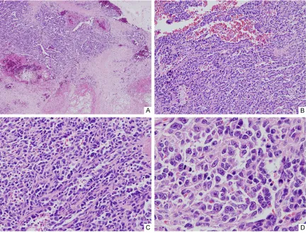
Figure 2. Images from histological examination after H&E staining of specimens of the spleen at different
mag-nification. A. 40× magnification; note hypercellular area with slit-like spaces, necrosis, and hemorrhage. B. 100×
[image:2.612.90.525.265.594.2]long axis and weighed 466 g. Bisection revealed that the cut surface was variegated, with con-gestions, hemorrhages, multifocal necrosis, and ill-defined solid lesions (Figure 1C). Tissue sections were collected from various parts of the spleen and stained with H&E for histologi-cal examination under a light microscope. Microscopic examination revealed hypercellu-lar lesions with slit-like spaces, and confirmed the presence of necrosis and hemorrhage (Figure 2A). Under high magnification (Figure 2B), it was apparent that the slit-like spaces were irregularly anastomosed with one another and filled with red blood cells. These complex anastomosing channels were lined with atypi-cal cells with short-spindle or epithelioid cyto-logical features, eosinophilic cytoplasm, irregu-lar nuclear shapes, and prominent nucleoli (Figure 2C, 2D). Mitotic cells were common: there was an average of approximately 50 mitotic cells in 10 high-power fields.
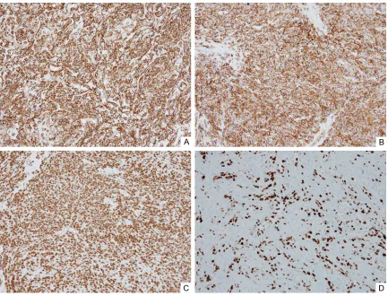
Immunohistochemical staining revealed that vascular markers CD31, CD34, and Fli-1 had a
strong positive signal in the tumor cells (Figure 3A-C). Staining for Ki-67 was positive in approx-imately 30% of tumor cells, revealing increased proliferative activity of tumor cells (Figure 3D). The wedge-shaped resected hepatic lesion had similar histological findings and immunohisto -chemical profiles as did the tumor cells.
These results allowed us to make a diagnosis of primary splenic angiosarcoma with multiple hepatic metastases. The patient was under-went a paclitaxel-based chemotherapy, and was scheduled for a 2-month follow-up radio-logical examination. This exam revealed that the disease status was stable. The patient died 12 months after the surgery.
Clinical features of pediatric splenic angiosar-coma
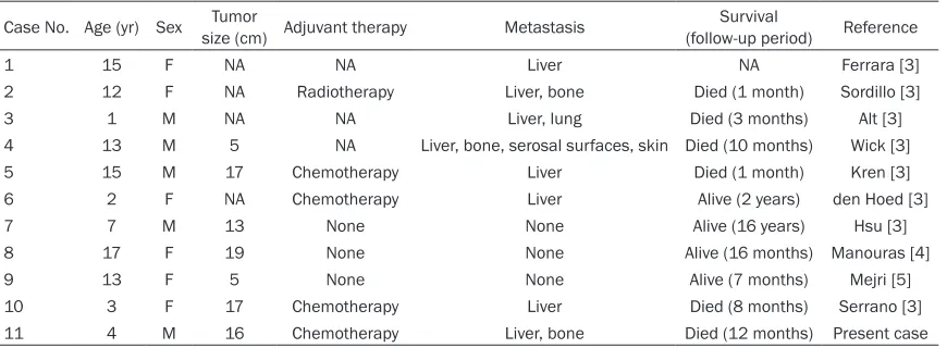
[image:3.612.91.524.69.399.2]The cases of pediatric splenic angiosarcoma that we collected from a literature search are presented in Table 1. If the single case dis-cussed here is included, there have been 11 reported cases of this condition. Patients
Figure 3. Results from immunohistochemical staining of specimens for the proteins: A. CD31; B. CD34; C. Fli-1; D.
ranged from 1 to 17 years old, with a mean age of 9.27 years. Pediatric splenic angiosarcoma affected males and females equally. The lesions ranged from 5 to 19 cm in diameter, with a mean of 13.14 cm. Most tumors metas-tasized to the liver and then to bone. The over-all survival rate was 40% (4 out of 10 patients survived; the follow-up period was 27.4 ± 58.27 months).
Discussion
Angiosarcomas are malignant neoplasms that arise in the endothelial cells of vascular tissue; primary splenic angiosarcomas occur in the spleen. In general, angiosarcomas have poor prognoses, but this is particularly true of pri-mary splenic angiosarcoma because of its high-ly aggressive behavior and frequent metasta-ses. The 6-month overall survival after dia- gnosis is less than 25% [3]. The rarity of this disease makes planning for treatment difficult. Splenectomy can be curative in early stages of the disease, and adjuvant chemotherapy with taxanes such as paclitaxel is also prescribed [3].
Primary splenic angiosarcomais extremely rare in pediatric patients, with only ten cases report-ed worldwide (Table 1) [3-5]. Among these cases, the most common clinical presentation was splenomegaly, and most patients had left upper abdominal pain. Anemia was a common laboratory finding in these patients [6]. Pediatric angiosarcoma of organs other than the spleen appears to occur slightly more often in males than in females [7, 8].
The pathogenesis of primary splenic angiosar-coma is not well characterized. Proposed
caus-al factors include exposure to ionizing radia-tion, previous chemotherapy, or chemical ag- ents such as arsenic [9]. A case report sug-gests that benign infantile hemangioma can be transformed into angiosarcoma by the muta-tion of KRAS gene [10].
[image:4.612.91.526.84.244.2]Our microscopic examination and immunohis-tochemical study supports the diagnosis of primary splenic angiosarcoma. Microscopic ex- amination revealed variably pleomorphic endo-thelial tumor cells lining irregularly anastomos-ing vascular spaces, well-known and typical histological features of angiosarcoma. Frequent mitoses, multifocal necroses, and hemorrhage were also identified in the tissue. Other diagno -ses that should be considered include heman-gioma, littoral cell anheman-gioma, angiosarcoma, ly- mphangioma, or lymphangiosarcoma [6]. He- mangioma is a benign vascular neoplasm with well-formed vascular structures that has no atypical cells, necrosis, or infiltrations. The ca-se under consideration had high-grade nuclear atypia, frequent mitoses, and multifocal necro-sis, which are presumed to be malignant. Li- ttoral cell angioma and angiosarcoma are com-posed of splenic littoral cells, which have posi-tive immunohistochemical staining for CD68 and CD163. Lymphangioma or lymph angiosar-coma are composed of lymphatic vessels and the endothelial cells of this tumor have variable immunohistochemical staining patterns with CD31 and CD34, but strongly positive staining with D2-40. We performed immunohistochemi-cal staining for CD68 and D2-40 on the tumor cells in the case presented here, with negative results (data not shown). Thus, we are confi -dent in our diagnosis.
Table 1. Cases of pediatric splenic angiosarcoma
Case No. Age (yr) Sex size (cm) Adjuvant therapyTumor Metastasis (follow-up period)Survival Reference
1 15 F NA NA Liver NA Ferrara [3]
2 12 F NA Radiotherapy Liver, bone Died (1 month) Sordillo [3]
3 1 M NA NA Liver, lung Died (3 months) Alt [3]
4 13 M 5 NA Liver, bone, serosal surfaces, skin Died (10 months) Wick [3]
5 15 M 17 Chemotherapy Liver Died (1 month) Kren [3]
6 2 F NA Chemotherapy Liver Alive (2 years) den Hoed [3]
7 7 M 13 None None Alive (16 years) Hsu [3]
8 17 F 19 None None Alive (16 months) Manouras [4]
9 13 F 5 None None Alive (7 months) Mejri [5]
10 3 F 17 Chemotherapy Liver Died (8 months) Serrano [3] 11 4 M 16 Chemotherapy Liver, bone Died (12 months) Present case
Angiosarcoma of the spleen is extremely uncommon in adults and even less common in children. It is an aggressive cancer, character-ized by fast growth, frequent metastasis, and a poor prognosis. We have described a case of primary splenic angiosarcoma in a young child. This case report presents a pediatric splenic angiosarcoma which has the characteristic his-tological and immunohistochemical features of this cancer.
Acknowledgements
This study was supported by a faculty research grant of Yonsei University College of Medicine (6-2015-0087 and 6-2016-0034).
Disclosure of conflict of interest
None.
Address correspondence to: Dr. Sang Kyum Kim, Department of Pathology, College of Medcine, Yonsei University, 50-1 Yonsei-ro, Seodaemun-gu, Seoul, Korea. Tel: 2228-6751; Fax: +82-2-362-0860; E-mail: [email protected]
References
[1] Neuhauser TS, Derringer GA, Thompson LD, Fanburg-Smith JC, Miettinen M, Saaristo A, Ab-bondanzo SL. Splenic angiosarcoma: a clinico-pathologic and immunophenotypic study of 28 cases. Mod Pathol 2000; 13: 978-987. [2] Falk S, Krishnan J and Meis JM. Primary
angio-sarcoma of the spleen: a clinicopathologic study of 40 cases. Am J Surg Pathol 1993; 17: 959-970.
[3] Serrano OK, Knapp E, Huang K, Baran G, Stat -ter M, McClain D, Gill J. Pediatric primary splenic angiosarcoma: an aggressive multidis-ciplinary approach to the oncologic manage-ment of a rare malignancy. World J Surg Oncol 2014; 12: 379.
[4] Manouras A, Giannopoulos P, Toufektzian L, Markogiannakis H, Lagoudianakis EE, Papadi-ma A, Papanikolaou D, Filis K, Kekis P. Splenic rupture as the presenting manifestation of pri-mary splenic angiosarcoma in a teenage wom-an: a case report. J Med Case Rep 2008; 2: 133.
[5] Mejri A, Ariane E, Mannai MH, Essoussi M. Pe-diatric primary splenic angiosarcoma: a very rare disease. Tunis Med 2015; 93: 266-8. [6] Auerbach A, Aguilera N, Hall E, Medeiros LJ,
Miranda RN, Vasef MA, Vos J, Wallentine JC, Wang G. Chapter 3: neoplastic splenic disor-ders. In: diagnostic pathology. Spleen Amirsys 2014; 40-45.
[7] Deyrup AT, Miettinen M, North PE, Khoury JD, Tighiouart M, Spunt SL, Parham D, Weiss SW,
Shehata BM. Angiosarcomas arising in the vis -cera and soft tissue of children and young adults: a clinicopathologic study of 15 cases. Am J Surg Pathol 2009; 33: 264-9.
[8] Ayadi L, Khabir A. Pediatric angiosarcoma of soft tissue: a rare clinicopathologic entity. Arch Pathol Lab Med 2010; 134: 481-5.
[9] Ozturk E, Mutlu H, Sonmez G and Sildiroglu HO. Primary angiosarcoma of the spleen. Turk J Gastroenterol 2007; 18: 272-275.
[10] Jeng MR, Fuh B, Blatt J, Gupta A, Merrow AC,
Hammill A, Adams D. Malignant transforma-tion of infantile hemangioma to angioma: re-sponse to chemotherapy with bevacizumab.