warwick.ac.uk/lib-publications
A Thesis Submitted for the Degree of PhD at the University of Warwick
Permanent WRAP URL:
http://wrap.warwick.ac.uk/106749
Copyright and reuse:
This thesis is made available online and is protected by original copyright.
Please scroll down to view the document itself.
Please refer to the repository record for this item for information to help you to cite it.
Our policy information is available from the repository home page.
Attention is drawn to the fact that the
copyright of this thesis rests with its author.
■
LIST OF CONTENTS
ii
INTRODUCTION
Part 1
-Transformation - An In Vitro Model for
Malignancy.
Section A
-Tissue Culture in the Study of Malignancy.
Establishment of Cells in Culture .
In Vitro Transformation.
Agents used for Transformation
Section B
-The Transformed Phenotype.
Contact Inhibition.
Density Dependent Inhibition of Growth.
Anchorage Dependence.
Morphology and Cytoskeleton.
Cell Surface Components .
Increased Agglutinability.
Induction of Cell Surface Antigens .
Increased Nutrient Uptake
Secretion
Cyclic AMP
Correlation of JJi Vitro Transformation
iii
Paqe N o .
In Vivo Tumourigenicity 28
Section C
-RNA Tumour Viruses - Tools for
Transformation.
Relationship Between Mammalian Sarcoma
29
and Lymphatic Leukaemia Viruses . 31
Structure of the Genome . 32
Src Gene and Transformation by R S V .
Transforming Genes of Murine Sarcoma
33
Viruses. 35
Part 2
-Interferon's Potential as an Antitumour
Agent.
«
Section A
-37
Historical Perspective. 37
Section B
-Properties of Interferon. 39
Types of Interferon. 39
Some Characteristics. 42
Section C
-Production and Purification. 43
Inducers . 43
iv
Paqe No.
Purification. 45
Section D
-Cellular Actions of Interferon. 46
Antitumour Activities Iri V i v o . 46
Spontaneous Tumours.
Inhibition of Growth of Normal Cells
48
In V i v o . 49
Clinical Trials . 49
Mechanisms of Antitumour Activity. 50
Inhibition of Growth In Vitro.
Growth Inhibition Studied in Synchronised
53
Cultures . 56
Other Effects on the Cellular Phenotype. 57
Immune Effects of Interferon.
Possible Mechanisms for Interferon's
61
Cellular Activities. 63
Reversion of the Transformed Phenotype. 69
MATERIALS AND METHODS 72
Cells . 75
Interferon Production and Purification. 76
Assay of Interferon Titres. 78
Measurement of Protein Content.
Measurement of Antiviral and Anticellular
78
DNA Synthesis at Different Cell
Densities .
Growth Curves and Saturation Density.
Focus Assay.
Anchorage Dependence.
Cloning Efficiency in Liquid Medium.
Concanavalin A Agglutination.
Morphological Studies .
Conjugation of FITC to DNase I .
Coupling of Affinity Adsorbents to
CNBr-Activated Sepharose 4 B .
Fibronectin Purification.
Preparation of Antiserum to Fibronectin.
Attachment of Cells to Glass .
Polyacrylamide Gel Electrophoresis of
Cell Lysates.
Immunoprécipitation of p 2 1 .
RESULTS
Chapter One
Production and Purification of Interferon.
A. Production of Interferon.
B. Purification.
C. Stability.
^ mggm. ’/'ii** * ■ **<•
Paqe n o .
Chapter Two
Effects of Interferon on Growth of Cells .
A. Sensitivities to Antiviral and Cell
112
Growth Inhibitory Activities. 112
B . Growth and Saturation Densities .
C. Effects of Cell Density on Interferon's
118
Inhibition of DNA Synthesis . 128
D. Focus Formation. 130
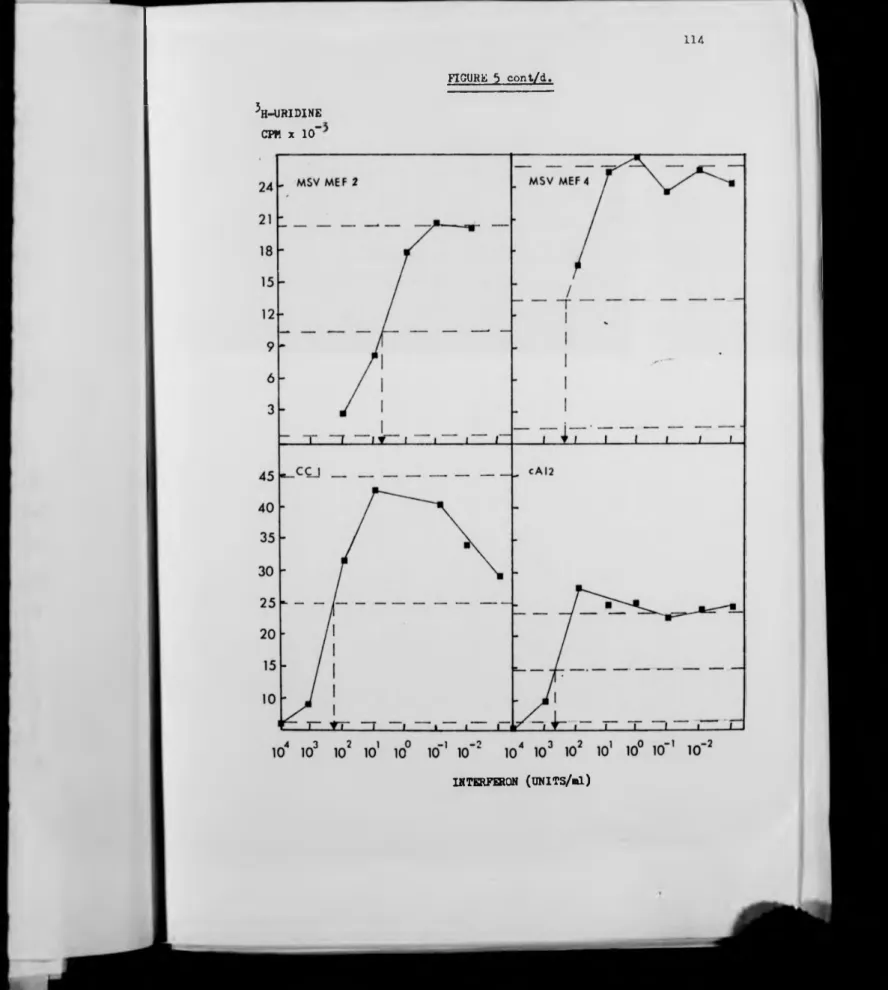
E . Anchorage Independence . 131
F. Cloning Efficiency in Liquid Medium.
G. Effects of Serum Concentration on
134
Interferon Activity. 135
H. Conclusions. 138
Chapter Three
Interactions Between Butyric Acid and
Interferon in Growth Inhibition.
A. Effects of Butyric Acid on Growth
140
Curves.
B . Effects of Butyric Acid on ^H-Thymidine
140
Incorporation into Cellular DNA. 142
C . Conclusions. 145
Chapter Four
Effects on Cell Surfaces and Morphology. 146
A. Agglutinability by Concanavalin A. 146
Page no
Chapter Two
Effects of Interferon on Growth of Cells .
A. Sensitivities to Antiviral and Cell
112
Growth Inhibitory Activities. 112
B. Growth and Saturation Densities.
C. Effects of Cell Density on Interferon's
118
Inhibition of DNA Synthesis. 128
D . Focus Formation. 130
E. Anchorage Independence. 131
F. Cloning Efficiency in Liquid Medium.
G. Effects of Serum Concentration on
134
Interferon Activity. 135
H . Conclusions . 138
Chapter Three
Interactions Between Butyric Acid and
Interferon in Growth Inhibition.
A. Effects of Butyric Acid on Growth
140
Curves.
B . Effects of Butyric Acid on ^H-Thymidine
140
Incorporation into Cellular DNA. 142
C . Conclusions . 145
Chapter Four
Effects on Cell Surfaces and Morphology. 146
A. Agglutinability b y Concanavalin A. 146
l l . i l W i m u ' i P M M l i Ü 9 W
vii Paqe n o .
C. Microfilament System. 158
D. Cell Surface Fibronectin. 163
E. Adhesion to Glass Surfaces. • 172
F. Polyacrylamide Gel Electrophoresis. 174
G . Conclusions. 176
Chapter Five
Effects of Interferon on p21 Levels. 179
A. Electrophoresis. 179
B. Quantitation. 182
DISCUSSION 184
Part I Use of Interferon. 184
1. Interferon Purity. 184
2 . Interferon Doses . 185
Part II The Effects of Interferon
1. Antiviral and Cell Growth Inhibitory
187
Activities . 187
2 . Growth and Saturation Densities. 189
3. Transformation-Specific Growth Parameters. 197
4. Agglutinability. 202
5 . Adhesiveness. 203
6 . Morphology and Fluorescence. 203
7 . Effects of Interferon p21 Levels . 214
Part III General Conclusions. 216
viii
LIST OF TABLES
1. Purification of Interferon by Affinity
Chromatography on BSA-Sepharose 4B
(Experiment 1). 98
2 . Purification of Interferon by Affinity
Chromatography on BSA-Sepharose 4B
(Experiment 2). 100
3. Purification of Interferon by Affinity
Chromatography on Affi-Gel 202. 103
4. Purification of Interferon by Affinity
Chromatography on Poly U-Sepharose 4B . 106
5. Interferon Doses which cause 50% Inhibition
of Cell Growth and Virus Replication. 117
6. Effect of Interferon on DNA Synthesis at
Different Cell Densities. 129
7. Effect of Interferon on Cloning Efficiency
in Liquid, on Focus Formation and on Growth
in Soft Agar of Two Normal and Six
MSV-Transformed Cell Clones . 132
8. Effects of Serum Content of Medium on
ix
9 . Agglutinability of CC1 and NIH 3T3 Cells
by Con A after Growth in the Presence or
Absence of Interferon.
10 . Comparison of p21 Levels in
Interferon-Treated and Control Cell Cultures .
Page n o .
147
■■HP
LIST OF FIGURES
1. Purification of Interferon by Affinity
Chromatography on BSA-Sepharose 4B
(Experiment 1).
2 . Purification of Interferon by Affinity
Chromatography on BSA-Sepharose 4B
(Experiment 2) .
3. Purification of Interferon by Affinity
Chromatography on Affi-Gel 202 .
4. Purification of Interferon by Affinity
Chromatography on Poly U-Sepharose 4 B .
5. Sensitivity of Cell Lines to Interferon's
Antiviral Activity.
6. Sensitivity of Cell Lines to the Growth
Inhibitory Activity of Interferon.
7 . Effects of Interferon on Growth and
Saturation Densities of Several Cell Lines .
8. Effect of Interferon on Growth of Late
Passage (30th passage) C3H10T*s Cells.
9. Lack of Effect of Mouse Interferon on Growth
Page n o .
99
101
104
107
113-114
115-116
119-121
125
■ -■ * ■ - v*' w r wm n n y y » ■ ■ ■m ■ ' •
10. Effect of Interferon on Growth and
Saturation Densities of Cells Grown
in Medium Containing a Very High Serum
Content.
11. Effects of Butyric Acid, in the Presence
or Absence of Interferon, on the Growth
of Cells .
12. Effect of Interferon and Butyric Acid on
DNA Synthesis of Cells at Different
Densities .
13. The Effects of Interferon and Butyric Acid
on Cell and Culture Morphology. i
14. The Effects of Interferon on the Micro
filament System of Normal and Transformed
Cells .
15 . Effects of Interferon andButyric Acid
on Fibronectin Distribution.
16. Effects of Interferon and Butyric Acid
on Distribution of Fibronectin in
Sub-cellular Matrix.
17 . Ability of Cells Grown in Interferon to
Page n o . xi
137
141
143-144
150-156
160-162
165-168
169-170
18. SDS Polyacrylamide Gel Electrophoresis
of Whole Cell Lysates Taken from Cells
4
Grown with or without Interferon (10 U/ml)
and/or Butyric Acid (0.5mM), and Labelled
with 35S-methionine.
19. Levels of Actin in Cells Treated with
Butyric Acid and Interferon for up to •
One Week .
20 . Levels of Fibronectin in Cells Treated
with Butyric Acid and Interferon.
21. SDS Polyacrylamide Gel Electrophoresis of
Cell Lysates Precipitated with Antisera
xii
Page no
175
177
178
m tiÊÊKÊÊÊÊÊÊÊH ÊÊÊÊÊÊÊÊÊÊÊtÊÊÊÊ
xiii
Acknowledgements
I wish to thank those many people who have beeninvolved
with my project, particularly my supervisors, Professor D. C.
Burke and Dr. A. G. Morris, for their endless discussion and
criticism of my work. I am also indebted to innumerable
people within the department for their frequent advice,
assistance and use of equipment, especially Mr. R. Bird for
maintaining a constant supply of interferon, Mrs. B. Wood
for maintaining a constant flow of growth medium, Dr. F. Cooke
for conducting the immunoprécipitation experiments and D r . K .
Logan for instruction in the use of the Reichert ultraviolet
fluorescent microscope. Dr. D. Bray and his colleagues of
the MRC Biophysics unit, Kings' College, London, gave me
valuable instruction in immunofluorescence techniques and > Dr. R. C. Hughes of the National Institute of Medical Research,
Mill Hill, London, gave me advice and donated a small amount
of antiserum to fibronectin. Finally I must thank Miss C.
Alderson for typing the final version of this thesis.
This work was undertaken by award from the Cancer Research
xiv
Declaration
Work from this thesis is presently in press with the
Journal of Cell Science, and is reported herein in Chapter 2
and the Morphology and Microfilament System sections of
Chapter 4. This work and all other studies in this thesis
were conducted b y myself except for the studies on p21 levels
in transformed cells which were conducted jointly between
1
XV
LIST OF ABBREVIATIONS
ALS Antilymphocyte Serum
ALV Acute Leukaemia Virus
AMD Actinomycin D
ASV Avian Sarcoma Virus
AT P Adenosine Triphosphate
BA Butyric Acid
BSA Bovine Serum Albumin
CAMP Cyclic Adenosine-3',5'-monophosphate
CAPS Cyclohexylaminopropanesulphonic Acid
CDNA Complementary Deoxyribonucleic Acid
CEA Carcinoembryonic Antigen
Ci Curie
cl Clone
CNBr Cyanogen Bromide
CM
O
O
Carbon Dioxide
Con A Concanavalin A
cpm Counts per minute
CSA Cell Surface Antigen
cyclic GMP Cyclic Guanosine-3', 5'-Monophosphate
DMEM Dulbecco's Modification of Eagle's Medium
DMSO Dimethylsulphoxide
DNA Deoxyribonucleic Acid
DNase I Deoxyribonuclease I
dsRNA Double-Stranded Ribonucleic Acid
xvi
EDTA Ethylenediamenetetraacetic Acid
EGF • Epidermal Growth Factor
eIF2 Eukaryotic protein synthesis Initiation
Factor 2
EMC Encephalonyocarditis Virus
FITC Flourescein Isothyocyanate
GI50 Interferon dose giving 50% inhibition of
DNA synthesis
GMEM Glasgow's Modification of Eagle's Medium
HaSV Harvey Sarcoma Virus
HAU Haemagglutinating Units
HCl Hydrochloric Acid
IFN Interferon
IUdR Iododeoxyuridine
KiLV Kirsten Leukaemia Virus
KiSV Kirsten Sarcoma Virus
KNRK Normal Rat Kidney Cells transformed by KiSV
LD50 A dose (of a substance, tumour cells etc.) that
will kill half of its target (cells, animals
etc .)
LETS Large External Transformation-Sensitive Protein
LLV Lymphatic Leukaemia Virus
MEF Mouse Embryo Fibroblasts
MEV Mouse Erythroblastosis Virus
met.tRNA.40s Methionyl-transfer ribonucleic acid-40s
xvii
MoLV Moloney Leukaemia Virus
MoSV Moloney Sarcoma Virus
MSV Murine Sarcoma Virus
NaCl Sodium Chloride
Na2C°3 Sodium Carbonate
NaHC03 Sodium Hydrogencarbonate
NaOH Sodium Hydroxide
MOV Newcastle Lisease Virus
63„.
Nl Nickel-63
NIH National Institutes of Health
NK Natural Killer
NRK Normal Rat Kidney
P71 Plasminogen Activator
pH -log^0 hydrogen ion concentration
PHA Phytohaemagglutinin
Poly(HEMA) Poly (2 -Hydroxyethyl me thacrylate)
Poly r l . poly rC . Polyriboinosinic A c i d . polyribocytidylic
acid
Poly U Polyuridylic acid
POPOP 1, 4-di(2(5-phenyl-oxazolyl))-benzene
pppA21p 5 1Ap5'A . (2.5A) .
5'-Triphospho-2' , 5'-Oligoadenylic acid
RSV Rous Sarcoma Virus
SDS Sodium Dodecyl Sulphate
SFV Semliki Forest Virus
• T i-» ? ' ~ " * * ~ * t f ' • « « #
KV
SRBC
TEMED
VI50
VSV
WGA
xviii
Sheep Red Blood Cells
N, N, N ' , N*-Tetramethylethylenediamine
Interferon dose that inhibits virus replication
by 50%
Vesicular Stomatitis Virus
0 H M B P m m m m m
ixx
SUMMARY
The aim of this research was to establish whether or not mouse interferon could reverse the phenotype of transformed cells so that they behaved in a more normal manner.
For this study, clonal isolates of transformed cells from two continuous cell lines and fibroblasts extracted from mouse embryos were used.
It was found that interferon could inhibit the growth of both normal and transformed cells. With several transformed
/
clones interferon also reduced their saturation densities, which were normally several fold higher than those of the
non-transformed parents. This suggested that interferon had induced a partial reversion to density-dependent growth control. Butyric acid also inhibited growth rate and acted additively with interferon when cells were treated with the two agents
together.
Interferon had a variable effect on the ability of dispersed cells to form colonies on plastic substrate in liquid media, but had a consistently greater effect on the ability of transformed cells to form foci on a monolayer of normal cells, and to grow suspended in agar, two growth
conditions specific to the transformed state. It was concluded that interferon had inhibited focus formation and growth in agar by a combination of its growth inhibitory activity and an effect specific for the transformed phenotype.
Interferon also affected the morphology of both normal and transformed cells. The cells became more spread-out, and in transformed cells there was a partial restoration of the microfilament bundle system. Despite these effects on the cytoskeleton, the extracellular matrix of fibronectin fibrils appeared to be little altered by interferon, except when added in conjunction with butyric acid. Under these circumstances, the fibronectin matrix became much more extensive.
1
INTRODUCTION
In the Western World cancer is a major disease. For
a number of years research has been conducted in order to
understand the mechanisms of tumour formation, to learn
how to detect and prevent the processes which lead to a
tumour, and to find methods to cure patients in which
tumours have already g r own.
At present a number of cancers can be treated
successfully by a range of techniques employing combinations
of chemotherapy, radiotherapy and surgery, though such
treatments can be toxic to the p atient.
Interferon was originally identified as an antiviral
agent, but in recent years has been shown to possess
considerable potential for the treatment of a range of
tumours, possibly with minimal side-effects. Experiments
with animals, mainly mice, found that it could prevent the
growth of transplanted tumours and has also caused some
well-developed tumours to regress. Trials with people,
however, have barely begun, having been used only in a
few isolated cases, using small numbers of patients.
Full-scale clinical trials are now being planned
both in Europe and the U.S.A., but one of the major
problems facing these trials is how to secure a sufficient
supply of interferon. The past eighteen months have seen
some dramatic advances towards improving the supply, which,
coupled wit h the planned trials and treatment of two
young patients in Glasgow, has resulted in considerable
publicity in the media .
To date almost all the interferon used in tests on
people has been produced in batch processes b y induction
of leucocytes or lymphoblastoid cells grown in suspension.
Such techniques are time-consuming, expensive and generate
relatively small amounts of interferon. Genetic manipulation
of human interferon genes into bacteria has resulted in
isolation of clones which constitutively produce quite
large amounts of interferon. This material has yet to be
fully characterised and it is not known if it will be
effective in humans. If it is found to be active it should
then become possible to continuously produce vast
quantities on a large industrial scale, thus overcoming
the problems of costs and supply for clinical application.
These developments have placed interferon firmly in
the vanguard of the 'revolution' in biotechnology and, if
successful, may point the way towards enabling large-scale
production of other biological agents for important medical,
agricultural and environmental applications.
Before interferon can be successfully employed as an
anti-cancer agent it is important to establish the
5
dosage, duration of treatment, mode of action, specificity
for tumours and what might result from effects on growth
and behaviour of normal tissues. Some of these can only
be established through the clinical trials, but others,
such as mode of action and effects on normal tissues can
be studied in the laboratory. This thesis was initiated,
as part of a programme in this laboratory, to study the
ways in which interferon affects the transformed phenotype,
and establish whether interferon can actually revert the
transformed phenotype towards a more normal behaviour.
The following introduction is divided into two parts .
The first discusses how the application of tissue culture
has advanced our understanding of neoplasia over the past
few years, and how in vitro transformation can be correlated
with in vivo malignancy. The second part introduces
interferon and surveys our present knowledge of its anti
_
PART I
TRANSFORMATION - AN IN VITRO MODEL FOR MALIGNANCY
4
"I
Section A Tissue Culture in the Study of Malignancy
The study of a range of tumour explants and normal
cells which have been transformed in vitro has significantly
advanced our understanding of the controls placed upon the
growth of normal cells and of the process whereby cells
free themselves from these controls to become tumourigenic
(4, 128, 189, 197, 204) . However, tissue culture does have
a number of limitations that stem from the way in which
cells of different species and different tissue origin
behave in widely differing fashions. This leads one to
question how closely the properties of cells in culture
reflect those prevalent in v ivo. This problem of how
relevant in vitro studies are to in vivo malignancy will be
considered later. This section will discuss the basic
properties and behaviour of different cell types grown in
vitro. with a view to establishing which criteria need to be
considered when selecting a cell system to study in vitro
transformation.
1. Establishment of Cells in Vitro
The most direct way to study tumourigenic cells in
vitro is to explant human or animal tumours and establish
their use suffers from a number of disadvantages which are
at least partly due to most tumours being a heterogeneous
collection of different cell types (204) . One particular
cell type may grow more vigorously than any of the others,
and thereby come to dominate the culture, making it un
representative of the original tumour (ref 204 p403), and
indeed may not even be the malignant cell type. Moreover,
rodent cells, for reasons discussed below, when in vitro
often spontaneously undergo changes which may greatly alter
their characteristics (ref 204 p403) . Finally, in a
heterogeneous tumour it may be impossible to determine from
which cell type the tumour originated and so there can be
no normal cell type with whose behaviour it can be compared.
A more closely controlled experimental system can be
established ¿n vitro by explanting normal adult or
embryonic tissues and then transforming these. The trans
formation process yields transformed cells whose properties
can be directly compared with the normal parental line.
Apart from being diploid normal fibroblasts when
explanted possess a number of characteristic behavioural
properties;
1. they show contact inhibition of movement, i.e. migration
of a cell in one particular direction is halted when contact
is made with another cell, but movement in other directions
is unhindered (3, 4).
2. their growth shows density dependent inhibition, i.e.
growth is arrested (158, 241, ref 115 p451) .
3. their growth is anchorage dependent, i .e. they can
only grow when attached to a solid substrate, not in
suspension (242, ref 115 p452-3) .
These properties will be discussed in greater detail
later.
Cells of human and avian origin retain these properties
for between ten and over 100 cell doublings, after which
time the cultures die out (ref 204 p 4 02). Such cells are
said to be stable, and possess finite growth potential (204) .
Cell cultures of rodent origin, however, frequently
produce cultures which are aneuploid and have acquired
infinite growth potential (ref 204, pp 402 & 409) - they
will grow in culture forever. These cells are said to be
of unstable species origin (204). The resultant aneuploid
cultures invariably eventually transform spontaneously,
and become tumourigenic in vivo (ref 204, pp402 & 408).
However, it is possible to isolate by careful culture
techniques lines which retain the phenotype of normal cells,
even though they are aneuploid.
In this way the established 'normal' cell lines have
been developed, such as the mouse 3T3's (2, ref 204 p409)
and C3H 10T*5 (210) and the baby hamster kidney (BHK) cells
(240) . These too tend to undergo spontaneous transformation
7
2 . In Vitro Transformation
Ponten has defined transformation as the acquisition / of permanent disturbance of growth and/or locomotion
control (204) . Thus, transformed cells tend to lose
density-dependent growth (241, ref 115 p451, ref 204 pp 406
& 409), contact inhibition of movement (3, 4, ref 204
pp405-6) and anchorage dependence (242, ref 115 pp 452-3) .
The final and most fundamental criterion for tranformation,
however, is tumourigenicity in vivo. The correlation
between in vitro transformation and iji vivo tumourigenicity
is not total, a problem that will be discussed later.
3 . Agents used for Transformation
Agents which have been used for in vitro transformation
are all known to cause tumours in vivo. They fall into
three main categories;
1) Ultraviolet or X-ray irradiation (50, ref 204 p410) ,
2) Chemicals such as polycyclic aromatic hydrocarbons,
alkylating agents and nitrosamides (62, 117) .
3) Oncogenic viruses, such as the DNA viruses SV40 and
polyoma, plus the RNA tumour viruses, or retroviruses
(115, 204) .
Chemical and radiation-induced transformation probably
occurs by mutagenesis . In vivo many carcinogenic chemicals
must be activated by oxidases . When activated such
target cells, and it is possible to detect chromosome
alterations a few cell doublings after treatment (20, 21, 181).
Onset of transformation, however, can vary from just
a few up to 80 or more cell doublings following treatment.
Rodent cell lines have proved to be the easiest to transform
by chemicals and radiation, but only quite recently has it
been possible to transform human cells (140, 148, 211) .
It has been suggested that resistance to chemical
and radiation-induced tranformation by human cells may be
due to a highly efficient DNA-repair mechanism (62).
Oncogenic viruses transform cells by introducing new
genes into the cellular genome which code for transformation.
The whole process of infection and transformation is quite
rapid and c.an be completed within a few cell doublings
(204). Retroviruses will readily transform murine and avian
cells, but not human, for which SV40 is the usual trans
forming agent (251-3) . Maintenance of the transformed
phenotype requires the constant presence of the product(s)
of the transforming gene. Studies with mutants of Avian
Sarcoma Virus (ASV) and Kirsten Murine Sarcoma Virus (KiSV)
temperature sensitive for transformation have found that
transformed cells rapidly revert to normal upon shift from
the permissive to the non-permissive temperature (228, 232,
ref 115 p423) , thus suggesting rapid inactivation of the
transforming gene's product(s).
9
cells by activating endogenous oncornaviruses has been
discounted since no viral synthesis can be detected in
chemically transformed cells, whilst agents which do
activate endogenous viruses, such as iododeoxyuridine (IUdR),
do not transform cells (117) .
The tendency for an established cell line to continue
through a series of changes, progressing towards a more
transformed state, from which clones of spontaneous trans
formants can frequently be isolated (204), creates problems
when comparing the behaviour of transformed clones with
the 'normal' parental line, since the 'normal' line may
itself be progressively becoming more transformed.
The use of cells of stable species (i.e. human and
avian) may overcome these problems since their behaviour
remains constant throughout and a culture of normal cells
is more likely to be representative of its in vivo
ancestors.
The study of human cancer should ideally use human cells
in vitro, but, as already described, transformation of
human cells b y chemical carcinogens and radiation has only
recently been achieved. Transformation of human cells by
retroviruses is also difficult, and the only really successful
agent routinely used is SV40 (234, ref 253 pp360-l). The
ease with which avian and rodent cells transform with
retroviruses has made these cells the choice system for many
It could be argued that the study of viral transform
ation is irrelevant to human cancer since no human tumour,
with the exception of papillomas, has yet been found to
be virus-induced. This argument is strengthened by three
points:-a) The initial stages of transformation by chemicals (the
main human carcinogens) are different from those with
oncogenic viruses,
b) The latent period before transformation by oncogenic
viruses is complete is only a few days, but can be up to
several weeks for chemicals (20, 21, 53, 140, 181, 211),
c) Some human tumour explants may still show some normal
growth and movement control which is only lost when the
cells are infected and transformed by oncogenic viruses
(ref 204 p411) . /
On the other hand, many naturally occurring animal
tumours are caused by endogenous and exogenous retroviruses,
and explants of animal tumours behave in a manner very
similar to cells transformed by viruses in vitro (204) .
Furthermore, though there have been considerably fewer studies
of the transformed phenotype resulting from chemically- and
radiation-induced transformation, the resulting phenotype
seems to be quite similar to that of the virally transformed
cells, suggesting that whatever the mechanisms of transform
ation may be, the end result in terms of phenotype is
■> w* '
Thus, in studies where only the behaviour of transformed
cells is being studied, not the mechanisms whereby trans
formation occurred, choice of transforming agent may not
be so crucial. The next section describes the transformed
phenotype using data drawn mainly from studies of viral
transformation, though some data obtained from chemically
transformed cells are also included.
11
Section B The Transformed Phenotype
Upon transformation, either spontaneous or induced,
cells acquire a number of characteristics which usually
reflect the generally decreased control of growth and
movement, such as loss of contact inhibition of movement,
loss of density dependent growth control and loss of anchorage
dependence.
Contact Inhibition
Contact inhibition was first described by Abercrombie
and Heaysman (3) . As defined earlier, it is the property
by which a normal cell tends to stop moving in a particular
direction if it comes into contact with another cell, but
is able to move away in other directions. This leads to
confluent cultures becoming highly ordered with elongated
cells running parallel to each other and forming complex
12
fibroblast cultures, at least, cell movement does not
stop necessarily, but a continuous cell streaming occurs
(204) .
Transformed cultures rarely show much order. Usually
the cells grow in a random criss-crossed fashion, with cells
piling up on top of each other.
It has been possible to quantitate contact inhibition,
and generally it is found that transformed and tumour cells
have greatly reduced contact inhibition, which may account
for the disorder in transformed cultures (4) . Bell (22) ,
however, concluded that the criss-crossed pattern he
observed with a culture of polyoma-transformed 3T3 cells was
due to the random extension of pseudopodial arms from the
cells, not loss of contact inhibition. However, it is
probably partly loss of contact inhibition that enables
transformed cells to grow and move on top of a monolayer of
normal cells. This has enabled identification and selection
of newly transformed clones from such foci growing amidst
a background of normal cells (ref 115 pp418-420) . Efficiency
with which dispersed transformed cells are able to grow to
form foci on a normal monolayer is a simple quantitative
assay for loss of contact inhibition and density independent
growth.
Density Dependent Inhibition of Growth
15
little below that of transformed cells, but when a confluent
monolayer of cells forms growth often abruptly ceases or
slows greatly (158, 197, ref 115 p451, ref 204 pp406 & 409) .
Transformed cells, on the other hand continue to grow
rapidly until their density is 10 to 20 times greater than
normal cells, forming cultures which may be several cell
layers thick, even in the presence of only very low levels
of serum (197, ref 204 pp409-410, ref 115 p451) .
It was at one time thought that contact inhibition
prevented growth of normal confluent cultures (158, 159,
241, ref 115 p451); contact with cells on all sides inhibited
a cell's further growth. However, many nontransformed cell
lines may actually grow to quite high density if the supply
of serum or growth factors is increased (ref 115 p451) . In
such dense cultures of normal cells, unlike transformed
cultures, the orderly arrangement, which is attributed to
contact inhibition, is retained. Stoker and Rubin (241)
proposed the term 'density dependent inhibition' to describe
this growth control phenomenon, which may be somehow linked
to the supply of serum growth factors (ref 115 p451) .
Folkman and Greenspan (72) proposed that cell shape may be
important in determining a normal cell's ability to grow.
This was supported by Folkman and Moscona (73) w h o coated
plastic Petri dishes with a nontoxic substance called poly
(2-hydroxyethylmethacrylate) (poly(HEMA) ) at a range of
14
of the plastic so that on dishes bearing no poly(HEMA)
normal cells spread well, whereas with increasing concen
trations of poly(HEMA) cells became progressively more round.
They found that the rate of DNA synthesis in any sparse
culture was inversely proportional to the vertical thickness
of each cell. In confluent cultures growing on untreated
plastic the cells became more rounded, presumably due to
cell crowding, and DNA synthesis slowed considerably, to a
level very similar to sparse cells which were rounded by
poly(HEMA) treatment.
SV40-transformed 3T3 cells grew equally rapidly on all
treated and untreated plates, showing that their growth rate
was not regulated with cell shape.
Folkman and Moscona suggested that sensitivity to serum
growth factors in normal cells is controlled in some way by
cell shape, possibly due to changes in surface area, so
that flat cells are able to respond to serum and so grow,
whereas rounded cells do not grow because they are less
sensitive. Increasing the serum concentration should at
least partially overcome this and allow the cells to grow.
Cell shape and sensitivity to serum may be uncoupled b y
transformation and thus allow transformed cells to grow to
very high densities regardless of morphology and serum
concentration.
Anchorage Dependence
15
dependence' to describe the property of rton-transformed cells
whereby they are obliged to attach to a solid support in
order to grow, unlike transformed cells which are able to
grow freely in suspension. Macpherson and Montagnier (173)
developed a method to grow single transformed cells into
colonies suspended in semi-solid agar. This technique has
proved very useful for isolation of transformed clones from
a background of normal cells, and for quantitation of the
degree of anchorage independence possessed by different
transformed clones.
How cells lose anchorage dependence is not known but
it may be explained b y the coupling of cell shape to growth
control, outlined above. Cells in suspension are spherical
and therefore under the theory put forward by Folkman and
Greenspan (72) normal cells will be unable to grow, possibly
due to insensitivity to serum growth factors. Transformed
cells will grow due to uncoupling of cell shape from growth
control. A role of cell shape in growth control is supported
by the observations that normal cells will grow suspended
in agar if glass beads large enough for the cells to attach
and flatten out are included in the a g a r . Silica particles
to which the cells can anchor but are too small for them
to spread out will not support their growth (72). This
would suggest that anchorage per se is not sufficient for
cell growth, rather that cell flattening may b e .
■ w jM p g jp W n
behaviour are profoundly changed by transformation. These
changes involve cell-cell communication, cell morphology
and nutrient requirements, all of which are closely related
to cell surface phenomena. It is not surprising, therefore,
that many of the biochemical changes so far identified in
transformed cells involve, either directly or indirectly,
the cell surface. It is still not clear, however, which
changes may be primary lesions causing the transformed
phenotype and which are secondary changes occurring as a
result of transformation.
The changes so far identified include rounded cellular
morphology and partial dissolution of the cytoskeleton
(197, 203, ref 115 pp444-447) , altered plasma membrane lipids
and glycolipids (ref 189 pp5-6, ref 115 pp436-9), decreased
cell surface fibronectin (259, 274, ref 128 pp78-82, ref
189 pp7-8), collagen (ref 128 p85) and glycosaminoglycans
(263, ref 189 p 5 ) , new cell surface antigens (150,ref 189
pp34-43), increased cell agglutinability (ref 115 p443,
ref 189 ppl9-34), increased uptake of certain amino acids
(188, ref 189 pl4) and sugars (ref 115 pp441-3, ref 189 pl4 ) ,
secretion of proteases (ref 115 p439) and other proteins
(88, 226) and reduced levels of cyclic AMP (197, ref 115
p447) .
Very few transformed clones possess all these character
istics; usually only a few are expressed. Some properties
but may manifest themselves some time later, vivo
tumourigenicity is the ultimate test of a transformed
clone. No single phenotypic marker for transformation
correlates completely with iji vivo tumourigenicity, but
some, such as growth in agar, are better indicators than
others (75, 139) . This will be discussed in detail later.
Normal cells under certain conditions can actually be
made to temporarily mimic the transformed state. For
instance,cells in mitosis or when mildly treated with protease
are rounded, have lost the microfilament system and cell
surface fibronectin, and are more agglutinable (ref 189 p l 2 ) .
Morphology and Cytoskeleton
Normal fibroblasts usually have a well-spread flattened
morphology and are firmly adherent to the substratum. Cell
shape, adhesion and motility are largely controlled by the
cytoskeleton (39, 149, 156), which consists of microtubules,
intermediate filaments and microfilaments.
Microtubules, consisting principally of tubulin, and
intermediate filaments, consisting of desmin, form two
separate lattices throughout the cytoplasm (130, 193, 266,
267). Microfilaments, containing mainly actin, exist in
two arrangements (149):
a) as single filaments dispersed throughout the cytoplasm,
in the perinuclear region and associated with the leading
as bundles (sometimes called stress fibres) running in
straight parallel arrays along the lower surface of the
plasma membrane ( 87, 155, 156, 203).
Upon transformation the cells usually become smaller,
rounded and less adherent to the substrate. Some observations
have suggested that the microtubule system may break down
upon transformation (37, ref 115 pp444-7), but in the
majority of cases at least, it now seems that the micro
tubules remain intact (60, 192). Similarly, the intermediate
filaments remain essentially unchanged (130) . The micro
filaments, however, particularly the stress fibres in most
transformed lines are either lost or at least greatly
reduced . The total amount of cellular actin may or may not
be reduced (70, 215, ref 128 p 8 8 ) , but is probably present
in the cytoplasm in the monomeric form instead of the poly
merised state (ref 128 p88) .
Loss of the stress fibres probably at least partially
explains the rounded morphology and weaker adhesion (ref
128 pl22) .
Cell Surface Components
Alterations in synthesis of various plasma membrane
lipids, glycolipids, glycosaminoglycans and phospholipids
have been reported but much of the data are conflicting
and few generalisations can be drawn (115, 189, 262).
19
fluidity of the plasma membrane is increased after
transformation, but the weight of evidence which has
accumulated against this possibility now makes it seem
unlikely (ref 115 p444) .
Normal cells secrete onto their outer surfaces a
matrix which aids adhesion to the substrate (259, ref 128
p70) . This matrix, or glycocalyx, is known to consist of
at least three principal components; fibronectin, collagen
and glycosaminoglycans (259), and seems to be involved in
adhesion of cells to the substrate.
In transformed cells this glycocalyx is greatly reduced.
The glycosaminoglycans are still produced and secreted in
large quantities but they are not retained on the cell
surface (ref 259 pl6) . Synthesis of collagen and fibronectin
is reduced, and in addition, fibronectin which is synthesised
and secreted is not retained on the cell surface as efficiently
as on normal cells (191, ref 259 pl4 & 17).
Fibronectin, LETS (Large External
Transformation-Sensitive protein) or CSP (Cell Surface Protein) as it may
be known (amongst other titles), is a large glycoprotein
with subunit molecular weight of 2.2 to 2.5 x 10^ daltons
(259, 274, 275, 278).
Primary explants of both epithelial and fibroblastic
cells synthesise and secrete onto their surfaces large
amounts of fibronectin while established cell lines produce
20
if any, fibronectin. Its absence was thought to correlate
very closely with in vivo tumourigenicity (259, 274), but
several tumourigenic transformed lines have recently been
found to possess levels of fibronectin similar to the
parental non-transformed line (274) .
Antisera raised to fibronectin of one species
cross-reacts with fibronectin of a range of species, suggesting
a conserved structure (2 79, 286) . Immunofluorescence using
such antisera reveals several different patterns on cells
of primary expiants (177, 274) . Confluent cultures possess
a complex intercellular filamentous matrix running across
the upper surfaces of the cells . Subconfluent cultures
show mainly 'stitches' of fibronectin between adjacent cells,
with very little matrix running across the cells. Extraction
with NP40 allows the antisera to stain fibronectin beneath
the cells which forms a filamentous network across each
cell, with some intercellular connections (131, 177).
Addition of fibronectin to cultures of transformed
cells often results in their adopting a more normal
phenotype-flattened, more orderly alignment and containing micro
filament bundles (6, 274, 277), and also increased cell
motility (7) . Again, fibronectin from one species is
active in a range of other species of cells (6, 277). There
is no effect, however, on growth rate, saturation density
or nutrient transport (6, 274, 277) .
21
and microfilaments. In normal cells immunofluorescent
staining patterns for actin and fibronectin reveal con
tiguous fibres with microfilaments inside the cells and
fibronectin outside (131), treatment with proteases or
cytochalasin B disaggregates both microfilaments and fibro
nectin fibres, and mitotic cells too lack both structures
(128, 189, 259) .
For interactions between the microfilaments and fibro
nectin fibres to occur there must be a third component in
the plasma membrane since neither actin nor fibronectin have
yet been identified as intramembranous proteins . Evidence
for such a component is increasing, though its identity is
still a mystery (128, 131, 230) . Thus it seems probable that
microfilaments and fibronectin together play a major role
in facilitating cellular adhesion and controlling cell
morphology, but apparently having little effect on control
of growth.
Disruption of this microfilament-fibronectin complex
in transformation would clearly result in considerable
changes in cellular behaviour.
Increased Agglutinabilitv
Transformation is usually associated with increased
agglutinability of suspended cells by lectins such as
con-conavalin A (con A) and wheat germ agglutinin (WGA) (51,
22
mitosis or when subjected to mild protease treatment mimic
this increased agglutinability (ref 115 p443, ref 189 p 2 4 ) .
Transformed cells do not bind more lectin, rather
cross-linking of bound lectin molecules with each other on the
same and adjacent cells is somehow facilitated (51, 195,
ref 189 p 2 2 ) . This facilitation is probably achieved by
improved mobility of lectin-bound receptors within the plasma
membrane. Control of mobility of surface receptors is thought
to be by a structure called the Surface Modulating Assembly,
consisting of microtubules, microfilaments and possibly
fibronectin (65, 276, ref 128 pp95-99, ref 189 pp26, 29-31).
Partial disruption of the SMA by transformation through loss
of microfilaments and fibronectin would free the receptors
from limitations on their mobility. No direct experimehts
have proved the existence of the SMA, but experiments with
drugs which disaggregate microtubules and microfilaments
resulting in increased agglutinability argue strongly in its
favour (65, 66, 178, 179, ref 189 p 3 0 ) .
A number of reports have shown that raised agglutin
ability with con A, but not WGA, correlated quite well with
in vivo tumourigenicity (ref 189 p22) . Recently a 100000
dalton cell surface glycoprotein was found to bind proportion
ally more con A, but less WGA, following transformation than
before (273) . A range of transformed cells which showed
this alteration were also tumourigenic in v i v o , whereas those
23
so was used for selecting normal revertants . Such revertants
showed decreased con A binding to this 100K glycoprotein,
as well as being non-tumourigenic (273).
Induction of Cell Surface Antigens
Both chemically transformed cells and some human tumour
cells express on their surfaces common embryonic antigens
and tumour-specific transplantation antigens which vary with
cell type and carcinogen (ref 115 p 4 4 0 , ref 189 pp34-36).
Retrovirus-transformed cells also express cell surface'
antigens (CSA) which appear to be mainly virus-coded,
consisting of, or at least associated with, precursor poly
proteins of the gag and env structural genes (150, 235).
These CSA's of course make such cells antigenically
distinguishable from normal cells and will thus elicit an
immune response iji vivo which will limit the spread of
tumours caused by these cells (150) .
Increased Nutrient Uptake
The uptake of glucose and its nonmetabolisable analogues
2-deoxyglucose and 3-0-methylglucose as well as mannose,
galactose and glucosamine are all stimulated following
transformation, whereas the uptake of other carbohydrates
is unaffected (ref 115 p442, ref 189 p l4). Similarly the
amino acids glutamine, arginine glutamic acid and alpha
24
readily (ref 189 pl4), though a recent report by Nakamura
and Weber (188) found no differences in amino acid uptake
between normal and chick embryo fibroblasts transformed by
Rous Sarcoma Virus (RSV) unless they were starved of amino
acids first.
The reduced requirement for serum by transformed cells
may be attributable to increased uptake of nutrients from
the medium, or perhaps to increased sensitivity to growth
factors in the serum.
Secretion
Transformed cells and tumours both in vivo and in vitro
secrete proteases which, it has been proposed, may be
partly responsible for the transformed phenotype, for instance
by cleaving away cell surface fibronectin (ref 115 p439,
ref 128 pp91-92, ref 259 pll) .
One such protease is plasminogen activator which
converts plasminogen (normally found in the serum in culture
media) to plasmin, to which fibronectin is very sensitive
(ref 128 p 9 1 ) . This may be one cause of the loss of
fibronectin from cell surfaces, but the evidence so far
suggests that it is at best only a minor cause (ref 128 pp91-92)
Senger et al^. (226) have recently identified two classes
of transformation-specific proteins, both with molecular
weights of about 60000 daltons, and one of which is
25
different mammalian species transformed by a range of type
C viruses. However, it is still not clear just how
general these proteins are throughout the whole range of
transformed and tumour cells, nor is their role in trans
formation understood .
Cyclic AMP
Cyclic adenosine-3',5'-monophosphate (cAMP) has been
implicated many times in the control of the growth of cells
(197, ref 115 p447) . Several normal cell types, e .g.
Swiss 3T3 and NRK, when growing rapidly have quite low
levels of cAMP, but as confluence is reached the amount
present rises by about two fold or more (197) . In trans
formants of these cells cAMP levels always remain low (197) .
There are, however, several normal cell lines which do not
show this fluctuation, such as BALB/c 3T3 and chick embryo
fibroblasts (197), so the significance of cAMP is
questionable.
The best evidence for a role of cAMP in control of
cell behaviour comes from experiments on the effects of
treatment of transformed cells with cAMP or dibutyryl cAMP.
Transformed cells treated with dibutyryl oAMP became flatter,
more adherent and arranged in orderly parallel arrays,
cytoskeleton is restored, growth is slower and the cells
are less agglutinable (197, 205, ref 115 p448). This effect
The physiological significance of results obtained
with dibutyryl cAMP is in doubt since butyric acid has
similar effects on morphology (8, 83, 85, 171, 271), though
Storrie et al. (243) found that the effects were not
identical. Butyric acid-treated transformed cells become
flatter, have a restored cytoskeleton, raised intracellular
cAMP levels and grow more slowly (as do normal cells in the
presence of butyric acid) , but density dependent growth
control is not restored (8) . Altenberg et al_. (8) found
that butyric acid did not restore adherens junctions
between cells, a type of junction which is common between
normal cells . They proposed that this may be why growth
control was not restored.
Corrélation of In Vitro Transformation with In Vivo
Malignancy
Many of the observations regarding behaviour of
transformed cells often apply well to cells of naturally
occurring tumours explanted into tissue culture. They are
small, rounded, generally lack a microfilament system and
cell surface fibronectin, grow in agar, grow to high
densities on a plastic substrate and are highly agglutinable.
Thus it would seem that the in vitro transformed phenotype
is a realistic equivalent to in vivo tumours. It is probable
that certain of the i_n vitro properties correspond with
27
inhibition of movement in vitro may correspond to invasive
ness ^ n vivo (4, ref 204 pp405-6), loss of density
dependent growth control to uncontrolled irregular growth
in vivo (ref 189 p22, ref 204 p410) and anchorage independence
to ability to metastasise (50).
Similarly the expression of a number of in vitro
phenotypic characteristics of transformed cells have been
shown to correspond quite well with their ability to form
tumours in v i v o . Such characteristics include growth in
agar (50, 75, 139), loss of cell surface fibronectin (274),
growth to high saturation densities (1, ref 189 p22, ref
204 p410) and raised agglutinability by con A (272, 273,
ref 189 p 2 2 ) . None of these correlates fully with in vivo
tumourigenicity (23, 44, 137), but the combination of the
four is generally sufficient to identify a tumourigenic clone.
Growth in agar is probably the most widely used method,
reportedly correlating very closely with in vivo tumourigen
icity (50, 53, 75, 139, 229). Loss of cell surface fibronectin
was thought to correlate completely with in vivo tumourigen
icity, but a number of exceptions have been discovered
(274) . It is now thought to correlate more closely with
the ability to metastasise in vivo or in vitro growth in
agar (274) . Growth to high saturation density was probably
one of the first phenotypes to be equated with in vivo
tumourigenicity (ref 189 p22, ref 204 p410) . Many
exceptions have been found (ref 204 p410). though it is still
Con A agglutinability generally parallels loss of
growth control (ref 189 p22), so its consequent correlation
with tumourigenicity is not surprising. This may also
relate to the 100000 dalton marker for malignancy mentioned
earlier (273) . Agglutination by other lectins, such as
soybean and wheat germ agglutinins, does not correlate
with tumourigenicity.
In Vivo Tumourigenicity
The ultimate test to equate .in vitro transformation
with in vivo carcinogenesis is to inoculate animals with
transformed cells and watch for the growth of tumours.
The need for histocompatibility of cells and hosts
restricts iji vitro work to use of cells explanted from
inbred mice. Transformed cells can then be inoculated into
syngeneic hosts . This limitation has been partly overcome
by the introduction of nude mice which are athymic and so
are unable to mount T cell-mediated immunity (196, ref 204
p413) . These mice are able to take allogeneic grafts,
including those from other species (207) . However, these
mice can mount humoral immunity and Natural Killer (NK) cell
activity which could destroy allogeneic grafts.
Due to the lack of T lymphocytes these mice are very
susceptible to infections which may prove lethal, and so
are very difficult to handle. It is at least partly for
this reason that the majority of studies are still conducted