Bacterial sialic acid degradation:
membrane transport and enzymology
A thesis submitted in partial fulfilment of the
requirements for the degree of
Doctor of Philosophy in Biochemistry
in the School of Biological Sciences
by Rachel Aimee North
University of Canterbury
Abstract
The overarching aim of this thesis is to understand how sialic acids are transported into the bacterial cell and then degraded by bacterial pathogens. In heavily sialylated environments, bacterial pathogens utilise host-derived sialic acid as a nutrient source, and this pathway constitutes a novel and unexploited target for antibiotic drug design.
Three sialic acid transporters, from two distinct gene families were investigated to probe how sialic acid is transported across the cytoplasmic membrane. The Yersinia pestis sugar proton symporter (NanT) exists as a single species in solution, while the Staphylococcus aureus sodium solute symporter (SSS) and Proteus mirabilis SSS appear to be in a self-association. It is likely that the SSS sialic acid transporters exist in a monomer-dimer equilibrium, although higher oligomeric structures cannot be ruled out. The structure of the P. mirabilis SSS sialic acid transporter was solved, which is the first known structure of a sialic acid transporter to be presented. It was solved in complex with sialic acid and two sodium ions, providing insight into how this transporter mediates the movement of sialic acid across the membrane. In addition, the structure presents a novel conformation among sodium symporters and the structural basis for the movement of sialic acid across the membrane is elucidated.
The structure, function and catalytic mechanism of the third enzyme involved in sialic acid degradation, N-acetylmannosamine-6-phosphate 2-epimerase, were explored from MRSA. This enzyme was demonstrated to adopt a dimeric architecture in solution, consistent with the crystal structure that was solved and presented. A multi-enzyme coupled assay was developed to assess N-acetylmannosamine-6-phosphate 2-epimerase activity in real time. Residues were probed that may be important for catalysis and tested by mutagenesis and kinetic analysis. A novel mechanism of carbohydrate epimerisation is proposed for this enzyme, whereby catalysis occurs via a proton displacement mechanism mediated by the substrate.
Acknowledgements
My biggest thank you is owed to my supervisor, Renwick Dobson, whose enthusiasm and passion for science is inspiring. Thanks for all of the incredible opportunities and for making the last four years enjoyable. But most of all, thank you for your unwavering support, guidance and encouragement. Thanks also to the other members of my PhD committee; Sarah Kessans, Andrew Muscroft-Taylor and Juliet Gerrard for your interest, advice and input into my project.
Thanks to Richard Neutze for inviting me to work on part of my project in your lab, which unknowingly was the exact same project as the group next door to you. Thus, an incredibly big thank you goes to Rosmarie Friemann, for allowing me to work so closely with your group. Thanks to all of your group members for sharing the project and for teaching me.
A special thank you goes to Kat, for your friendship and support during these challenging four years. I don’t know how I would have got through it without you. Thank you to all the people that have been a part of the Dobson, Gerrard and Pearce labs over the last four years. In particular, thanks to Jen, Chris, Jeremy, Amy and Arvind. I feel privileged to have worked with such an amazing group of people and to form such great friendships with you all. Thank you to Grant for your graphical assistance and thanks to Jackie for making sure the lab was always running smoothly.
Table of contents
Title page ... i
Abstract ... iii
Acknowledgements ... vi
Table of contents ... viii
Abbreviations ... xiv
Chapter One: Introduction ... 1
1.1 Sialic acid ... 1
1.2 Sialic acid utilisation by bacterial pathogens ... 2
1.3 Bacterial pathogens evade the host immune response by molecular mimicry ... 3
1.3.1 Precursor scavenging ... 4
1.3.2 De novo biosynthesis ... 4
1.3.3 Donor scavenging ... 4
1.3.4 Trans-neuraminidase activity ... 5
1.4 Importation of sialic acid into the bacterial pathogens ... 6
1.4.1 ATP binding cassette transporters for sialic acid transport ... 7
1.4.2 Tripartite ATP independent periplasmic transporters for sialic acid transport ... 8
1.4.3 Sugar proton symporters for sialic acid transport ... 9
1.4.4 Sodium solute symporters for sialic acid transport ... 10
1.4.5 Sialic acid is transported using a specific transporter ... 10
1.5 Sialic acid degradation by bacterial pathogens ... 11
1.5.1 N-Acetylneuraminate lyase ... 12
1.5.2 N-Acetylmannosamine kinase ... 13
1.5.3 N-Acetylmannosamine-6-phosphate 2-epimerase ... 14
1.5.4 N-Acetylglucosamine-6-phosphate deacetylase ... 15
1.5.5 Glucosamine-6-phosphate deaminase ... 16
1.6 Regulation of the nan nag cluster ... 18
1.8 Sialic acid degradation is essential for pathogen colonisation and
persistence ... 21
1.9 Aims and objectives of this thesis ... 22
1.10 References ... 24
Chapter Two: The structure, function and inhibition of methicillin-resistant Staphylococcus aureus N-acetylneuraminate lyase ... 35
2.1 Introduction ... 35
2.1.1 N-Acetylneuraminate lyase is a target for drug design ... 35
2.1.2 The reaction catalysed by N-acetylneuraminate lyase ... 36
2.1.3 The catalytic mechanism of N-acetylneuraminate lyase ... 37
2.1.4 The structure of N-acetylneuraminate lyase ... 39
2.1.5 Overview of this chapter ... 40
2.2 Results and discussion ... 42
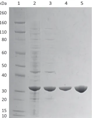
2.2.1 Purification of MRSA N-acetylneuraminate lyase ... 42
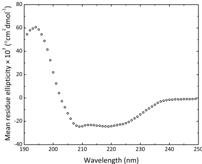
2.2.2 Solution structure of MRSA N-acetylneuraminate lyase ... 43
2.2.3 Kinetic analysis of MRSA N-acetylneuraminate lyase ... 46
2.2.4 Inhibition of MRSA N-acetylneuraminate lyase ... 49
2.2.5 Crystallisation of MRSA N-acetylneuraminate lyase ... 51
2.2.6 Data processing and structure refinement ... 52
2.2.7 The structure of MRSA N-acetylneuraminate lyase ... 55
2.2.8 A sulfate ion residing in the active site ... 57
2.2.9 A comparison of S. aureus and MRSA N-acetylneuraminate lyase structures ... 58
2.2.10 The structure of MRSA N-acetylneuraminate lyase in complex with a strong inhibitor ... 61
2.2.11 How inhibition is achieved by sialic acid alditol ... 63
2.3 Summary ... 65
2.4 References ... 66
Chapter Three: The structure and mechanism of N-acetylmannosamine-6-phosphate 2-epimerase from methicillin-resistant Staphylococcus aureus ... 73
3.1 Introduction ... 73
3.1.2 Epimerase enzymes implicated in sialic acid utilisation ... 74 3.1.3 The catalytic mechanism of N-acetylmannosamine-6-phosphate 2-epimerase ... 75 3.1.4 Kinetic analysis of N-acetylmannosamine-6-phosphate
2-epimerase ... 77 3.1.5 N-Acetylmannosamine-6-phosphate 2-epimerase is an antibiotic drug target ... 78 3.1.6 Overview of this chapter ... 79 3.2 Results and discussion ... 80
3.2.1 Cloning of acetylmannosamine-6-phosphate 2-epimerase, N-acetylphosphate deacetylase and
glucosamine-6-phosphate deaminase from MRSA ... 80 3.2.2 Purification of N-acetylmannosamine-6-phosphate 2-epimerase, N-acetylphosphate deacetylase and
glucosamine-6-phosphate deaminase from MRSA ... 81 3.2.3 The solution structure of wild type MRSA
N-acetylmannosamine-6-phosphate 2-epimerase ... 83 3.2.4 Development of a multi-enzyme kinetic assay for
N-acetylmannosamine-6-phosphate 2-epimerase ... 86 3.2.5 Michaelis-Menten kinetic analysis of wild type MRSA
N-acetylmannosamine-6-phosphate 2-epimerase ... 94 3.2.6 Crystallisation of MRSA N-acetylmannosamine-6-phosphate 2-epimerase ... 95 3.2.7 Data processing and structure refinement of MRSA
N-acetylmannosamine-6-phosphate 2-epimerase ... 95 3.2.8 The structure of MRSA N-acetylmannosamine-6-phosphate 2-epimerase ... 97 3.2.9 Michaelis-Menten kinetic analysis of mutant MRSA
N-acetylmannosamine-6-phosphate 2-epimerase enzymes ... 100 3.2.10 The structure and proposed mechanism of C. perfringens
N-acetylmannosamine-6-phosphate 2-epimerase ... 103 3.2.11 Alternative catalytic mechanisms ... 103 3.2.12 Sodium borohydride reduction of wild type MRSA
3.3 Summary ... 108
3.4 References ... 110
Chapter Four: Towards a structural and functional understanding of bacterial sialic acid transporters ... 115
4.1 Introduction ... 116
4.1.1 Sialic acid transporters ... 116
4.1.2 Sialic acid is transported using specific transporters ... 116
4.1.3 Sugar proton symporters for sialic acid transport ... 117
4.1.4 Sodium solute symporters for sialic acid transport ... 118
4.1.5 A proposed mechanism of sodium and solute symport ... 120
4.1.6 Overview of this chapter ... 122
4.2 Results and discussion 4.2.1 Cloning and overexpression of the Y. pestis NanT, S. aureus SSS and P. mirabilis SSS sialic acid transporters ... 123
4.2.2 Detergent screening of the Y. pestis NanT sialic acid transporter . 127 4.2.3 Purification of the Y. pestis NanT, S. aureus SSS and P. mirabilis SSS sialic acid transporters ... 129
4.2.4 The oligomeric structure of the Y. pestis NanT, S. aureus SSS and P. mirabilis SSS sialic acid transporters ... 132
4.2.5 A potential protein-protein interaction ... 135
4.2.6 Crystallisation studies ... 138
4.2.7 The structure of the P. mirabilis SSS sialic acid transporter ... 139
4.3 Summary ... 147
4.4 References ... 148
Chapter Five: Conclusions and future perspectives ... 153
5.1 Overview ... 153
5.1.1 The first reported structure of a sialic acid transporter ... 154
5.1.2 Structure, function and inhibition of MRSA N-acetylneuraminate lyase ... 156
5.1.3 A novel mechanism for carbohydrate epimerase enzymes ... 157
Chapter Six: Experimental procedures ... 161
6.1 Experimental Reagents ... 161
6.1.1 Chemical reagents ... 161
6.1.2 Biological reagents ... 161
6.1.3 General materials ... 162
6.2 General methods ... 163
6.2.1 Bioinformatic analyses of nucleotide and amino acid sequences .. 163
6.2.2 Centrifugation ... 163
6.2.3 Measurement of molecular weight ... 163
6.2.4 pH measurement ... 163
6.2.5 Sterilisation of media and equipment ... 164
6.3 Microbiology ... 165
6.3.1 Bacterial strains ... 165
6.3.2 Antibiotics ... 165
6.3.3 Media ... 166
6.3.4 Transformation of chemically competent bacterial cells ... 167
6.3.5 Bacterial culturing ... 167
6.3.6 Preparation of glycerol stocks ... 167
6.4 Molecular biology ... 168
6.4.1 Plasmids ... 168
6.4.2 Plasmid extraction and isolation ... 168
6.4.3 Dideoxynucleotide sequencing ... 169
6.4.4 Primers ... 169
6.4.5 Polymerase chain reaction ... 169
6.4.6 Restriction digestion ... 170
6.4.7 Agarose gel electrophoresis for DNA preparation ... 170
6.4.8 Ligation reactions ... 171
6.5 Electrophoresis ... 172
6.5.1 Agarose gel electrophoresis ... 172
6.5.2 Sodium dodecyl sulfate polyacrylamide gel electrophoresis ... 172
6.6 Protein biochemistry ... 173
6.6.1 Recombinant protein expression ... 173
6.6.2 Harvesting of cells ... 173
6.6.4 Whole cell fluorescence ... 174
6.6.5 Harvesting of membranes ... 174
6.6.6 Membrane protein solubilisation ... 175
6.6.7 Detergent screening ... 175
6.6.8 Protein purification ... 175
6.6.9 Protein quantitation ... 180
6.7 X-ray crystallography ... 181
6.7.1 Crystallisation ... 181
6.7.2 Data collection, processing and structure refinement ... 182
6.8 Biophysical techniques ... 184
6.8.1 Circular dichroism spectroscopy ... 184
6.8.2 Differential scanning fluorimetry ... 184
6.8.3 Analytical ultracentrifugation ... 184
6.8.4 Small angle X-ray scattering ... 185
6.9 Enzyme kinetics ... 186
6.9.1 Coupled assay for N-acetylneuraminate lyase ... 186
6.9.2 Multi-enzyme coupled assay for N-acetylmannosamine-6-phosphate 2-epimerase ... 188
6.9.3 Sodium borohydride reduction ... 189
6.10 References ... 191
Abbreviations
ADP adenosine diphosphate
ATP adenosine triphosphate
ABC adenosine triphosphate binding cassette transporter BetP sodium glycine betaine symporter
BLAST basic local alignment search tool C3 Collaborative Crystallisation Centre
CD circular dichroism
CHAPS 3-[(3-cholamidopropyl)dimethylammonio]-1-propanesulfonate
cm centimetre
c(M) continuous mass distribution
c(s) continuous sedimentation coefficient distribution
CMP cytidine monophosphate
Da dalton
DDM n-dodecyl-β-D-maltoside
DHDPS dihydrodipicolinate synthase
DM n-decyl-β-D-maltoside
Dmax maximum particle diameter
DNA deoxyribonucleic acid
dNTP deoxynucleotide triphosphate EDTA ethylenediaminetetraacetic acid
EMBO European Molecular Biology Organisation
F fluorescence
f/f0 frictional ratio
FN normalised fluorescence
FSEC fluorescence detection size exclusion chromatography
g gram
GFP green fluorescent protein His-tag Histidine tag
1H NMR proton nuclear magnetic resonance spectroscopy
IMAC immobilised metal affinity chromatography IPTG isopropyl β-D-1-thiogalactopyranoside kcat catalytic turnover number
kDa kilodalton
Ki inhibition constant
Kic competitive inhibition constant Kinc non-competitive inhibition constant Kiuc un-competitive inhibition constant
KM Michaelis-Menten constant
koff rate constant for dissociation kon rate constant for association
L litre
lacUV5 isopropyl β-D-1-thiogalactopyranoside-inducible promoter
LB luria bertani
LDAO n-dodecyl-N,N-dimethylamine-N-oxide
LDH lactate dehydrogenase
LeuT sodium leucine symporter
M molar
mg milligram
Mhp1 sodium hydantoin symporter
min minute
mL millilitre
mm millimetre
mM millimolar
MNG 2,2-didecylpropane-1,3-bis-β-D-maltopyranoside
mol moles
MRE mean residue ellipticity
MRSA methicillin-resistant Staphylococcus aureus
msmK shared ATPase domain
NAD+ nicotinamide adenine dinucleotide
NADH nicotinamide adenine dinucleotide, reduced form NADP+ nicotinamide adenine dinucleotide
NADPH nicotinamide adenine dinucleotide, reduced form
NanT sugar proton symporter
NG n-octyl-β-D-glucopyranoside
Ni-NTA nickel nitrilotriacetic acid
nm nanometre
OD600 optical density at 600 nm
P(r) real space distance distribution function
PBS phosphate buffered saline
PCR polymerase chain reaction
r radius
Rfactor residual factor
Rfree free Rfactor
RFU relative fluorescent units
Rg radius of gyration
rhaBAD L-rhamnose promoter rmsd root-mean-square deviation
RNA ribonucleic acid
ROK repressor, open reading frame, kinase
rpm revolutions per minute
S sedimentation coefficient, Svedberg
s second
s20,w standardised sedimentation coefficient relative to water at 20 °C
[S] substrate concentration
SatA periplasmic substrate binding protein A SatB/SatC integral membrane permease domain SatC periplasmic substrate binding protein C SatD adenosine triphosphatase domain SAXS small angle X-ray scattering
SDS sodium dodecyl sulfate
SDS-PAGE sodium dodecyl sulfate polyacrylamide gel electrophoresis SGLT sodium galactose transporter
SOC catabolite repression super optimal broth
SSS sodium solute symporter
TB terrific broth
TM melting temperature
TRAP tripartite adenosine triphosphate independent periplasmic transporter Tris 2-amino-2-hydroxymethyl-propane-1,3-diol
UDP uridine diphosphate
UV ultraviolet
V0 initial velocity
VM (Å3 Da-1) wilson B value
v/v volume to volume
w/v weight to volume
Å angstrom
° degree
°C degree celcius
µg microgram
µL microlitre
µM micromolar
% percent
π pi
σ sigma
θ theta
(β/α)8-barrel triosephosphate isomerase barrel
Chapter One
Introduction
The following is a general introduction to the field of sialobiology in the context of bacterial sialic acid metabolism. Subsequent chapters include more detailed, specific introductions to the chapter topics, as these are intended to be independent bodies of work.
1.1
Sialic acid
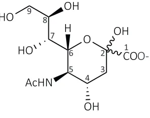
[image:17.595.229.381.559.679.2]Sialic acid is the designation given to a large family comprising over 50 structurally distinct nine carbon amino sugars. All are derived from the most common compound 2-keto-3-deoxy-5-acetamido-D-glycero-D-galacto-nonulosonic acid, more commonly known as N-acetylneuraminic acid (Figure 1.1) (Vimr et al., 2004; Almagro-Moreno & Boyd, 2009a). Derivatives of this molecule carry various substituents at the amino or hydroxyl groups (Varki, 1992; Schauer, 2000; Angata & Varki, 2002). Interestingly, N-acetylneuraminic acid is the only sialic acid that is ubiquitous in all organisms (Schauer, 2004). Throughout this thesis, the term sialic acid refers to N-acetylneuraminic acid.
Figure 1.1. Chemical structure of N-acetylneuraminic acid. Carbons are numbered one through nine.
O
OH
CO
2-OH
HO
HO
OH
AcHN
H
1 2 3 4 5 6 7 8 9
O-1.2
Sialic acid utilisation by bacterial pathogens
Eukaryotic cell surfaces are decorated with a complex array of glycoconjugates. Negatively charged sialic acids are found attached to the terminal sugar positions of these cell surface glycoconjugates where they mediate a diverse array of cellular interactions, recognition and adhesion (Varki, 1993; Schauer, 2000; Vimr et al., 2004). In the human respiratory and gastrointestinal tract, sialic acid coated glycoconjugates are highly abundant (Vimr, 2013) and glucose tends to be limited in supply (Jeong et al., 2009). Thus, bacterial pathogens that colonise these environments, along with commensal competitors, have evolved mechanisms by which they can utilise host-derived sialic acid (Vimr et al., 2004; Severi et al., 2007; Almagro-Moreno & Boyd, 2009a).
Although the concentration of sialic acid present in human serum is relatively high at approximately 1.6 to 2.2 mM/L, it is bound to glycoconjugates under normal physiological conditions (Sillanaukee et al., 1999). By definition, these sialoglycoconjugates are unavailable for utilisation by bacterial pathogens. In order to make sialic acid available, it needs to be released by a neuraminidase, which hydrolyses the linkage between the sialic acid molecule and the sub-terminal sugar of the glycoconjugate (Vimr, 1994; Vimr, 2013). Neuraminidases are produced either endogenously by the host in response to inflammation (Sohanpal et al., 2004; Sohanpal et al., 2007) or exogenously by neuraminidase expressing bacteria present in the heavily sialylated niche (Shakhnovich et al., 2002; Vimr, 2013).
1.3
Bacterial pathogens evade the host immune
response by molecular mimicry
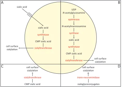
Some bacterial pathogens have evolved the capacity to incorporate sialic acid into cell surface macromolecules known as lipopolysaccharides and lipooligosaccharides or to produce capsules containing homopolymers of sialic acid (Vimr et al., 2004). This allows for the bacterial pathogen to circumvent the host immune response through molecular mimicry. The evolutionary advantage of such mimicry is so pronounced that at least four pathways of cell surface sialylation have evolved (Vimr & Lichtensteiger, 2002). These include precursor scavenging, de novo biosynthesis, donor scavenging and trans-neuraminidase activity (Figure 1.2).
Figure 1.2. The pathways of bacterial cell surface sialylation. Four pathways have evolved that allow sialic acid to be incorporated into cell surface macromolecules. (A) The precursor scavenging pathway. (B) The de novo biosynthesis pathway. (C) The donor scavenging pathway. (D) The trans-neuraminidase pathway.
CMP sialic acid sialyltransferase
cell surface sialyla!on
sialic acid
CMP sialic acid cell surface sialyla!on cell surface sialyla!on sialic acid N-acetylmannosamine UDP N-acetylglucosamine
[image:19.595.104.505.353.639.2]1.3.1
Precursor scavenging
A recently discovered method of cell surface sialylation known as precursor scavenging (Figure 1.2 A) has been revealed in Haemophilus influenzae. This organism scavenges host-derived sialic acid by importing it into its cell. Once imported, H. influenzae activates the sialic acid by converting it into cytidine monophosphate (CMP) sialic acid, which is incorporated into appropriate acceptors by a sialyltransferase, with the resultant sialoconjugate exported to the cell surface (Vimr & Lichtensteiger, 2002). Appropriate acceptors include lipopolysaccharides, lipooligosaccharides or capsules containing homopolymers of sialic acid. Alternatively, the imported sialic acid can be degraded into a carbon, nitrogen and energy source (Section 1.5) (Almagro-Moreno & Boyd, 2009a). Thus, H. influenzae must make an important metabolic decision between cell surface sialylation or sialic acid degradation so that a fine balance is maintained between the need to evade the host’s immune response and any nutritional requirements (Vimr & Lichtensteiger, 2002).
1.3.2
De novo biosynthesis
De novo biosynthesis of sialic acid (Figure 1.2 B) is used by a number of bacteria including Neisseria meningitides and Escherichia coli (Vimr et al., 2004). This route involves synthesising sialic acid from uridine diphosphate (UDP)N-acetylglucosamine, a simple metabolite produced by most cells. Once synthesised, sialic acid is converted into its activated form, CMP sialic acid, which is incorporated into appropriate acceptors by a sialyltransferase, with the resultant sialoconjugate exported to the cell surface (Vimr & Lichtensteiger, 2002).
1.3.3
Donor scavenging
incorporated into appropriate acceptors by a sialyltransferase, with the resultant sialoconjugate exported to the cell surface (Shell et al., 2002).
1.3.4
Trans-neuraminidase activity
1.4
Importation of sialic acid into bacterial
pathogens
For bacterial pathogens to utilise the precursor scavenging mechanism of cell surface sialylation or degrade host-derived sialic acid into a carbon, nitrogen and energy source (Section 1.5), sialic acid must be transported across the cytoplasmic membrane and into the bacterial cell. For gram negative bacterial pathogens, sialic acid must first cross the outer cell membrane into the periplasmic space before it can be imported into the cell. Sialic acid transport across the outer cell membrane of bacterial pathogens is not well understood (Severi et al., 2007). However, a sialic acid inducible outer membrane porin known as NanC has been well characterised in E. coli (Condemine et al., 2005; Wirth et al., 2009).
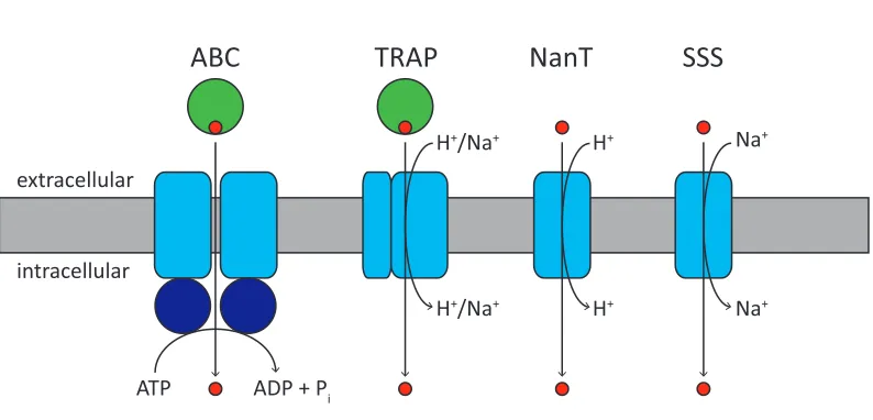
Figure 1.3. Sialic acid transporter types. In ABC and TRAP transporter systems, a periplasmic or cell surface associated substrate binding protein (green circles) interacts with two membrane protein domains (blue rectangles) to transport the sialic acid (red circles) across the membrane. ABC transporters are primary transporters that couple solute translocation with the hydrolysis of ATP by the ATPase domains harbouring the Walker motifs (dark blue circles). TRAP, NanT and SSS transporters are secondary transporters that couple solute translocation with an electrochemical gradient. It is unclear whether TRAP sialic acid transporters are proton or sodium ion dependent; but NanT sialic acid transporters have been shown to be proton dependent (Mulligan et al., 2012), while SSS sialic acid transporters have been shown to be sodium ion dependent (Severi et al., 2010). The stoichiometry of ions required for transport is not well understood.
1.4.1
ATP binding cassette transporters for sialic acid
transport
The ABC superfamily of transporters is one of the largest families of transporters with representatives in all phyla (Jones & George, 2004). Containing uptake and efflux systems, this superfamily is made up of dozens of families that vary in their substrate specificity (Saurin et al., 1999). ABC transporters are known as primary transporters because they utilise the energy generated from ATP hydrolysis to transport solutes across a membrane (Higgins, 1992). They transport a variety of substrates across both extracellular and intracellular membranes, including ions, sugars, amino acids, peptides, polysaccharides and even proteins.
All ABC transporters are composed of four domains, two transmembrane domains that form the translocation pathway and two cytoplasmic nucleotide binding domains that
extracellular
intracellular
ATP ADP + Pi
H+/Na+
ABC
TRAP
H+
H+
NanT
Na+
Na+
SSS
hydrolyse ATP (Higgins, 1992). Prokaryotic ABC transporters contain an additional periplasmic or cell surface associated binding protein that bind substrates with high affinity and deliver them to the transmembrane domains. Since the first high resolution structure of the E. coli vitamin B12 ABC transporter (Locher et al., 2002), crystal structures of a handful of others have been solved (Oldham et al., 2007; Hohl et al., 2012; Woo et al., 2012; Shintre et al., 2013), some of which have been solved in complex with their substrate binding proteins (Hollenstein et al., 2007; Oldham et al., 2007).
The first sialic acid ABC transport system was identified and characterised in Haemophilus ducreyi (Post et al., 2005). More recently, an ABC transporter system for sialic acid has been identified in Streptococcus pneumoniae (Marion et al., 2011a; Marion et al., 2011b). Although the import of sialic acid by ABC transporters is not well characterised in other organisms, Streptococcus agalactiae, Streptococcus gordonii, Streptococcus pyogenes and Streptococcus sanguinis are predicted to utilise this type of sialic acid transport system (Almagro-Moreno & Boyd, 2009a). The components that make up the H. ducreyi ABC transporter include a periplasmic substrate binding protein (SatA), integral membrane permease domains (SatB/SatC) and an ATPase domain (SatD). S. pneumoniae has a similar structural organisation, except that the ATPase domain is a shared ATPase (msmK) that is responsible for energising multiple carbohydrate transporters (Marion et al., 2011a).
1.4.2
Tripartite ATP independent periplasmic transporters
for sialic acid transport
on the direction and magnitude of the concentration and/or electrochemical gradient (Poolman & Konings, 1993; Severi et al., 2010).
TRAP transporters are composed of three protein domains; these include the substrate binding protein, a small membrane spanning domain and a large membrane spanning domain. The purpose of the substrate binding protein is to bind substrate with high affinity and specificity and present it to the membrane spanning domains of the transporter for transport across the membrane (Doeven et al., 2004; Mulligan et al., 2009). Conveniently, it is found either free in the periplasm of gram negative bacteria or anchored to the cytoplasmic membrane in gram positive bacteria and archaea (Kelly & Thomas, 2001). For drug discovery, TRAP transporters are of particular interest because they are widespread across bacteria and archaea but are not found in eukaryotes (Kelly & Thomas, 2001).
The first C4-dicarboxylate TRAP transport system to be described was from Rhodobacter capsulatus (Forward et al., 1997). Although TRAP transporters are not well characterised, the large membrane spanning domain is predicted to form the membrane translocation pathway through which the substrate passes (Mulligan et al., 2012). Whereas the small membrane spanning domain has been shown to be essential for transport, but the function remains unknown (Forward et al., 1997; Mulligan et al., 2012). It is unclear whether TRAP sialic acid transporters are proton or sodium ion dependent (Mulligan et al., 2012).
1.4.3
Sugar proton symporters for sialic acid transport
1.4.4
Sodium solute symporters for sialic acid transport
More recently, a novel type of sialic acid transporter has been discovered from Salmonella enterica (Severi et al., 2010), belonging to the sodium solute symporter (SSS) family of secondary transporters that co-transport sodium ions with sugars, amino acids, inorganic ions or vitamins (Wright et al., 2004). Like NanT, the SSS transporters are also single component systems. The S. enterica sialic acid transporter was established as a typical member of the SSS family because its functionality was dependent upon the presence of sodium ions (Severi et al., 2010). The genes encoding the SSS sialic acid transporters are widespread among both gram positive and gram negative species of bacteria, whereas other types of sialic acid transporters do not appear to be as widespread (Severi et al., 2010). An SSS sialic acid transporter from S. aureus and Proteus mirabilis will be explored in more detail in Chapter Four.
1.4.5
Sialic acid is transported using a specific transporter
1.5
Sialic acid degradation by bacterial
pathogens
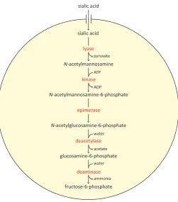
Following the transport of host-derived sialic acid into the cell, some bacterial pathogens will utilise it by degrading it into a carbon, nitrogen and energy source (Vimr & Troy, 1985; Olson et al., 2013). The genes required for the sequestration and subsequent degradation of host-derived sialic acid are known as the ‘nan nag cluster,’ and are confined to pathogenic species of bacteria and mammalian commensals (Almagro-Moreno & Boyd, 2009a). The nan nag cluster encodes the transporter responsible for importing sialic acid into the bacterial cell and five catalytic enzymes that successively degrade it into fructose-6-phosphate, a key metabolite for glycolysis.
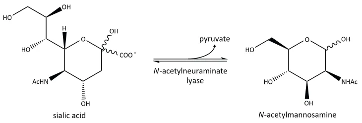
Figure 1.4. The sialic acid degradation pathway. Following the import of sialic acid into the bacterial cell, it is successively degraded into fructose-6-phosphate by N-acetylneuraminate lyase, N-acetylmannosamine-6-phosphate, N-acetylmannosamine-6-phosphate 2-epimerase, N-acetylglucosamine-6-phosphate deacetylase and glucosamine-6-N-acetylglucosamine-6-phosphate deaminase.
1.5.1
N
-Acetylneuraminate lyase
N-Acetylneuraminate lyase (which will be discussed in more detail in Chapter Two) catalyses the retro-aldol cleavage of open chain sialic acid to form N-acetylmannosamine and pyruvate (Figure 1.5) via a Schiff base intermediate (Izard et al., 1994; Lawrence et al., 1997; Barbosa et al., 2000). This reaction represents the first and the committed step of the degradation pathway. Structurally, N-acetylneuraminate lyase is classified within the N-acetylneuraminate lyase sub-family of triosephosphate isomerase barrel [(β/α)8-barrel] enzymes. Members of this sub-family identified thus far include the archetype N-acetylneuraminate lyase, 4-hydroxy-tetrahydrodipicolinic acid synthase [formally known as dihydrodipicolinate synthase (DHDPS)] (Mirwaldt et al., 1995), D-5-keto-4-deoxyglucarate dehydratase (Jeffcoat et al., 1969b; Jeffcoat et al., 1969a), 2-keto-3-deoxygluconate aldolase (Buchanan et al., 1999), trans-o-hydroxybenzylidenepyruvate
N-acetylmannosamine-6-phosphate
N-acetylmannosamine sialic acid
epimerase
sialic acid
N-acetylglucosamine-6-phosphate
glucosamine-6-phosphate
fructose-6-phosphate
kinase
deacetylase
deaminase lyase
ATP
ADP pyruvate
acetate
ammonia water
hydratase-aldolase (Eaton, 1994) and trans-2’-carboxybenzalpyruvate hydratase-aldolase (Iwabuchi & Harayama, 1998). All of which share a common structural framework, but catalyse reactions in separate biochemical pathways (Lawrence et al., 1997).
Figure 1.5. The reaction catalysed by N-acetylneuraminate lyase. N-Acetylneuraminate lyase catalyses the retro-aldol cleavage of open chain sialic acid to form N-acetylmannosamine and pyruvate.
The structure of N-acetylneuraminate lyase has been well characterised in various organisms including, E. coli (Izard et al., 1994; Lawrence et al., 1997; Joerger et al., 2003; Campeotto et al., 2009; Campeotto et al., 2010; Daniels et al., 2014), H. influenzae
(Barbosa et al., 2000), Pasteurella multocida (Huynh et al., 2013) and S. aureus (Timms et al., 2013). Further analysis of N-acetylneuraminate lyase in complex with substrate analogues has revealed the mechanism of substrate binding within the active site (Lawrence et al., 1997; Barbosa et al., 2000; Huynh et al., 2013; Daniels et al., 2014). As with all members of the N-acetylneuraminate lyase sub-family of (β/α)8-barrel enzymes, catalysis is proposed to involve the formation of a Schiff base between the amine of a highly conserved lysine residue and the C2 carbon of the α-keto acid moiety on the substrate (Izard et al., 1994; Blickling et al., 1997; Lawrence et al., 1997). In recent years,
N-acetylneuraminate lyase has received considerable attention from both mechanistic and structural viewpoints and has been recognised as a viable antibiotic drug target (Severi et al., 2007; von Itzstein, 2007).
1.5.2
N-Acetylmannosamine kinase
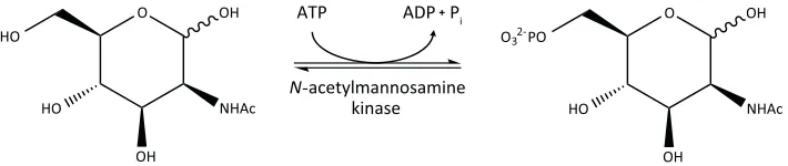
Following the reaction catalysed by N-acetylneuraminate lyase, N-acetylmannosamine kinase transfers a phosphate group from ATP to the C6 position of N-acetylmannosamine,
O
OH
COO -OH HO
HO
OH
AcHN H
O
OH HO
HO NHAc
OH
sialic acid N-acetylmannosamine
pyruvate
generating N-acetylmannosamine-6-phosphate and ADP (Figure 1.6). N-Acetylmannosamine kinase is a phosphotransferase belonging to the repressor, open reading frame, kinase (ROK) superfamily. ROK kinases usually contain a conserved N-terminal ATP binding motif and a zinc binding motif (Holmes et al., 1993; Larion et al., 2007). Zinc is proposed to play a key structural role in the active site of many ROK kinases (Larion et al., 2007). However, this zinc binding motif is not obvious in N-acetylmannosamine kinase from S. aureus and Streptococcus mitis (North et al., 2014b). The lack of a zinc binding motif in these organisms suggests that the geometry and stability of the active site may be affected in some other way. This difference could be explored as a target for inhibitory molecules.
Figure 1.6. The reaction catalysed by N-acetylmannosamine kinase. N-Acetylmannosamine kinase transfers a phosphate group from ATP to the C6 position of N-acetylmannosamine, generating N -acetylmannosamine-6-phosphate and ADP.
There is a lack of literature published on the bacterial N-acetylmannosamine kinase, but much is known about its bifunctional eukaryotic homologue UDP N-acetylglucosamine 2-epimerase/N-acetylmannosamine kinase. In mammals, this enzyme is proposed to be involved in sialic acid biosynthesis (Hinderlich et al., 1997). Crystal structures of the Homo sapiens N-acetylmannosamine kinase domain of UDP N-acetylglucosamine 2-epimerase/N-acetylmannosamine kinase have been solved (Martinez et al., 2012). Yet, only one bacterial crystal structure of N-acetylmannosamine kinase has been determined from E. coli (New York Structural Genomics Research Centre, unpublished work).
1.5.3
N-Acetylmannosamine-6-phosphate 2-epimerase
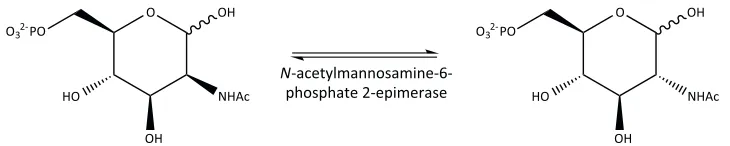
N-Acetylmannosamine-6-phosphate 2-epimerase (which will be investigated in more detail in Chapter Three) converts N-acetylmannosamine-6-phosphate into
N-acetylglucosamine-O
OH HO
HO NHAc
OH
N-acetylmannosamine
O
OH O3 PO
HO NHAc
OH
N-acetylmannosamine-6-phosphate N-acetylmannosamine
kinase
ATP ADP Pi
2-6-phosphate (Figure 1.7). N-acetylmannosamine-6-phosphate 2-epimerase and two other amino sugar 2-epimerase enzymes have been implicated in pathways that utilise sialic acid in both eukaryotes and prokaryotes, all of which use N-acetylmannosamine as the essential sugar precursor (Ferrero et al., 2007). In eukaryotes, N-acetylglucosamine 2-epimerase is responsible for the degradation of sialic acid, whereas the previously discussed bifunctional UDP-N-acetylglucosamine 2-epimerase/N-acetylmannosamine kinase is involved in the biosynthesis of sialic acid. An evolutionarily and mechanistically related UDP N-acetylglucosamine 2-epimerase has been implicated in the biosynthesis of sialic acid in prokaryotes (Tanner, 2005), but it lacks the bifunctional kinase activity.
Figure 1.7. The reaction catalysed by N-acetylmannosamine-6-phosphate 2-epimerase. N-Acetylmannosamine-6-phosphate 2-epimerase epimerises N-acetylmannosamine-6-phosphate into N-acetylglucosamine-6-phosphate.
Crystal structures of N-acetylmannosamine-6-phosphate 2-epimerase from methicillin-resistant S. aureus (MRSA) (Midwest Centre for Structural Genomics, unpublished work),
S. enterica (Centre for Structural Genomics of Infectious Diseases, unpublished work) and
S. pyogenes (Midwest Centre for Structural Genomics, unpublished work) have been solved. However, these structures have not yet been published in the literature. Recently however, the structure of Clostridium perfringens N-acetylmannosamine-6-phosphate 2-epimerase has been solved and published (Pelissier et al., 2014).
1.5.4
N-Acetylglucosamine-6-phosphate deacetylase
Following the reaction catalysed by N-acetylmannosamine-6-phosphate 2-epimerase, N -acetylglucosamine-6-phosphate deacetylase catalyses the deacetylation of N -acetylglucosamine-6-phosphate with water to form glucosamine-6-phosphate and acetate (Figure 1.8). Crystal structures of N-acetylglucosamine-6-phosphate deacetylase have been
O
OH
HO NHAc
OH O
OH
HO NHAc
OH
N-acetylmannosamine-6-phosphate 2-epimerase
N-acetylmannosamine-6-phosphate N-acetylglucosamine-6-phosphate
[image:31.595.122.487.289.362.2]reported from E. coli in a metal free state (Ferreira et al., 2006), E. coli in a metal bound state (Hall et al., 2007), Bacillus subtilis in a metal bound state (Vincent et al., 2004) and Thermotoga maritima in a metal bound state (Joint Centre for Structural Genomics, unpublished work). These structures show that the N-acetylglucosamine-6-phosphate deacetylases belong to the metal dependent amidohydrolase superfamily (Vincent et al., 2004). This family of enzymes can bind one or more divalent metal ions in the active site, including Fe2+, Zn2+, Co2+ and Cu2+ (Ferreira et al., 2006). A comparison of these crystal structures reveals different active site motifs for the complexation of divalent metal ions. The structure for B. subtilis N-acetylglucosamine-6-phosphate deacetylase contains a binuclear Fe2+ centre, whereas the T. maritima structure N-acetylglucosamine-6-phosphate deacetylase has a mononuclear Fe2+ centre (Vincent et al., 2004). The structure of the E. coli enzyme in a metal bound state has a mononuclear Fe2+ centre (Hall et al., 2007), yet this enzyme has also been shown to bind other metal ions (Ferreira et al., 2006).
Figure 1.8. The reaction catalysed by N-acetylglucosamine-6-phosphate deacetylase. N-Acetylglucosamine-6-phosphate deacetylase catalyses the deacetylation of N-acetylglucosamine-6-phosphate with water to form glucosamine-6-phosphate and acetate.
1.5.5
Glucosamine-6-phosphate deaminase
The final enzyme involved in the degradation of sialic acid is glucosamine-6-phosphate deaminase. This enzyme catalyses the isomerisation and deamination of glucosamine-6-phosphate with water into fructose-6-glucosamine-6-phosphate and ammonia (Figure 1.9). Fructose-6-phosphate is an essential metabolite that is used in glycolysis (Almagro-Moreno & Boyd, 2009a). Glucosamine-6-phosphate deaminase belongs to an aldose ketose isomerase class of proteins, all of which catalyse the removal of one hydrogen from the C2 position of the aldose and add back a hydrogen to the C1 position of the ketose through a cis-enediol intermediate (Oliva et al., 1995). This enzyme has been identified in several species of
O OH HO NHAc OH N-acetylglucosamine-6-phosphate deacetylase N-acetylglucosamine-6-phosphate
O32-PO O
OH
HO NH
OH O32-PO
2
glucosamine-6-phosphate acetate
animal, fungus and bacteria, where it is involved in sialic acid degradation for nutritional purposes or amino sugar synthesis for peptidoglycan formation (Liu et al., 2008). The equilibrium of the reaction favours the degradation reaction (Calcagno et al., 1984).
Figure 1.9. The reaction catalysed by glucosamine-6-phosphate deaminase. Glucosamine-6-phosphate deaminase catalyses the isomerisation and deamination of glucosamine-6-phosphate with water into fructose-6-phosphate and ammonia.
Crystal structures of glucosamine-6-phosphate deaminase have been published from a range of bacterial species including, B. subtilis (Vincent et al., 2005), E. coli (Oliva et al., 1995; Horjales et al., 1999; Rudino-Pinera et al., 2002) and Streptococcus mutans (Liu et al., 2008). The crystal structure of glucosamine-6-phosphate deaminase from E. coli shows that the enzyme is hexameric and allosterically activated by N -acetylglucosamine-6-phosphate (Horjales et al., 1999). In contrast, glucosamine-6-phosphate deaminase from B. subtilis and S. mutans is monomeric and not allosterically regulated (Vincent et al., 2005; Liu et al., 2008). It has been proposed that through evolution, glucosamine-6-phosphate deaminase from gram positive bacteria has diverged into a monomeric family, causing it to lose the property of allosteric regulation (Liu et al., 2008).
O
OH O3-2PO
HO NH2
OH
glucosamine-6-phosphate deaminase
glucosamine-6-phosphate
O O3-2PO
HO OH
OH OH
fructose-6-phosphate ammonia
1.6
Regulation of the
nan nag
cluster
Interestingly, sialic acid degradation does not appear to be under strict allosteric control, apart from the activation of glucosamine-6-phosphate deaminase by N-acetylglucosamine-6-phosphate. Instead, expression of the enzymes involved in the transport and degradation of sialic acid is controlled by a transcriptional regulator, known as the nanRepressor (Kalivoda et al., 2003; Johnston et al., 2007; Hwang et al., 2013; Kalivoda et al., 2013; Olson et al., 2013). The nanRepressor belongs to the RpiR family of transcriptional regulators and contains a helix-turn-helix motif and a sugar isomerase domain (Jaeger & Mayer, 2008). An exception to the RpiR family of transcriptional regulators is the nanRepressor found in E. coli, which belongs to the GntR superfamily. Despite this exception, all nanRepressor proteins are responsible for regulating the expression of the sialic acid degradation operon, thus they are also known as ‘sialoregulators’.
Helix-turn-helix motifs are often found in proteins that are known to bind deoxyribonucleic acid (DNA) and regulate gene expression. The motif is characterised by two α-helices that make intimate contacts with the DNA, one of which is important for sequence recognition, while the other assists in stabilising the structure (Sauer et al., 1982; Brennan & Matthews, 1989). For nanRepressor proteins belonging to the RpiR superfamily, positively charged amino acid residues of the helix-turn-helix domain are speculated to interact with the nan promoter. For the S. aureus nanRepressor in particular, the positively charged amino acid residues of the helix-turn-helix domain specifically interact with the nan promoter at the nanAT and nanE transcripts (Olson et al., 2013).
N-acetylmannosamine-6-phosphate alone acts as the inducer for V. vulnificus (Kim et al., 2011)and S. aureus (Olson et al., 2013). For H. influenzae, glucosamine-6-phosphate is responsible for both the induction and the repression of nan expression (Johnston et al., 2007).
1.7
Antibiotic resistance in bacterial pathogens
The discovery of potent antibiotic agents was one of the greatest advances in the control of bacterial diseases in the 20th century (Cohen, 1992). In the United States of America alone, it is estimated that more than 2 million people are infected with antibiotic resistant bacteria resulting in 23 000 deaths annually (Hampton, 2013). There are no reliable estimates of the number of infection cases globally, but there is evidence that antibiotic resistance is significantly worse in developing countries (Toner et al., 2015). The rise in antibiotic resistant bacteria stems from a multitude of factors including the widespread and sometimes inappropriate administration of antibiotics in both human medicine and agriculture (Lowy, 2003; Blair et al., 2015). Frighteningly, this increasing resistance has resulted in a pronounced lack in the development of new antibiotics (Blair et al., 2015). Thus, the search for antibiotic compounds has recently taken on a new urgency.
1.8
Sialic acid degradation is essential for
pathogen colonisation and persistence
The sialic acid degradation pathway has been well documented in several bacterial pathogens that colonise heavily sialylated niches, including E. coli, H. influenzae, S. aureus, V. cholerae, V. vulnificus and Y. pestis (Chang et al., 2004; Almagro-Moreno & Boyd, 2009b; Almagro-Moreno & Boyd, 2009a; Jeong et al., 2009; Olson et al., 2013). As described in Section 1.2, glucose is limited in heavily sialylated niches, such as the human respiratory and gastrointestinal tract (Jeong et al., 2009). Thus, the capability of these organisms to utilise sialic acid as an alternate source of carbon, nitrogen and energy is important for their survival (Almagro-Moreno & Boyd, 2009b).
Various in vitro and in vivo studies demonstrate that the degradation of sialic acid is crucial for the colonisation and persistence of several human bacterial pathogens. Deletion of one or more of the genes encoded by the nan nag cluster rendered E. coli, S. aureus, V. cholerae, V. vulnificus and S. aureus incapable of growing on sialic acid as a sole carbon, nitrogen and energy source in vitro (Vimr & Troy, 1985; Almagro-Moreno & Boyd, 2009b; Jeong et al., 2009; Vogel-Scheel et al., 2010; Olson et al., 2013), which suggests that this is the only pathway for sialic degradation in these bacteria. More importantly, the ability to degrade sialic acid has proven necessary for pathogen colonisation and persistence in vivo, with mouse models for E. coli (Chang et al., 2004), V. cholerae
1.9
Aims and objectives of this thesis
The overall aim of this research is to understand how sialic acid is transported into the bacterial cell and subsequently degraded. Following the import of sialic acid into the bacterial pathogen, five successive enzymes degrade it into a carbon, nitrogen and energy source. Given the importance of sialic acid transport and degradation for the colonisation and persistence of clinically important human pathogens, surprisingly little structural data exists for this pathway. Enzymology and structural biology is used to gain valuable information on how this pathway functions. These data underpin and inform future drug design and expand our understanding of this important, but poorly understood, metabolic pathway.
Chapter Two investigates the structure, function and inhibition of MRSA N -acetylneuraminate lyase. Biophysical characterisation, kinetic analysis and a structural analysis of MRSA N-acetylneuraminate lyase are presented. A previously characterised molecule that has been shown to inhibit C. perfringens N-acetylneuraminate lyase was synthesised in collaboration with Professor Antony Fairbanks (University of Canterbury). The ability of this molecule to inhibit MRSA N-acetylneuraminate lyase was tested by kinetic analysis. To probe how inhibition is achieved in this organism, the structure was solved in complex with this molecule.
may occur via the formation of a Schiff base or a proton displacement mechanism mediated by the substrate.
1.10 References
Allen, S., Zaleski, A., Johnston, J. W., Gibson, B. W. & Apicella, M. A. (2005). Novel sialic acid transporter of Haemophilus influenzae. Infection and Immunity, 73, 5291-5300.
Almagro-Moreno, S. & Boyd, E. F. (2009a). Insights into the evolution of sialic acid catabolism among bacteria. BioMed Central Evolutionary Biology, 9, 118-133. Almagro-Moreno, S. & Boyd, E. F. (2009b). Sialic acid catabolism confers a competitive
advantage to pathogenic Vibrio cholerae in the mouse intestine. Infection and Immunity, 77, 3807-3816.
Angata, T. & Varki, A. (2002). Chemical diversity in the sialic acids and related alpha-keto acids: an evolutionary perspective. Chemical Reviews, 102, 439-469.
Barbosa, J., Smith, B., DeGori, R., Ooi, H., Marcuccio, S., Campi, E., Jackson, W., Brossmer, R., Sommer, M. & Lawrence, M. (2000). Active site modulation in the N-acetylneuraminate lyase sub-family as revealed by the structure of the inhibitor-complexed Haemophilus influenzae enzyme. Journal of Molecular Biology, 303, 405-421.
Blair, J. M., Webber, M. A., Baylay, A. J., Ogbolu, D. O. & Piddock, L. J. (2015). Molecular mechanisms of antibiotic resistance. Nature Reviews Microbiology, 13, 42-51.
Blanco, A. G., Sola, M., Gomis-Ruth, F. X. & Coll, M. (2002). Tandem DNA recognition by PhoB, a two-component signal transduction transcriptional activator. Structure, 10, 701-713.
Blickling, S., Renner, C., Laber, B., Pohlenz, H. D., Holak, T. A. & Huber, R. (1997). Reaction mechanism of Escherichia coli dihydrodipicolinate synthase investigated by X-ray crystallography and NMR spectroscopy. Biochemistry, 36, 24-33.
Boyce, J. M., Cookson, B., Christiansen, K., Hori, S., Vuopio-Varkila, J., Kocagoz, S., Oztop, A. Y., Christiansen, K., Hori, S., Vuopio-Varkila, J., Kocagoz, S., Oztop, A. Y., Vandenbroucke-Grauls, C. M. J. E., Harbarth, S. & Pittet, D. (2005). Meticillin-resistant Staphylococcus aureus. Lancet Infectious Diseases, 5, 653-663.
Brennan, R. G. (1993). The winged-helix DNA-binding motif - another helix-turn-helix takeoff. Cell, 74, 773-776.
Brennan, R. G. & Matthews, B. W. (1989). The helix-turn-helix DNA-binding motif.
Journal of Biological Chemistry, 264, 1903-1906.
Buchanan, C. L., Connaris, H., Danson, M. J., Reeve, C. D. & Hough, D. W. (1999). An extremely thermostable aldolase from Sulfolobus solfataricus with specificity for non-phosphorylated substrates. The Biochemical Journal, 343, 563-570.
Busby, S. & Ebright, R. H. (1999). Transcription activation by catabolite activator protein (CAP). Journal of Molecular Biology, 293, 199-213.
Calcagno, M., Campos, P. J., Mulliert, G. & Suastegui, J. (1984). Purification, molecular and kinetic properties of glucosamine-6-phosphate isomerase (deaminase) from
Escherichia coli. Biochimica et Biophysica Acta, 787, 165-173.
Campeotto, I., Bolt, A. H., Harman, T. A., Dennis, C., Trinh, C. H., Phillips, S. E. V., Nelson, A., Pearson, A. R. & Berry, A. (2010). Structural insights into substrate specificity in variants of N-acetylneuraminic acid lyase produced by directed evolution. Journal of Molecular Biology, 404, 56-69.
Campeotto, I., Carr, S. B., Trinh, C. H., Nelson, A. S., Berry, A., Phillips, S. E. & Pearson, A. R. (2009). Structure of an Escherichia coli N-acetyl-D-neuraminic acid lyase mutant, E192N, in complex with pyruvate at 1.45 Å resolution. Acta crystallographica Section F, Structural Biology and Crystallisation Communications, 65, 1088-1090.
Chambers, H. F. (1988). Methicillin-resistant staphylococci. Clinical and Microbiology Reviews, 1, 173-186.
TRAP transporters (SiaPQM) and constitute the sole route of sialic acid uptake in the human pathogen Vibrio cholerae. Microbiology, 158, 2158-2167.
Cohen, M. L. (1992). Epidemiology of drug resistance: implications for a post-antimicrobial era. Science, 257, 1050-1055.
Condemine, G., Berrier, C., Plumbridge, J. & Ghazi, A. (2005). Function and expression of an N-acetylneuraminic acid-inducible outer membrane channel in Escherichia coli.
Journal of Bacteriology, 187, 1959-1965.
Daniels, A. D., Campeotto, I., van der Kamp, M. W., Bolt, A. H., Trinh, C. H., Phillips, S. E., Pearson, A. R., Nelson, A., Mulholland, A. J. & Berry, A. (2014). Reaction mechanism of N-acetylneuraminic acid lyase revealed by a combination of crystallography, QM/MM simulation, and mutagenesis. American Chemical Society Chemical Biology, 9, 1025-1032.
David, M. Z. & Daum, R. S. (2010). Community-associated methicillin-resistant
Staphylococcus aureus: epidemiology and clinical consequences of an emerging epidemic. Clinical and Microbiology Reviews, 23, 616-687.
Doeven, M. K., Abele, R., Tampe, R. & Poolman, B. (2004). The binding specificity of OppA determines the selectivity of the oligopeptide ATP-binding cassette transporter. Journal of Biological Chemistry, 279, 32301-32307.
Eaton, R. W. (1994). Organisation and evolution of naphthalene catabolic pathways: sequence of the DNA encoding 2-hydroxychromene-2-carboxylate isomerase and
trans-o-hydroxybenzylidenepyruvate hydratase-aldolase from the NAH7 plasmid.
Journal of Bacteriology, 176, 7757-7762.
Ferreira, F. M., Mendoza-Hernandez, G., Castaneda-Bueno, M., Aparicio, R., Fischer, H., Calcagno, M. L. & Oliva, G. (2006). Structural analysis of N -acetylglucosamine-6-phosphate deacetylase apoenzyme from Escherichia coli. Journal of Molecular Biology, 359, 308-321.
Ferrero, M. A., Martinez-Blanco, H., Lopez-Velasco, F. F., Ezquerro-Saenz, C., Navasa, N., Lozano, S. & Rodriguez-Aparicio, L. B. (2007). Purification and characterisation of GlcNAc-6-P 2-epimerase from Escherichia coli K92. Acta Biochimica Polonica, 54, 387-399.
Fuller, T. E., Kennedy, M. J. & Lowery, D. E. (2000). Identification of Pasteurella multocida virulence genes in a septicemic mouse model using signature-tagged mutagenesis. Microbial Pathogenesis, 29, 25-38.
Furuya, E. Y. & Lowy, F. D. (2006). Antimicrobial-resistant bacteria in the community setting. Nature Reviews Microbiology, 4, 36-45.
Gualdi, L., Hayre, J. K., Gerlini, A., Bidossi, A., Colomba, L., Trappetti, C., Pozzi, G., Docquier, J. D., Andrew, P., Ricci, S. & Oggioni, M. R. (2012). Regulation of neuraminidase expression in Streptococcus pneumoniae. BioMed Central Microbiology, 12, 200-211.
Hall, R. S., Brown, S., Fedorov, A. A., Fedorov, E. V., Xu, C., Babbitt, P. C., Almo, S. C. & Raushel, F. M. (2007). Structural diversity within the mononuclear and binuclear active sites of N-acetyl-D-glucosamine-6-phosphate deacetylase. Biochemistry, 46, 7953-7962.
Hampton, T. (2013). Report reveals scope of US antibiotic resistance threat. Journal of the American Medical Association, 310, 1661-1663.
Hare, R. (1982). New light on the history of penicillin. Medical History, 26, 1-24.
Higgins, C. F. (1992). ABC Transporters - from microorganisms to man. Annual Review of Cell Biology, 8, 67-113.
Hinderlich, S., Stasche, R., Zeitler, R. & Reutter, W. (1997). A bifunctional enzyme catalyses the first two steps in N-acetylneuraminic acid biosynthesis of rat liver - purification and characterisation of UDP-N-acetylglucosamine 2-epimerase/N-acetylmannosamine kinase. Journal of Biological Chemistry, 272, 24313-24318. Hohl, M., Briand, C., Grutter, M. G. & Seeger, M. A. (2012). Crystal structure of a
heterodimeric ABC transporter in its inward-facing conformation. Nature Structural & Molecular Biology, 19, 395-402.
Hollenstein, K., Frei, D. C. & Locher, K. P. (2007). Structure of an ABC transporter in complex with its binding protein. Nature, 446, 213-216.
Holmes, K. C., Sander, C. & Valencia, A. (1993). A new ATP-binding fold in actin, hexokinase and Hsc70. Trends in Cell Biology, 3, 53-59.
Horjales, E., Altamirano, M. M., Calcagno, M. L., Garratt, R. C. & Oliva, G. (1999). The allosteric transition of glucosamine-6-phosphate deaminase: the structure of the T state at 2.3 angstrom resolution. Structure with Folding and Design, 7, 527-537. Huynh, N., Aye, A., Li, Y., Yu, H., Cao, H., Tiwari, V. K., Shin, D. W., Chen, X. &
N-acetyl-D-neuraminic acid lyase from Pasteurella multocida. Biochemistry, 52, 8570-8579.
Hwang, J., Kim, B. S., Jang, S. Y., Lim, J. G., You, D. J., Jung, H. S., Oh, T. K., Lee, J. O., Choi, S. H. & Kim, M. H. (2013). Structural insights into the regulation of sialic acid catabolism by the Vibrio vulnificus transcriptional repressor NanR.
Proceedings of the National Academy of Sciences of the United States of America, 110, 2829-2837.
Iwabuchi, T. & Harayama, S. (1998). Biochemical and genetic characterisation of trans -2'-carboxybenzalpyruvate hydratase-aldolase from a phenanthrene-degrading
Nocardioides strain. Journal of Bacteriology, 180, 945-949.
Izard, T., Lawrence, M. C., Malby, R. L., Lilley, G. G. & Colman, P. M. (1994). The three-dimensional structure of N-acetylneuraminate lyase from Escherichia coli.
Structure, 2, 361-369.
Jaeger, T. & Mayer, C. (2008). The transcriptional factors MurR and catabolite activator protein regulate N-acetylmuramic acid catabolism in Escherichia coli. Journal of Bacteriology, 190, 6598-6608.
Jeffcoat, R., Hassall, H. & Dagley, S. (1969a). The metabolism of D-glucarate by
Pseudomonas acidovorans. The Biochemical Journal, 115, 969-976.
Jeffcoat, R., Hassall, H. & Dagley, S. (1969b). Purification and properties of D-4-deoxy-5-oxoglucarate hydrolyase (decarboxylating). The Biochemical Journal, 115, 977-983.
Jeong, H. G., Oh, M. H., Kim, B. S., Lee, M. Y., Han, H. J. & Choi, S. H. (2009). The capability of catabolic utilisation of N-acetylneuraminic acid, a sialic acid, is essential for Vibrio vulnificus pathogenesis. Infection and Immunity, 77, 3209-3217.
Jevons, M. P. (1961). Celbenin-resistant staphylococci. British Medical Journal, 1, 124-125.
Joerger, A. C., Mayer, S. & Fersht, A. R. (2003). Mimicking natural evolution in vitro: an
N-acetylneuraminate lyase mutant with an increased dihydrodipicolinate synthase activity. Proceedings of the National Academy of Sciences of the United States of America, 100, 5694-5699.
Jones, P. M. & George, A. M. (2004). The ABC transporter structure and mechanism: perspectives on recent research. Cellular and Molecualr Life Science, 61, 682-699. Kalivoda, K. A., Steenbergen, S. M. & Vimr, E. R. (2013). Control of the Escherichia coli
sialoregulon by transcriptional repressor NanR. Journal of Bacteriology, 195, 4689-4701.
Kalivoda, K. A., Steenbergen, S. M., Vimr, E. R. & Plumbridge, J. (2003). Regulation of sialic acid catabolism by the DNA binding protein NanR in Escherichia coli.
Journal of Bacteriology, 185, 4806-4815.
Kelly, D. J. & Thomas, G. H. (2001). The tripartite ATP-independent periplasmic (TRAP) transporters of bacteria and archaea. Federation of European Microbiological Societies Microbiology Reviews, 25, 405-424.
Kim, B. S., Hwang, J., Kim, M. H. & Choi, S. H. (2011). Cooperative regulation of the
Vibrio vulnificus nan gene cluster by NanR protein, cAMP receptor protein, and N -acetylmannosamine 6-phosphate. Journal of Biological Chemistry, 286, 40889-40899.
Larion, M., Moore, L. B., Thompson, S. M. & Miller, B. G. (2007). Divergent evolution of function in the ROK sugar kinase superfamily: role of enzyme loops in substrate specificity. Biochemistry, 46, 13564-13572.
Lawrence, M., Barbosa, J., Smith, B., Hall, N., Pilling, P., Ooi, H. & Marcuccio, S. (1997). Structure and mechanism of a sub-family of enzymes related to N -acetylneuraminate lyase. Journal of Molecular Biology, 266, 381-399.
Liu, C., Li, D., Liang, Y. H., Li, L. F. & Su, X. D. (2008). Ring-opening mechanism revealed by crystal structures of NagB and its ES intermediate complex. Journal of Molecular Biology, 379, 73-81.
Locher, K. P., Lee, A. T. & Rees, D. C. (2002). The E. coli BtuCD structure: a framework for ABC transporter architecture and mechanism. Science, 296, 1091-1098.
Lowy, F. D. (2003). Antimicrobial resistance: the example of Staphylococcus aureus. The Journal of Clinical Investigation, 111, 1265-1273.
Marion, C., Aten, A. E., Woodiga, S. A. & King, S. J. (2011a). Identification of an ATPase, MsmK, which energises multiple carbohydrate ABC transporters in
Streptococcus pneumoniae. Infection and Immunity, 79, 4193-4200.
Martinez, J., Nguyen, L. D., Hinderlich, S., Zimmer, R., Tauberger, E., Reutter, W., Saenger, W., Fan, H. & Moniot, S. (2012). Crystal structures of N-acetylmannosamine kinase provide insights into enzyme activity and inhibition. The Journal of Biological Chemistry, 287, 13656-13665.
Martinez, J., Steenbergen, S. & Vimr, E. (1995). Derived structure of the putative sialic acid transporter from Escherichia coli predicts a novel sugar permease domain. Journal of Bacteriology, 177, 6005-6010.
McDonald, M., Hurse, A. & Sim, K. N. (1981). Methicillin-resistant Staphylococcus aureus bacteraemia. Medical Journal of Australia, 2, 191-194.
Mirwaldt, C., Korndorfer, I. & Huber, R. (1995). The crystal structure of dihydrodipicolinate synthase from Escherichia coli at 2.5 Å resolution. Journal of Molecular Biology, 246, 227-239.
Mulligan, C., Fischer, M. & Thomas, G. H. (2011). Tripartite ATP-independent periplasmic (TRAP) transporters in bacteria and archaea. Federation of European Microbiological Societies Microbiology Reviews, 35, 68-86.
Mulligan, C., Geertsma, E. R., Severi, E., Kelly, D. J., Poolman, B. & Thomas, G. H. (2009). The substrate-binding protein imposes directionality on an electrochemical sodium gradient-driven TRAP transporter. Proceedings of the National Academy of Sciences of the United States of America, 106, 1778-1783.
Mulligan, C., Leech, A. P., Kelly, D. J. & Thomas, G. H. (2012). The membrane proteins SiaQ and SiaM form an essential stoichiometric complex in the sialic acid tripartite ATP-independent periplasmic (TRAP) transporter SiaPQM (VC1777–1779) from Vibrio cholerae. Journal of Biological Chemistry, 287, 3598-3608.
North, R. A., Kessans, S. A., Griffin, M. D., Watson, A. J., Fairbanks, A. J. & Dobson, R. C. (2014a). Cloning, expression, purification, crystallisation and preliminary X-ray diffraction analysis of N-acetylmannosamine-6-phosphate 2-epimerase from methicillin-resistant Staphylococcus aureus. Acta crystallographica Section F, Structural Biology and Crystallisation Communications, 70, 650-655.