Human Eye Iris Recognition Using Discrete 2d
Reverse Biorthogonal Wavelet 6.8
Deepika Prashar, Mnupreet Kaur
Abstract: Based on unique features possessed by an individual, the biometric system provides automatic identification of the person. There have been various implementations using biometric especially for identification and verification cases. In general, typical iris recognition follows the approach of image processing and computer vision. This approach contains various stages-image segmentation, image normalization, feature extraction and image recognition. Iris Biometry has been proposed as sound measure. In this paper, an iris recognition system is presented with four steps. First, image segmentation is performed using Canny Edge Detector followed by iris Circular Hough transformation (CHT) ,and is able to localize the iris and pupil regions. The segmented iris is further normalized. Then features are extracted using discrete 2D reverse biorthogonal wavelet 6.8. Finally, the iris codes are compared. The proposed system gives a high recognition rate of 99.82% whereas the FAR and FRR values are calculated the lowest as compared to existing systems. The proposed method is simple and effective. The system is implemented in MATLAB.
Keywords: Iris Recognition, Human eye, Normalization, Segmentation, Wavelet.
————————————————————
.
1
INTRODUCTION
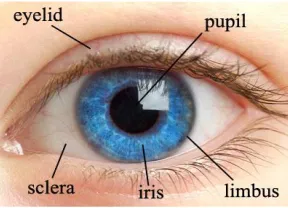
The rapid development in technology and services in life, have increased the human activities and transactions, where reliable personal identification is necessary. The chances of unauthorized access are more due to the advancements in the field of information technology. The person’s identity is becoming more and more important with the enlargement and enhancement of mankind activity range. There are traditional and knowledge based methods to restore person’s identity which include ID cards, keys and passwords for authentication. But, these methods are usually not reliable. A new method named as biometrics is more revealing and promising [16]. A Biometric system provides recognition of an individual based on some sort of unique characteristics possessed by the individual [3]. The imposters are denied access and genuine is granted access. Thus, a higher degree of confidence can be achieved by using unique physical or behavioral characteristics such as fingerprints, voice, face and iris, of the person. The human eye as shown in Fig. 1 is almost a spherical ball with a slight bulge in the front part. Iris is the part of the human eye. Iris, as shown in Fig. 2 is the best biometric trait than other traits as these get altered with the age and diseases but iris pattern remains unaltered throughout the life. Iris is gaining lot of attention due to its permanence and measurability. Located between cornea and eye lens, iris is internally protected yet externally visible.
Fig. 1 The human eye
The iris consists of distinct characteristics such as the freckles, coronas, strips, furrows and so on.
Fig. 2 The human iris
Extensive Research over the decade has increased the reputation of iris. Most commercial iris recognition systems [3], use the algorithms developed by Daugman and these algorithms are able to produce perfect recognition rates [1][2]. The published results have usually been produced under favorable condition. The problems with iris identification reside in the structure of organ itself. Iris images captured are occluded by eyelids and eyelashes. The structure of iris and pupil is not definitely circular and concentric. The iris boundaries are presumed to be circular, which leads to improper localization of iris. Therefore, effective localization and normalization methods are essential. There are four stages of iris recognition; segmentation, normalization, feature extraction and recognition [14]. Each stage applies different techniques. Iris segmentation [10] [12] refers to the process of __________________
Deepika Prashar is currently pursuing masters degree program in computer Science department in S.V.I.E.T, Punjab Technical University, India, E-mail: [email protected]
extracting region that provide information of iris pattern. It is the process of locating the inner and outer boundaries of iris. Normalization refers to the preparing of enhanced iris image. It converts the Cartesian coordinates to polar coordinates. The next stage is feature extraction, to extract features from normalized iris image and creating unique iris codes [15]. The iris codes are further used for comparisons. Our focus will be on these four stages and the recognition rate to show the accurate performance. The parameters which measure the performance are False Acceptance rate and false rejection rate. The FAR is the frequency that a non authorized person is accepted as authorized. FRR is the frequency that an authorized person is rejected access. The overview of iris recognition system is shown in Fig. 3. The iris images from UBIRISv1 [6] database is used for the experimental analysis.
Fig.3 Iris recognition System Overview
2
RELATED
WORK
Daugman [1] was the first to propose the iris recognition algorithm in 1990’s. His work consists of Iris codes. In his work, he proposed a methodology consisting of the four stages. The segmentation is done through operator and 1-D Gabor filter is used for iris extraction and hamming distance for matching. His algorithm was not able to deal with noises like reflections on iris [2]. Khan, M.T; Arora, D. & Shukla, S. in their work performed segmentation in two steps. In the first step, they detected pupil center and radius. The pupil region is extracted through the binary thresholding. Edges of pupil are obtained by two imaginary orthogonal lines passing through centroid of the region. In the second step, they detected the edges of iris through the average window vector. The feature extraction was performed by Gabor filters [7] and matching through hamming distance. Their recognition rate to the maximum on UBIRISv1 came out to be 93.445% [4]. Steve Zhou and Junping Sun gave an iris recognition system in which the iris localization is done with histogram analysis technique by converting the grayscale eye image into the binary form which contains only two values either 0 or 1. Normalization was carried out using Daugman’s Rubber Sheet Model. Feature Extraction was done by applying 1-D Log Gabor Wavelet. K-Dimensional tree is used for matching of the unique iris code. Algorithm was tested on CASIA database (version not mentioned). Recognition rate was 99.64%. Major drawback of their algorithm is that only limited number of iris codes can be loaded in the tree because the search efficiency decreases with increase in the tree size. Moreover, they have not
mentioned the FRR, FAR and EER, which are very important parameters to judge the performance of any algorithm [5]. Jimenez Lopez, F.R., Pardo Beainy, C.E. & Umana Mendez, O. E, described the segmentation and normalization process for automatic biometric iris recognition system, implemented in Mat lab. They used grayscale database images and performed Hough Transform as the segmentation technique. They have worked on one stage of iris recognition system that is the segmentation of the iris region from various eye images and were able to bring 78.5% accuracy [8]. Wagdarikar, A.M.U., Patel B.G. and Subbaraman, S. mainly focused on the feature extraction and matching iris patterns, and did not introduce the information about segmentation and normalization [11].
3
PROPOSED
METHODOLOGY
The proposed methodology in this section consists of four stages – Segmentation, Normalization, Feature Extraction and Matching or recognition. The various parameters- FAR, FRR and recognition rates are measured. The algorithm is proposed for iris identification and is tested on the UBIRISv1 noisy datasets [6]. The proposed method as shown in Fig. 4 proceeds as follows. The eye image is processed by first segmenting the iris region which is done by the canny edge detector for mapping the edges and then using Circular Hough Transformation. Then, Daugman’s rubber sheet model is applied for the normalization. 2D Reverse Biorthogonal Wavelet is used for extracting the features. The iris codes are then compared.
3.1 SEGMENTATION
The first motive in our proposed system is the localization of the iris outer and inner boundary. The iris outer boundary (limbus boundary) is present between the sclera (the white area) and the iris whereas the iris inner boundary (also known as pupil boundary) is present between the iris and the pupil (the darkest area). Both the limbus and pupil boundaries are assumed to be the circular model in the iris recognition system. The canny edge detector is used to generate the edge map. after doing a circular Hough transformation [10], the maximum value in the Hough space corresponds to the center and the radius of the circle. In our method, the pupil and iris boundaries are localized in the same way.
3.1.1 CANNY EDGE DETECTION
The canny edge detector [9] is the powerful tool for detecting edges in noisy environment. Based on the smoothed image, derivative in both x and y direction are computed, these in turn are used to compute the gradient magnitude of the image. After then, the non maximum suppression is performed in which the pixels are suppressed if they do not contain local maximum. The final step in this detector is to use the hysteresis operator, in which pixels are marked as either edges, non edges and in- between, this is based on the threshold values. The next step is to consider each of the pixels that are in-between, if they are connected to edge pixels these are marked as edge pixels as well. The result of this edge detection is the binary image where 1- valued pixel will symbolize the true edges.
3.1.2 CIRCULAR HOUGH TRANSFORM
In our proposed system, the CHT aims to find the circles in the eye image. After generating the edge image, we will consider the edge points as
(
x
k,
y
k)
, k=1, 2, 3……n. Theweights in a circular Hough space are analyzed for estimating the three parameters of one circle (𝑥𝑐 , 𝑦𝑐, 𝑟) A
Hough space can be written as:
)
,
,
,
,
(
)
,
,
(
1r
y
x
y
x
h
r
y
x
H
k c cn k k c c
(1)Where
ℎ 𝑥𝑘, 𝑦𝑘, 𝑥𝑐, 𝑦𝑐, 𝑟 = 1 𝑖𝑓 𝑥𝑘, 𝑦𝑘 is on the circle xc , 𝑦𝑐, 𝑟 ;
0 otherwise ;
(𝑥𝑘 , 𝑦𝑘) k=1, 2, 3……n are the edge pixels. The position
(𝑥𝑐, 𝑦𝑐, 𝑟) with the maximum value of 𝐻(𝑥𝑐, 𝑦𝑐, 𝑟) is chosen
as the parameter vector for the strongest circular boundary. However, in our proposed system, CHT is used for detecting the pupil and iris outer boundaries. The final segmented image of iris is shown in Fig. 5. (b)
Fig. 5 (a) The original image (b) The segmented Iris image
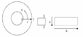
3.2 NORMALIZATION
The localized iris is then normalized to a rectangular block with constant size radius being in correspondence to the width of the block and the angular displacement Ɵ being in correspondence with the length of the block as shown in the Fig.6. The normalization is performed to overcome pupil dilation as there is different pupil for different eye images. So, there is a need to convert the circular model into the rectangular block. The Daugman’s rubber sheet model is the linear model that assigns to each pixel of the iris, a pair of real coordinates
(
r
,
)
, where r is on the unit interval [0, 1] and
is an angle in between [0, 2π] .Fig. 6 – Daugman’s rubber sheet model .
The remapping of iris image
P
(
u
,
v
)
is done from theCartesian coordinates
(
u
,
v
)
to the dimensionless non concentric polar coordinate system (r,).Thus the following set of equations are used to transform annular iris into polar equivalent
P
(
u
(
r
,
),
v
(
r
,
))
P
(
r
,
)
(2)Here in equation 2,
u
(
r
,
)
andv
(
r
,
)
are defined as linear combinations of both the set of pupil boundary points))
(
),
((
p
p
v
u
and set of the iris boundary points alongthe outer perimeter of the iris
(
u
i(
),
v
i(
))
:)
(
*
)
(
*
)
1
(
)
,
(
r
r
u
p
r
u
i
u
(3)
v
(
r
,
)
(
1
r
)
*
v
p(
)
r
*
v
i(
)
Here in equation 3,
P
(
u
,
v
)
is the iris region,(
u
,
v
)
are theoriginal Cartesian coordinates and
(
r
,
)
are the corresponding normalized polar coordinates. After, performing the normalization, the enhancement is done to remove the noises from the image. The result of normalization is shown in Fig.7.3.3 FEATURE EXTRACTION
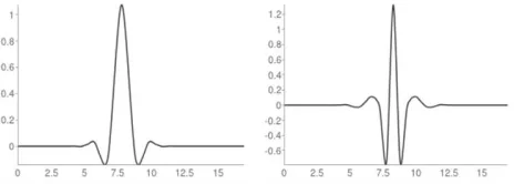
Feature extraction is used to extract the useful features from the normalized iris image. Various wavelet Transformation techniques [13] can be used for feature extraction. Φ is the scaling function and Ψ is the wavelet function. The family of wavelets exhibits the property of linear phase. The 2D rbio 6.8 wavelet scaling and wavelet functions are shown in Fig. 8. . In our proposed system, we have used discrete 2D reverse biorthogonal wavelet (rbio 6.8) to form the iris code.
Fig. 8 Two dimensional rbio 6.8 wavelet decomposition scaling and wavelet function
The rbio 6.8 wavelet is employed to each enhanced normalized image for obtaining the detailed coefficients. The three level of decomposition is used. To reduce the size of the template, the vertical detailed coefficients, dv are extracted by the wavelet, and then binarized. Thus, the feature codes are generated for each normalized enhanced iris image and are used, further, in making comparisons. We have implemented this method effectively in MATLAB and generated the iris codes for every normalized iris image.
3.4 MATCHING
The comparisons are performed using the hamming distance. The equation to calculate the hamming distance is:
(4)
Where Xi and Yi are the two templates compared. The two templates can be same i. e. from the same subject or can be from different subjects.
4
EXPERIMENTS
AND
RESULTS
In the experiments, we implemented the method described by the Daugman [2] which comprises of the four main stages. In the segmentation, we implemented the canny edge detection and the circular Hough transformation that detects the circular iris and pupil boundaries. We have used the same way for detecting both the limbus and pupil boundaries. Normalization is performed through Daugman Rubber Sheet Model. We have performed feature extraction through 2D reverse biorthogonal wavelet 6.8 which extracts the vertical detail coefficients at a particular level of decomposition to obtain the iris codes for each normalized iris image. The iris code comparisons were made through
inter and intra hamming distances, and finally the recognition rate of the system was calculated. The UBIRIS v1 dataset is taken which consists of grayscale images. We have taken 50 unique subjects and 5 image of each subject. Total number of images is 250 images. We carried out our experiments in MATLAB 7.10.0 (R2010a).
TABLE 1: FAR and FRR
Here, the False Acceptance Rate (FAR) and the False Rejection Rate (FRR), are defined as accepting a false person and rejecting a genuine person, respectively. The Recognition rate or the accuracy is calculated on the basis of FAR and FRR (Recognition Rate =100-(FAR+FRR)/2). Table 1, shows the accuracy of our proposed system.
5
CONCLUSIONS
Various researchers have contributed significant amount of research in developing a restricted free iris recognition system and lots of researches are going on accurately. Accuracy of the results depends on how effective the segmentation and normalization is done so that normalization and other later stages can be handled with effectiveness. Center and boundaries of the iris are detected even in the presence of eyelashes, eyelids and noise. The proposed methodology can serve as the essential measure. Angular deflections affect the recognition performance of the system which is not considered in the proposed work and can be taken as future scope.
REFERENCES
[1]. J.Daugman, ―New Methods in iris recognition‖, IEEE Trans. Syst., Man, Cybern.B, Cybern., Vol. 37, no. 5 ,pp. 1168-1176, Oct. 2007.
[2]. J.Daugman, ―How iris recognition works,‖ IEEE Trans. on Circuits and Systems for Video Technology, vol. 14, pp. 21-30, 2004.
[3]. http://www.wisegeek.com/what-is-iris-recognitiontechnology.htm
[4]. Khan, M.T; Arora, D. & Shukla, S., ―Feature Extraction through Iris Images using 1-D Gabor Filter,‖ 2013 Sixth International Conference on Contemporary Computing (IC3), pp. 445-450, 8-10 Aug.2013.
[6]. Im proceedings Proenca. Hugo and Alexandre, Luis A., ―UBIRIS: A noisy iris image database,‖ Proceedings of ICIAP 2005- International Conference on Image Analysis and Processing, vol. 1, pp. 970-977.
[7]. David Carr, ―Iris Recognition Gabor Filtering‖, Vol. 1.4, Dec18, 2004
[8]. Jimenez Lopez, F.R., Pardo Beainy, C.E. & Umana Mendez, O. E. , ―Biometric Iris Recognition Using Hough Transform,‖ 2013 XVIII Symposium of Image, Signal Processing, and Artificial Vision(STSIVA),pp. 1-6, 11-12 Sept. 2013.
[9]. J.Canny, ―A Computational approach to edge detection,‖ IEEE Transactions on Pattern Analysis and Machine Intelligence, Vol. 8, no. 6, pp. 679-698, 1986.
[10]. R.C.Gonzalez and R.E, woods, Digital Image Processing, in J.Houseman (2nd Ed.), Handbook of physiology, 4 (New Jersey: Upper Saddle River, 2002).
[11]. Wagdarikar, A.M.U., Patel B.G. and Subbaraman, S., ―Performance Evaluation of Iris Recognition using Neural Network Classifier,‖ 2010 3rd
IEEE International Conference on Computer Science and Information Technology(ICCSIT), Vol. 1, pp. 146-149, 9-11 July, 2010.
[12]. Z.He, T.Tan, Z.Sun, and X.Qui, ―Towards accurate and fast iris segmentation for iris biometrics,‖ IEEE Trans, On PAMI, Vol. 31, no. 9, pp.1670-1684, Sept. 2009.
[13]. D.M.Monro, S.Rakshit, and D, Zhang, ―DCT- based iris recognition,‖ IEEE Trans. Pattern Anal. Mach. In tell, Vol. 29, no. 4, pp. 586-596, Apr. 2007.
[14]. A.S Tuama, ―Iris Image Segmentation and Recognition,‖ International Journal of Computer Science & Emerging Technologies, IJCSET, Vol.3, no. 2,E- ISSN-2044-6004, April, 2012.
[15]. C.Sanchez-Avilla, R. Sanchez- Reillo, D. de Martin-Roche, ―Iris Based Biometric Recognition using Dyadic Wavelet Transform,‖ IEEE AESS Systems Magazine, Oct. 2002.