O R I G I N A L R E S E A R C H A R T I C L E
Open Access
In house virtual surgery and 3D complex
head and neck reconstruction
Kimberly Luu
*, Amirreza Pakdel, Edward Wang and Eitan Prisman
Abstract
Background:3-Dimensional (3D) printing can be applied to virtual planning and creation of surgical guides for mandibular reconstruction. Such systems are becoming increasingly prevalent in head and neck reconstruction. However, third party access to this technology is costly and removes the opportunity to design, create, and modify the bony reconstructions, as third party technology is a black box. This series is a pilot study to document the feasibility of an in-house software tool. The objectives of this study are to describe the design of an automated in house system and assess the accuracy of this in house automated software tool for mandibular reconstruction in a simulated environment.
Methods:Software was written to automate the preoperative planning and surgical guide creation process. In a simulation lab, Otolaryngology residents were tasked with resecting and reconstructing a simulated mandible using the 3D-printed cutting guides. A control group of residents performed resection and reconstruction using the traditional method without cutting guides. T-test analysis was performed to compare specific aspects of the final reconstructions including: change from native mandibular width and projection, segment gap distance, and reconstruction time.
Results:Mandibular reconstruction was successful in all participants using the 3D printed system.
The guided group performed significantly better on the measurement of change in Mandibular overlap, projection, segment gap volume. There was a non-significant trend towards better mandibular width and operative time for the guided group.
Conclusions:This study confirms functionality and feasibility of using an in house automated software for planning and creating surgical guides.
Keywords:Stereolithography, Mandibular reconstruction, 3D printing, Surgical cutting guide
Background
Advanced head and neck malignancies with underlying bony involvement often require aggressive oncological resection of large segments of the oral cavity including the mandible or maxilla. Reconstruction following these ablations is provided by microvascular transplantation of osseo-cutaneous tissue such as a fibular or scapula free flap [1]. These transplanted free flaps require multiple osteotomies in complex 3-dimensional (3D) orientations in order to reconstitute the premorbid bony and soft tissue anatomical structures of the head and neck. The accuracy of the reconstruction has significant impact on
cosmetic and functional outcomes such as mastication, articulation, deglutition and breathing, as well as consid-erable effect on the patient’s quality of life.
Traditional intraoperative execution of these complex reconstructions continues to be challenging and time consuming. This technique may be improved with ad-vanced preoperative planning. One such approach, which has been used in a variety of craniofacial recon-structions utilizes 3D printed objects that are generated with patient-specific geometrical data from computed tomography (CT) scans [2]. Potential benefits of pre-operative planning include reducing pre-operative time and cost, improving patient understanding, resident educa-tion, and refining surgical predictability and outcomes [3]. Ultimately, this innovation has the potential to lead * Correspondence:[email protected]
Division of Otolaryngology, Head & Neck Surgery, University of British Columbia, 2775 Laurel Street, DHCC 4th. Floor, Vancouver, BC V5Z 1M9, Canada
to a decrease in patient morbidity and an improved quality of life after having undergone head and neck reconstruction.
Studies on the use of 3D printing in head and neck reconstruction currently uses third party commercial software. Commercial software is expensive and the lack of direct access to the technology and immediate feed-back creates a tedious preoperative planning process. We postulate that technology designed and created in house will provide the benefits of commercial software in terms of preoperative virtual planning and accurate reconstructions, while reducing the cost. Additionally, continuous improvements can be made on the technol-ogy, guided by direct user experience. Future innova-tions, such as the ability to design one step dental implants, can be planned and implemented. The purpose of this study is to describe and pilot an in-house soft-ware to produce three-dimensional surgical guides for use in mandibular reconstruction. The objectives of this study are to describe the design of an automated in house system and assess the accuracy of an in house automated software tool for mandibular reconstruction in a simulated environment.
Methods
Mandibular guide development
In house software for preoperative virtual planning and creation of surgical guides was designed and developed through collaboration between the principal investigator, a head and neck reconstructive surgeon and a biomech-anical engineer. The preoperative surgical planning tool was developed with Amira v5 software (FEI Visualization Sciences Group, Berlin, Germany) for visualization of CT data, which was bridged with the automation algorithm written in MATLAB (Mathworks, Natick, MA)[4]. The process is based on the predetermined principles and begins with the input of a DICOM CT scan image. A 3D reconstruction of the mandible is generated by automatic segmentation based on Hounsfield unit thresholds of cortical bone (~ 400 HU), with subsequent refinement input from the user to remove any aberrations (e.g. due to tooth filling metal artefacts, segmentation defects due cartilaginous TMJ tissue etc.).
Resection margins are then indicated with 3D cutting planes on the virtual model through an interactive user interface. Next, the desired location of the mandibular plate is drawn free-hand on the outer cortical surface of the mandible, extending from the subcondyle surface, though the angle and body, and ending at the contralat-eral para-symphyseal surface. The reconstruction is then automatically planned based on a custom implementa-tion of the Ramer–Douglas–Peucker contour simplifica-tion algorithm. This process optimizes the mandibular contour and bony apposition of the osteotomies by
reconstructing the 3D curved geometry of the resected mandibular segment as a series of straight segments. A number of constraints can be defined, such as the desired number of osteotomies, the minimum and max-imum length of each segment. A critical component of the algorithm is automatic determination of the cutting angle for each segment of the donor bone to achieve maximal opposition when the segments are linearly arranged.
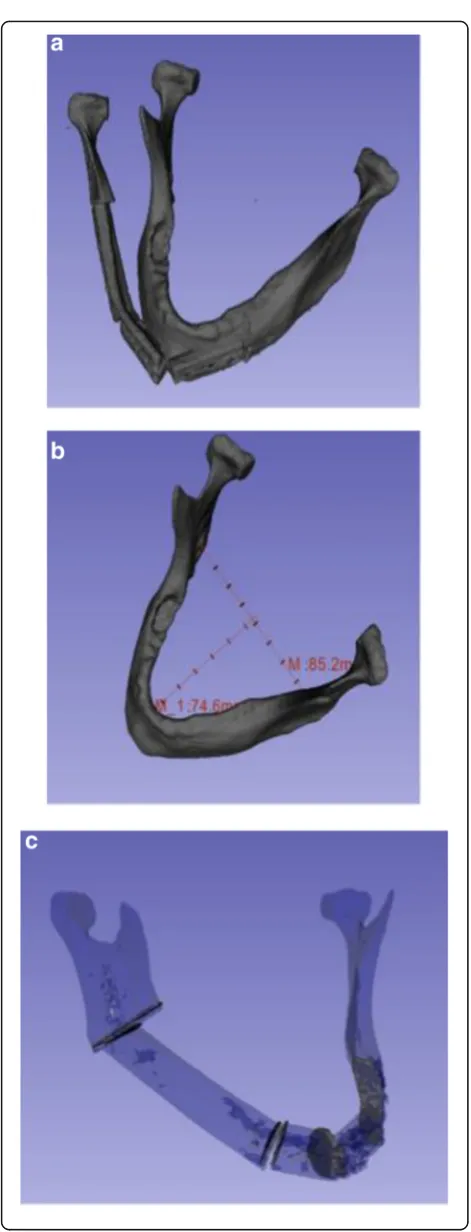
Once the resection and reconstruction is planned, the software displays the reconstructed mandible with the fibular segments for a final check. Virtual models of the surgical cutting guides are then created with the reverse image of the mandibular model so the guide can snap on at the appropriate place during the operation. Each guide houses a slit that corresponds to the cutting plane that will reproduce the virtually planned resection on the mandible, and the planned sectioning on the fibula. Figure 1 shows the 3D printed mandible and surgical cutting guides.
Experimental method
The institutional research ethics board approved the study protocol. Otolaryngology residents from the Uni-versity of British Columbia were invited to participate in the study during a bone-plating simulation day. The residents all received a demonstration and practice time for performing mandibular reconstruction on the simu-lated models.
Residents were randomized into a control group, which performed the reconstructions by a traditional free-hand method and a guided group, which performed the reconstructions with surgical cutting guides. The randomization was blocked by residency training year. Each resident had their own workstation and were not able to see the reconstructions of the other participants.
Both groups were tasked with resecting and reconstruct-ing a real case of an oral squamous cell carcinoma in-vading the mandible.
All participants were provided with a 3D print out of the diseased mandible and a generic plastic fibula. The free-hand group designed and performed the mandibular resection and reconstruction using a ruler as a guide [5]. The participants marked out the osteotomy sites on the fibula, including the angle at which to make the cuts. The group was allowed to make as many adjustments as they felt were needed. The guided group was provided plastic guides designed to lock onto the mandible and generic fibula with slots that guided the resection and osteotomies. Once they were all made, the participants plated the fibular sections. The reconstructed mandibles were then anonymized.
Analysis
All reconstructions were classified as successful if mandibu-lar continuity was achieved. All participants recorded the amount of time required to perform the reconstructions.
CT scans were taken of each reconstruction and a 3D print out of the original mandible. Using the open source medical imaging software 3DSlicer version 4.5 [6], triangu-lar mesh models were rendered from the CT scans. Accur-acy was assessed by a number of measurements shown in Fig. 2 including: overlap of the reconstruction with the native mandible, volume of each gap between osteotomies, the change in width and projection of the mandible.
Volume overlap
The volume overlap was calculated using the Model-to-Model Distance extension in 3DSlicer [7]. The mean distance between the two models on a point-to-point basis was used as the value of comparison.
Gap analysis
The volume of the gap was estimated by subtracting the convex hull (volume) of each individual segment from the convex hull of the two opposing segments. This algorithm was provided in 3DSlicer.
Width and projection
The width was defined as the distance between each condyle (point A and B) and the projection was defined
as the distance from the midpoint of AB and the most anterior part of the reconstruction.
Data was analyzed using SPSS. The mean results of the above measurements between the guided and non-guided groups was compared with a 2-sample paired t test. Comparison of the time to completion for each mandicular reconstruction between the guided and un-guided group was done with another 2-sample t test. Statistical significance was set atp< 0.05.
Results
A total of 10 residents participated in the study. There were two residents in each training year from PGY1 – PGY5. The residents in each year had a comparable amount of experience in mandibular reconstruction and
all participated in the morning plating course. All resi-dents completed reconstruction in the group they were allocated. All mandibular reconstructions achieved con-tinuity. Figure 3 displays the final reconstruction results for each participant.
Time to completion
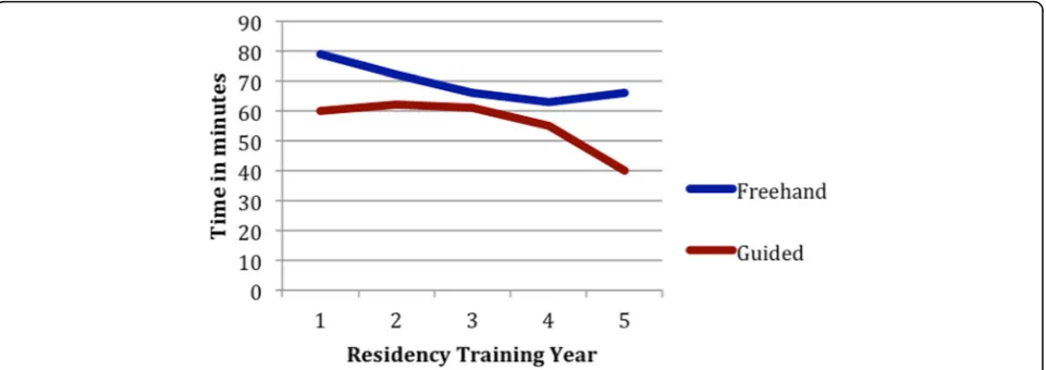
Average time for completion of reconstruction was less for the guided group but not statistically significant. Figure 4 shows the time for completion of each resi-dent There is an inverse relationship between time to completion and residency training year, as expected. Average time to completion without bone plating time for freehand and guided group was 51 min and 46 min respectively with p= 0.172.
Computed tomography analysis
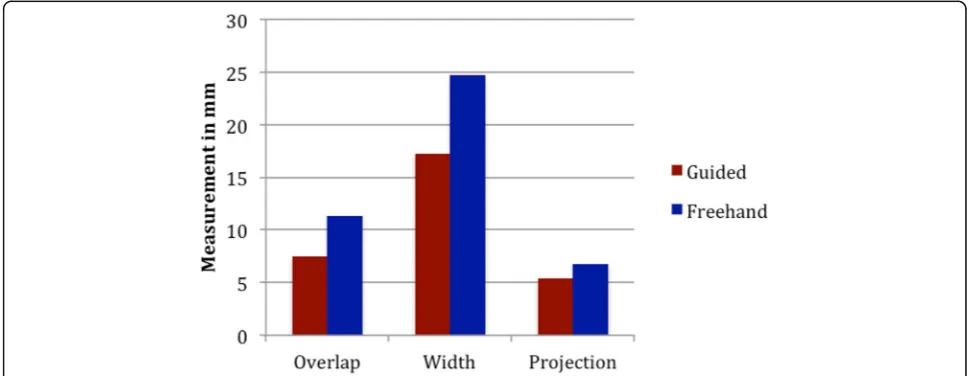
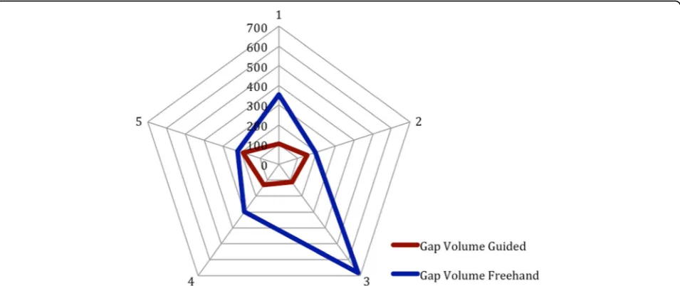
Results show that the guided group performed better in all categories. Table 1details the results of the measure-ments for each participant. Figures 5 and 6 graphically depicts the significant improvement in gap volume, over-lap, and projection.
Discussion
This study aimed to demonstrate feasibility and accuracy of surgical cutting guides created with an in-house ware for mandibular reconstruction. The in house soft-ware was able to create surgical guides with minimal intervention from the operating surgeon. Time to com-pletion of the guided versus freehand group was faster, but not statistically significant. The guided group per-formed significantly better in creating reconstructions with appropriate mandibular projection and lesser segment gap distant. There was a non-significant trend towards better mandibular width.
Fig. 4A non-significant decrease in time to completion was shown between the freehand and guided group. This graph shows the time to completion of the reconstructions with 1 line for the freehand group and 1 line for the guided group. There is a non-significant trend to decrease in time for the guided group. Additionally, residents in higher training years performed the reconstruction in less time as expected
The majority of 3D printing for mandibular recon-struction is done with third party software [8, 9]. The cost of commercial software from KLS Martin Group or Synthes can range from $4000 - $15,000 per case compared to $100 - $200 per case using this in house software. In addition to cost, the workflow of commercial software is often not ideal. It may take several interactions and face-to-face meetings the com-pany to finalize a preoperative plan, making additional changes difficult and time consuming. Dupre-Bories et al. have also made an effort to create the own in house technology to overcome this inefficient. The group has shown a reduction of cutting guide delivery time from 21 to 5 days [10]. In contrast, the technol-ogy described in this paper delivers a cutting guide in 2–3 days. The time gain stems from improved work-flow as well as increased automation in the guide creation process. This automation removes multiple steps in which the user has to interact with the software or a designer. In the future, this automation can allow for intraoperative changes to be made if unexpected changes, such as tumour progression, are encountered.
Multiple studies have shown the use of 3D printed surgical cutting guides for mandibular reconstruction is feasible [11–13]. The most common measurable benefit that has been shown is intraoperative time gain [14–16]. In the simulated environment, our study did not show a significant decrease in operative time. This could be explained by a decrease in complexity from a simulated environment that lacks soft tissue and challenges with exposure, bleeding, or access and the small number of participants in the study. Many additional benefits of virtual planning have been hypothesized, but have not been consistently proven. This pilot study demonstrates the use of appropriate objective measurements that can assess the accuracy of mandibular reconstruction and demonstrated an improvement in accuracy. A clinical trial can use the same accuracy assessment protocol.
The design and development of the in house software resulted in a number of learning points. There was discussion around what factors should be optimized in the reconstruction when developing the surgical guides. Lower contour of the mandible provided the best cos-metic outcome, but optimizing for upper alveolus allowed the design to remain flexible for the future
Fig. 5A significant decrease in the differences between the native and reconstructed mandible was shown for overlap and width. This graph compares the accuracy measurements for the reconstructions performed freehand with the reconstructions performed with the guide. There shows a decrease in the change of projection and width with the guided group
Table 1Results of accuracy measurements of the guided and nonguided groups
Overlap Guided Overlap Nonguided Gap Guided Gap Nonguided Width Guided Width Nonguided Projection Guided Projection Nonguided
(mm) (mm) (mm3) (mm3) (mm) (mm) (mm) (mm)
7.89 9.96 104.53 354.73 20.1 20.9 2.3 5.3
10.47 12.22 150.37 194.33 24.2 33.5 4.3 6.8
8.65 12.15 112.52 683.24 21.6 23.7 9.8 7.1
2.49 6.89 129.31 297.27 7.3 13.1 7.1 4.1
7.75 15.45 191.58 220.04 13.2 32.2 3.6 10.3
implementation of one-stage dental implants. The optimization of contour would most theoretically lead to an infinite number of osteotomies. The authors had to decide what practical constraints to include in the algo-rithm in order to consider bone survival, operative diffi-culty, and operative time. The use of an in-house software affords the author the ability to consider these variables and systematically test their influence on re-construction outcome.
This paper has a number of limitations. The partici-pants are residents who are not the primary surgeons in many of these complex reconstructive cases. Addition-ally, they are of varying years and a low number, both of which would influence the significance of the results. Novice surgeons would likely benefit more from the use of cutting guides so the increase in accuracy may be exaggerated. The residents were given time to practice plating skills to introduce the simulated environment and provide all participants with baseline skills. However, more practice would have put the residents at an equal skill level and improved the validity of the comparison. Given these limitations however, the guided group were all still able to complete the reconstruction, showing feasibility of the technology that is generalizable to real practice.
Conclusion
This pilot study confirmed the feasibility of designing and developing an in-house software to automate mandibular reconstruction for head and neck cancer resections. Mandibles reconstructed with the surgical guides have comparable accuracy when compared to traditional reconstruction in terms of overlap, volume
between each osteotomy gap, and the width and projec-tion of the mandible.
Abbreviations
3D:3 dimensional; CT: Computed tomography
Acknowledgments
Not applicable.
Funding
This project was partially funded by a VCHRI (Vancouver Coastal Health Research Institute) Innovation and Translational Research Award.
Availability of data and materials
Data and material are available upon request.
Authors’contributions
KL contributed to the design of the in house software, lead the experimental design, data collection, analysis, and was the primary author of the manuscript. EW and AP were the principle engineers of the software and contributed to editing of the manuscript. EP was involved in all aspect of the software design, experiment, and manuscript writing. All authors read and approved the final manuscript.
Ethics approval and consent to participate
Ethics approval was obtained from the Ethics Board at the University of British Columbia. All participants read and signed a consent form prior to participation.
Consent for publication
Not applicable.
Competing interests
The authors declare that they have no competing interests.
Publisher’s Note
Springer Nature remains neutral with regard to jurisdictional claims in published maps and institutional affiliations.
Received: 21 April 2017 Accepted: 22 November 2018
References
1. Mehta RP, Deschler DG. Mandibular reconstruction in 2004: an analysis of different techniques. Curr Opin Otolaryngol Head Neck Surg. 2004;12(4): 288–93.
2. Anuja A, Chen W, Kolokythas A, et al. Use of virtual surgery and Stereolithography-guided osteotomy for mandibular reconstruction with the free fibula. Plastic & Reconstructive Surgery. 2011;128(5):1080–4. 3. Chow LK, Cheung LK. The usefulness of stereomodels in maxillofacial
surgical management. J Oral Maxillofac Surg. 2007;65(11):2260–8. 4. Matlab by Mathworks.https://www.mathworks.com(2017).
Accessed Dec 2015.
5. Kang S, Old M, Teknos T. Contour and osteotomy of free fibula transplant using a ruler template. Laryngoscope. 2016;126(10):2288–90.
6. 3D Slicer.http://www.slicer.org(2017). Accessed 20 Feb 2016. 7. Model to Model Distance.https://www.slicer.org/wiki/Documentation/
Nightly/Extensions/ModelToModelDistance(2017). Accessed 23 July 2016. 8. Cohen A, Laviv A, Berman P, et al. Mandibular reconstruction using
stereolithogrpahic 3-dimensional printing modelling technology. Oral Surgery OralMedicine Oral Pathology Oral Radiology. 2009;108(5):661–6. 9. Sannomiya EK, Silva JV, Brito AA, Saez DM, Angelieri F, Dalben GS. Surgical
planning for resection of an ameloblastoma and reconstruction of the mandible using a selective laser sintering 3D biomodel. Oral Surg Oral Med Oral Pathol Oral Radiol Endod. 2008;106(1):e36–40.
10. Dupre-Boris A, Vergez S, Meresse T, Brouillet F, Bertrand G. Contribution of 3D printing to mandibular reconstruction after cancer. Eur Ann Otorhinolaryngol Head Neck Dis. 2018;135(2):133-36.
11. Cohen A, Laviv A, Berman P, Nashef R, Abu-Tair J. Mandibular reconstruction using stereolithographic 3-dimensional printing modeling. Technology Oral Surg Oral Med Oral Pathol Oral Radiol Endod. 2009;108(5):661–6. 12. Cunningham LL, Madsen MJ, Peterson G. Stereolithographic modeling
technology applied to tumor resection. J Oral Maxillofac Surg. 2005;63(6):873–8.
13. Thomas CV, McMillan KG, Jeynes P, Martin T, Parmar S. Use of a titanium cutting guide to assist raising the composite radial forearm free flap. Int J Oral Maxillofac Surg. 2013;42(11):1414–7.
14. Prisman E, Haerle SK, Irish JC, Daly M, Miles B, Chan H. Value of preoperative mandibular plating in reconstruction of the mandible. Head Neck. 2014;36(6):828–33.
15. Hanasono MM, Skoracki RJ. Computer-assisted design and rapid prototype modeling in microvascular mandible reconstruction. Laryngoscope. 2013; 123(3):597–64.