Volume 2013, Article ID 563019,4pages http://dx.doi.org/10.1155/2013/563019
Case Report
Parapharyngeal Neck Schwannomas with Unusual
Vascular Displacement
Kiran Sargar
Mallinckrodt Institute of Radiology, Washington University School of Medicine in St. Louis, MO, USA
Correspondence should be addressed to Kiran Sargar; kiran.sargar@gmail.com
Received 3 April 2013; Accepted 22 July 2013
Academic Editor: David W. Eisele
Copyright © 2013 Kiran Sargar. This is an open access article distributed under the Creative Commons Attribution License, which permits unrestricted use, distribution, and reproduction in any medium, provided the original work is properly cited.
This case report illustrates two unusual cases of parapharyngeal schwannomas mimicking carotid body tumors in terms of characteristic vascular displacement. Carotid body tumors classically cause splaying of internal and external carotid arteries demonstrating the Lyre sign on imaging. Also interestingly, both of these cases were seen in younger ages and include cervical sympathetic chain schwannoma and vagal schwannoma. However, these schwannomas revealed hypovascularity on imaging studies allowing differentiation from hypervascular carotid body tumors. Preoperative distinction between carotid body tumors and schwannomas is very important.
1. Introduction
Carotid body tumors, cervical sympathetic chain schwan-nomas, and vagal schwannomas have common location in neck that is the retrostyloid compartment of parapharyngeal space [1–3]. Among these, vagal schwannomas are most com-mon followed by carotid body tumors [1,3]. Various imaging features had been described to differentiate them, which con-sist of typical displacement pattern of adjacent vessels and internal vascularity. This case report includes two histopatho-logically proven rare cases of cervical sympathetic schwan-noma and vagal schwanschwan-noma having similar vascular dis-placement pattern as that of carotid body tumor. However, both of these tumors revealed hypovascularity on imaging unlike hypervascular carotid body tumors.
2. Case One
An 18-year-old female was referred to the otolaryngology department with a swelling on the left side of the neck since 4 months. On examination, nonpulsatile mass near angle of the left mandible was seen. Cranial nerve examination was normal. Doppler ultrasonography revealed hypoechoic mass in the left carotid space showing mild peripheral vascularity (Figure 1). Computed tomography of the neck showed a het-erogeneously enhancing hypodense mass in the left carotid
space causing splaying of carotid bifurcation (Figures 2 and3). On MRI examination, the mass was predominantly hypointense on T1- and hyperintense on T2-weighted images (Figures4and5). Also MR angiography confirmed splaying of external and internal carotid vessels on the left side (Figure 6). The patient underwent surgery. At surgery encap-sulated mass was found at the carotid bifurcation extending posteriorly which was separated by blunt dissection from carotid vessels. Vagal and glossopharyngeal nerves were sep-arately seen from the mass. At surgery, the tumor was easily peeled off from carotid and jugular vessels without significant bleeding. Histopathology confirmed diagnosis of cervical sympathetic chain schwannoma with distinct Antoni A areas (Figure 7). Postoperatively patient developed Horner’s syn-drome on the left side which recovered 3 months after surgery.
3. Case Two
2 Case Reports in Medicine
Figure 1: The Doppler ultrasonography of neck reveals a hetero-geneous predominantly hypoechoic mass on the left carotid space showing mild peripheral vascularity.
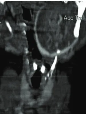
Figure 2: Computed tomography of the neck shows a heteroge-neously enhancing hypodense mass on the left carotid space causing splaying of carotid bifurcation.
Figure 3: Computed tomography of the neck shows heteroge-neously enhancing hypodense mass in left carotid space causing splaying of carotid bifurcation.
Figure 4: T1-weighted MR image reveals isointense mass on the left carotid space.
Figure 5: T2-weighted MR image shows hyperintense mass with central cystic component.
mild heterogeneous contrast enhancement (Figure 9). Mass was seen insinuating between internal and external carotid arteries with resultant splaying of internal and external carotid arteries (Figure 10). Digital subtraction angiography revealed hypovascular mass causing splaying of internal and external carotid arteries (Figure 11). The patient underwent surgery. Intraoperatively, tumour was seen arising from the left vagus nerve and was separating internal and external carotid arteries. On histopathology, mass was proven to be vagal schwannoma (Figure 12).
4. Discussion
Figure 6: MR angiography image is showing splaying of external and internal carotid vessels on the left side.
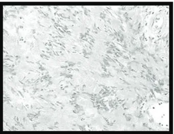
Figure 7: Histopathology slide of the surgical specimen is consistent with diagnosis of Antoni A schwannoma.
of the retrostyloid parapharyngeal space [1,2]. Carotid body tumors are rare neoplasms, although they represent about 65% of head and neck paragangliomas [1,2]. These tumors are derived from embryonic neural crest cells and arise in the chemoreceptoric, nonchromaffine paraganglia at carotid bifurcation [1–3]. Epidemiologically they are common in young people, with female preponderance [2,4]. Since these tumours are located at carotid bifurcation they typically cause splaying of internal and external carotid arteries which can be assessed with CT, MRI, Digital subtraction angiography and ultrasound colour Doppler [1]. This characteristic feature of carotid body tumors helps to distinguish it from other retro styloid pharyngeal mass lesions [1]. Carotid body tumours are hypervascular and enhance intensely on contrast enhanced CT and MRI scans [1].
Schwannomas or neurilemomas, are derived from schwann cells. They are common in middle age with female preponderance [1–3]. In the parapharyngeal space, common sites of the origin of schwannomas are the vagus nerve and the sympathetic chain. Anatomically, vagus nerve is situated within the carotid sheath posterior to the internal carotid
Figure 8: On USG neck, hypoechoic mass is seen on the left parapharyngeal space showing mild vascularity on Doppler study.
Figure 9: On CT scan, neck with contrast reveals a predominantly isodense mass on the left parapharyngeal space showing mild heterogeneous contrast enhancement. Note the insinuation of the mass between internal and external carotid arteries with resultant splaying of internal and external carotid arteries.
4 Case Reports in Medicine
Figure 11: Digital subtraction angiography reveals hypovascular mass causing splaying of internal and external carotid arteries.
Figure 12: Histopathology slide of the surgical specimen is consis-tent with diagnosis of Antoni B type of schwannoma.
artery. As a result, vagal schwannomas typically displace in-ternal carotid artery anteriorly and medially [1,3,5,6]. Cer-vical sympathetic chain is situated along medial and posterior borders of the carotid sheath, so cervical sympathetic chain schwannomas usually displace internal carotid artery ante-riorly [1, 3]. On CT and MRI, both vagal and cervical sympathetic chain schwannomas are hypodense or isodense to the muscles and may have internal cystic areas. Usually, these tumors are hypovascular and show mild contrast enhancement [1–3]. However, sometimes, these tumors also tend to be hypervascular and can show moderate hetero-geneous enhancement [1]. On MRI, these masses are hypo-intense on T1-weighted images and hyperhypo-intense on T2-weighted images [1].
Preoperative diagnosis is very important in these ret-rostyloid parapharyngeal masses as management of carotid body tumors varies from surgery to radiation to observation, while complete surgical excision is the therapy of choice in vagal and cervical sympathetic schwannomas [2]. Also, risk of intraoperative hemorrhage is high in carotid body tumors. In
carotid body tumor surgery is preferred in younger patients, and radiation is reserved for the elderly or unresectable cases [2].
Both of the described tumors displayed splaying of external and internal carotid arteries on CT contrast study, MRI, and DSA which was also confirmed at surgery. However both of these tumors demonstrated mild vascularity on the Doppler USG, CT, and DSA.
Although characteristic vascular displacement, that is, splaying of carotid bifurcation helps to distinguish carotid body tumor from other retrostyloid parapharyngeal masses, similar vascular displacement can be seen in vagal and cervi-cal sympathetic chain schwannomas. So, this imaging pitfall should be taken into account while considering differential diagnosis of retrostyloid parapharyngeal tumors.
Both of these tumors showed hypovascularity, hence favor schwannomas rather than carotid body tumors. Inter-nal vascularity and enhancement characteristics of the tumor should be given more importance while differentiating schwannomas from carotid body tumors. Hence enhance-ment pattern is the imaging pearl in differentiating carotid body tumors from schwannomas.
References
[1] P. M. Som and H. D. Curtin,Head and Neck Imaging, Mosby, St. Louis, Mo, USA, 4th edition.
[2] C. W. Cummings, B. H. Haughey, J. R. Thomas, L. A. Harker, and P. W. Flint,Cummings: Otolaryngology: Head and Neck Surgery, Mosby, St. Louis, Mo, USA, 4th edition.
[3] C. G. Gourin and J. T. Johnson, “Parapharyngeal space tumors,” eMedicine, Otolaryngology and Facial plastic surgery,http:// emedicine.medscape.com/article/849385-overview.
[4] M. Chaaban and K. M. Stenson, “Carotid body tumors,” eMed-icine, Otolaryngology and Facial plastic surgery, http://emed-icine.medscape.com/article/1575155-overview.
[5] D. T. Kehagias, E. C. Bourekas, and G. A. Christoforidis, “Schwannoma of the vagus nerve,” The American Journal of Roentgenology, vol. 177, no. 3, pp. 720–721, 2001.
Submit your manuscripts at
http://www.hindawi.com
Stem Cells
International
Hindawi Publishing Corporation
http://www.hindawi.com Volume 2014
Hindawi Publishing Corporation
http://www.hindawi.com Volume 2014
INFLAMMATION
Hindawi Publishing Corporation
http://www.hindawi.com Volume 2014
Behavioural
Neurology
Endocrinology
International Journal ofHindawi Publishing Corporation
http://www.hindawi.com Volume 2014
Hindawi Publishing Corporation
http://www.hindawi.com Volume 2014
Disease Markers
Hindawi Publishing Corporation
http://www.hindawi.com Volume 2014
BioMed
Research International
Oncology
Journal of Hindawi Publishing Corporationhttp://www.hindawi.com Volume 2014
Hindawi Publishing Corporation
http://www.hindawi.com Volume 2014
Oxidative Medicine and Cellular Longevity
Hindawi Publishing Corporation
http://www.hindawi.com Volume 2014
PPAR Research
The Scientific
World Journal
Hindawi Publishing Corporationhttp://www.hindawi.com Volume 2014
Immunology Research
Hindawi Publishing Corporation
http://www.hindawi.com Volume 2014
Journal of
Obesity
Journal ofHindawi Publishing Corporation
http://www.hindawi.com Volume 2014
Hindawi Publishing Corporation
http://www.hindawi.com Volume 2014
Computational and Mathematical Methods in Medicine
Ophthalmology
Journal ofHindawi Publishing Corporation
http://www.hindawi.com Volume 2014
Diabetes Research
Journal ofHindawi Publishing Corporation
http://www.hindawi.com Volume 2014
Hindawi Publishing Corporation
http://www.hindawi.com Volume 2014
Research and Treatment
AIDS
Hindawi Publishing Corporation
http://www.hindawi.com Volume 2014
Gastroenterology Research and Practice
Hindawi Publishing Corporation
http://www.hindawi.com Volume 2014
Parkinson’s
Disease
Evidence-Based Complementary and Alternative Medicine
Volume 2014 Hindawi Publishing Corporation