1535-9778/08/$08.00⫹0 doi:10.1128/EC.00161-08
Copyright © 2008, American Society for Microbiology. All Rights Reserved.
MINIREVIEW
What Do Genic Mutations Tell Us about the Structural Patterning of
a Complex Single-Celled Organism?
䌤
Joseph Frankel*
Department of Biology, University of Iowa, Iowa City, Iowa
PROLOGUE: FROM THE CYTOPLASM TO THE NUCLEUS
Structural inheritance in the ciliate cortex.Ciliates make up a distinctive group of unicellular organisms characterized by dualism of germ line and somatic nuclei, sex by reciprocal exchange of gametic nuclei with subsequent replacement of the somatic nucleus, and an extraordinarily complex organization of the cell surface layer. This organization is dominated by cytoskeletal structures, including rows of basal bodies, cilia, and accessory fibrillar structures, and is perpetuated by longi-tudinal extension of these structural ensembles during clonal growth.
This mode of perpetuation provides an ideal opportunity for the demonstration and analysis of cellular heredity at a non-genic level. This opportunity was seized by three gifted
biolo-gists, the comparative zoologist Emmanuel Faure´-Fremiet, the
experimental embryologist Vance Tartar, and the geneticist Tracy Sonneborn, who together led the way in establishing the principle that structural variations of nongenic origin in the ciliate cortex can indeed be faithfully transmitted to progeny over numerous cell generations.
Although this review is primarily devoted to the contribution of genic mutations to the understanding of the ciliate cortex, the interpretation of some of the more interesting of these mutations becomes more meaningful against a background of the three major arenas of nongenic structural inheritance in the ciliate cortex, all of which were solidly established before the genetic approach was initiated.
The best-known form of structural inheritance is the orga-nization of the longitudinal ciliary rows that cover the surfaces of most ciliates. This was first demonstrated by Beisson and Sonneborn (12), who showed that an inversion (180° rotation)
of one or more ciliary rows of Paramecium tetraurelia (then
Paramecium aurelia, syngen 4) (Fig. 1A) could be nongenically inherited; Sonneborn gave the name “cytotaxis” to “this order-ing and arrangorder-ing of new structures under the influence of pre-existing cell structure” (137). This demonstration was later
repeated forTetrahymena thermophila(thenTetrahymena
pyri-formis, syngen 1) (118) and for other ciliates (62, 63, 71). It was extended by Nanney’s observation that the preexisting number
of ciliary rows inT. thermophilatended to be conserved (103,
104), presumably due to the same structural constraints that conserve the geometrical organization of these rows.
A second form of structural inheritance is demonstrated by the propagation of the number of complete sets of cortical
structures. This was investigated in detail by Faure´-Fremiet
(31, 32), who noted that ciliates that became fused side by side, as a consequence of blockage of division followed by anterior sliding of the presumptive posterior daughter cell, could prop-agate their duality. Later, Vance Tartar (147) demonstrated that microsurgically constructed Siamese-twin doublets in the
large ciliateStentor coeruleus could perpetuate their doublet
condition. In both cases, the way in which the doublets were created virtually rules out the hypothesis that this condition had arisen from a genic mutation; instead, it was due to a “contrainte structurale” (32). Sonneborn (136) completed the
demonstration inParamecium tetraurelia(Fig. 1B) by proving
with results of appropriate crosses that the difference between the singlet and doublet conditions was not caused by differ-ences either in nuclear genes or in exchangeable internal cy-toplasm and hence had to reside in the cortical layer. There is good reason to believe that the same conclusion applies to
doublets induced in other ciliates, includingTetrahymena
ther-mophila(103, 109). This structural constraint within the cell cortex does not influence the number of macronuclei in dou-blets; these cells typically reverted from possession of two macronuclei to one, while the cortex continued to propagate its duality (14, 109).
The third form of structural inheritance was discovered by
Faure´-Fremiet (31). He noted that whereas the great majority
of the doublets that he studied manifested a twofold rotational symmetry in the organization of their two normal sets of
cor-tical structures, one exceptional clone of a ciliate named
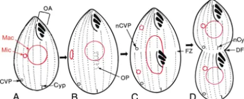
Uro-styla trichogaster displayed a mirror image symmetry in the arrangement of its two sets of cortical structures. This initial discovery was followed up by a Chinese group (153) and then was confirmed and extended by investigators worldwide for a variety of ciliates (44, 64, 70, 142, 155, 166). In these doublets, the arrangement of cortical structures in the “reversed” part-ner was close to a mirror image of that in the “normal” partpart-ner (Fig. 1C), but the internal organization of each individual cil-iary structure in the “reversed” component was normal, though sometimes rotationally permuted relative to the cellular axes (13, 64, 70, 131, 142). The mirror image arrangement could be reliably generated by certain microsurgical operations (132, 153) and was not caused by any relevant genic difference (114, 151). When such mirror image doublets were bisected
midlon-* Mailing address: Department of Biology, The University of Iowa, 143 Biology Bldg., Iowa City, IA 52242. Phone: (319) 335-1110. Fax: (319) 335-1069. E-mail: [email protected].
䌤Published ahead of print on 25 July 2008.
1617
on September 8, 2020 by guest
http://ec.asm.org/
gitudinally to yield two nucleated moieties, the normally ar-ranged moiety produced a clone of normal singlet cells, whereas the reversed moiety was unable to propagate itself despite repeated attempts to do so. This failure was caused by an inability to form food vacuoles, because the cilia of the rotationally permuted oral structures beat in the wrong direc-tion, away from the mouth (31, 61, 133, 152). Reverse-singlet
cells in the less complex hymenostome ciliates Glaucoma
scintillans(143) andTetrahymena thermophila(112, 114) could partially overcome this difficulty and produced slow-growing clones that inherited a reversed configuration of their major cortical landmarks.
While no investigator denied that genes could influence the structural organization of ciliates, the dominant paradigm be-fore about 1970 had been the “pattern and substance” view of Vance Tartar (147, 148), namely, that the nucleus supplies the “building blocks” for structural patterns, but “where and how the building blocks are put together in formed organelles is probably the work of the ectoplasm and its pre-formed struc-ture” (148). Sonneborn’s conclusion a decade later was more even-handed: “There is no evident escape from the conclusion
that essential aspects of development inParameciumare
en-coded partly by cortical geography, not solely by DNA” (138).
Genes and structural patterns in ciliates.In the middle of this period, Sonneborn’s long-time associate, Geoffrey Beale, pointed out that something important was largely missing from the study of the inheritance of ciliate structural patterns. So Beale proposed a research program: “In order to find and study gene-controlled surface variants, the same procedures as are used conventionally in genetics should be tried. A search among wild-races and varieties could be made to see if there are sufficiently definite and permanent variants, or changes induced by mutagenic agents could be studied” (9).
The first part of Beale’s research program was impeded by the circumstance that ciliates tend to evolve reproductive
iso-lating mechanisms that prevent genetic exchange (108, 135) and interspecific incompatibilities between nucleus and cyto-plasm (147) more rapidly than they evolve morphological dif-ferences. This part of Beale’s program nonetheless achieved
some success with two subspecies of the marine ciliateEuplotes
minuta, where a difference in the maximum number of ciliary rows was shown to be under long-term genic control (66). Other genically controlled structural variants have been dis-covered in aberrant clones isolated from nature (28, 72, 73, 74) or derived from inbreeding (34, 89, 122). Despite these suc-cesses, it became clear that the breeding of natural variants could not provide a sufficient foundation for the genetic anal-ysis of structural pattern in ciliates.
The second part of Beale’s program, the study of “changes induced by mutagenic agents,” was pursued intensively with
the two best-understood genetic models among ciliates,
Para-mecium tetraureliaandTetrahymena thermophila. The results of this research program first appeared in the 1970s (27, 139, 157) and were comprehensively reviewed in 1990 by Jerka-Dziadosz and Beisson (75). In the present review, I will focus on the large number of mutations affecting structural organization in
the cell cortex of Tetrahymena. This review has three main
objectives: first, to provide an inventory of genes known to affect structural organization in the ciliate cortex and of aspects of this organization influenced by gene products; second, to employ the altered phenotypes generated by genic mutations as analytical tools to improve our understanding of ciliate cortical organization; and third, to relate the genically con-trolled variants to the three forms of structural inheritance outlined above.
I should state at the outset that the analysis of mutations affecting structural organization in ciliates is still mostly in the
premolecular era. In this respect Paramecium is somewhat
ahead ofTetrahymena, owing to the discovery for the former
organism of a practical means of cloning by complementation
FIG. 1. Examples of forms of nongenic structural inheritance. (A) Inversion of a ciliary row. Shown are three adjacent ciliary rows on the surface ofParamecium tetraurelia, with basal bodies (BB), coated pits (CP), and striated rootlets (SR) indicated. N indicates normally oriented rows; I indicates an inverted row. (B) Siamese-twin doublet ofParamecium tetraurelia, indicating the locations of the two oral apparatuses (OA1 and OA2), the two cytoprocts (Cyp1 and Cyp2), and the two sets of contractile vacuole pores (CVP1 and CVP2). Dashed lines indicate structures behind the plane of view. (C) Ventral surface of a mirror image doublet of a spirotrich (formerly hypotrich) ciliate such asOxytrichaorStylonychia, with the normal and reversed oral apparatuses (nOA and rOA, respectively) labeled. The vertical dashed line indicates the approximate plane of the mirror image symmetry in the arrangement of structures. Panels A, B, and C were modified from Fig. 4.3a, 4.8b, and 8.3, respectively, from reference 38 with permission of the publisher.
on September 8, 2020 by guest
http://ec.asm.org/
genes previously identified only by classical genetic methods (65, 92). One gene with a mutation known to have pleiotropic
effects on cortical as well as nuclear organization,kin241(80),
has been cloned and found to encode a “cyclophilin-RNA
interacting protein” (94); simultaneously, thesmall-19 gene,
which controls cell size and the number of basal bodies (127), was found to encode a member of a new class of tubulin, eta-tubulin (126). Unfortunately, differences in the mecha-nisms of processing of macronuclear DNA (30) make the
spe-cific method of complementation cloning employed for
Para-meciumunfeasible forTetrahymena, and the available methods
of DNA-mediated transformation forTetrahymena(167) have
not been suitable or of sufficiently high efficiency to allow cloning by complementation yet. Nonetheless, recent techno-logical advances in the cloning and transformation of larger
TetrahymenaDNA inserts, subsequent to the sequencing of the macronuclear genome (29), suggest that cloning by comple-mentation of the more diverse array of “classical” genes
af-fecting cortical morphogenesis inTetrahymenamay be feasible
(R. Coyne and E. Orias, personal communication), inviting a molecular genetic analysis of intracellular patterning in this model organism.
THE ORGANISM: ANATOMY AND TOPOLOGY
Before describing mutations affecting cortical structure in
Tetrahymena, I must first introduce the “wild type” organism, as seen from a cortical perspective.
The cortical anatomy of Tetrahymena thermophila is
sche-matically illustrated in ventral (Fig. 2A) and polar (Fig. 2B)
views. This cell, which is about 40 to 50M long, is typically
covered by 18 to 21 longitudinally oriented ciliary rows (Fig. 2A). All but the two postoral ciliary rows originate anteriorly, near the anterior pole of the cell (Fig. 2B). Unlike the situation inParamecium(Fig. 1A), the structural units of the ciliary rows ofTetrahymenaare all made up of single basal bodies and their associated structures (“monokinetids” [99]), except for an asymmetrical crown of paired basal bodies (“dikinetids”) at the
anterior ends of the rows from the fifth row to the right of the oral apparatus (OA) to the second row to its left (Fig. 2B). (Throughout this review, “right” and “left” are used to mean the cell’s right and left, as seen by an imaginary observer standing inside the cell, aligned with its anteroposterior axis, and looking outward toward the surface. The cell’s right side is the actual viewer’s left side, and vice versa.)
Each ciliary unit (“kinetid”) of the ciliary rows has multiple accessory structures, including striated rootlets extending to
the anterior-right of basal bodies (shown in Fig. 1A for
Para-mecium), as well as microtubular transverse and postciliary microtubule bands (not shown here) (1; reviewed in reference 40). Longitudinal microtubule bands, not connected to the basal bodies, extend just under the plasma membrane parallel and to the right of the basal bodies.
The ciliary units are embedded within a structurally contin-uous membrane-skeletal layer, originally called the “epiplasm” (1). Subsequent analyses revealed that this layer has a complex substructure (162), with structurally and chemically distinctive basal-body domains (161) embedded within a continuous layer that itself exhibits a complex spatial distribution of its three major molecular components (163).
The ciliary rows do not meet and join at their anterior ends (Fig. 2B) or at their posterior ends (data not shown). Thus,
whileTetrahymenais topologically a sphere from the
perspec-tive of the plasma membrane and the membrane-skeletal layer below it, cytoskeletally it is more like a barrel bounded by curved staves.
Three major structural landmarks are superimposed on the relatively uniform ciliary rows. The most prominent by far is the OA, a complex structure that includes four compound ciliary elements, the three membranelles (Fig. 2A and 6A) and an undulating membrane (UM), as well as several other mi-crotubular and fibrillar structures, such as the ribbed wall and the deep fiber (see Fig. 6A). It is this organization that gives
Tetrahymena(58) its name. The UM, on the right of the OA, consists of a single row of cilia (whose bases are seen in Fig. 8A) underlain by a double row of basal bodies (8, 111, 159). The three membranelles, on the left of the OA, each consist of three rows of ciliated basal bodies modified into a “sculptured” pattern generated during late stages of oral development (see Fig. 8A) (8, 159).
The other two major structural landmarks are located near the posterior end of the cell. A slit-like cytoproct (Cyp) (Fig. 2), the site of defecation from spent food vacuoles (3), is located immediately to the left of the posterior end of the right postoral ciliary row. One or (more typically) two contractile vacuole pores (CVPs) (Fig. 2) are situated immediately to the left of the posterior ends of (typically) the fourth and fifth ciliary rows to the right of the right-postoral ciliary row, which is designated the oral meridian. Nanney (102) demonstrated that the distance between the oral meridian and the midpoint of the CVP meridians (measured in ciliary-row intervals) is not fixed; rather, it is somewhat less than one-quarter of the cir-cumference of a cell (or of a “semicell” in a Siamese-twin doublet).
The geometry of theTetrahymenacortex can be defined by
two orthogonal axes: an anteroposterior axis (Fig. 2A) and a circumferential axis (Fig. 2B). Both typically remain un-changed during clonal growth (see below). However, under
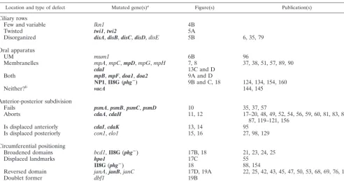
FIG. 2. Ventral view and polar projection of a wild-type Tetrahy-mena thermophilacell. (A) Schematic ventral view of a nondividing cell, showing ciliary rows (CR) with adjacent longitudinal microtubule bands, the bracketed oral apparatus (OA) including an undulating membrane (UM) and three membranelles (M1, M2, and M3), one of the two CVPs, and the cytoproct (Cyp). The anterior-posterior (A-P) axis is indicated in red, with an “X” marking the anterior pole. (B) Po-lar projection of the same cell. The anterior pole of the cell, marked by the “X,” is now in the center of the polar map, partly surrounded by an asymmetric anterior crown (AC) of basal-body couplets. The outer edge represents the posterior end of the cell. The circumferential axis (Circ.) is indicated in red. This figure was modified from Fig. 8.6a of reference 38 with permission of the publisher.
on September 8, 2020 by guest
http://ec.asm.org/
some unusual circumstances, the direction of the circumferen-tial axis may be reversed, so that the cell’s left-postoral ciliary row bears the cytoproct, and the CVPs become located four to five rows to the left of that oral meridian.
The dynamics of cortical structures through the cell cycle is shown schematically in Fig. 3 (see also the photographs in Fig. 15A to E), with the two types of nuclei, the smaller micronu-cleus and the larger macronumicronu-cleus, indicated. The ciliary rows elongate by the addition of new ciliary units anterior to old ones in the middle and posterior regions of cells that are preparing to divide (91, 107, 113). These rows are then subdi-vided transversely just before cytokinesis begins (Fig. 3C). The old OA is retained, and a new OA develops from an oral primordium (OP) located to the left of a subequatorial portion of the right-postoral ciliary row, at the same time that the micronucleus divides mitotically (Fig. 3B). New CVPs (Fig. 3C) and a new cytoproct (Fig. 3D) appear along the appropri-ate ciliary rows just anterior to the fission zone and the division furrow (Fig. 3D), which subsequently constricts along that zone. The macronucleus divides nonmitotically at the same time that the cell divides. The two daughter cells are similar in size (see Fig. 15A to E) (27).
The asymmetry of the developing fission zone has also been documented at the molecular level by the finding that one of the three major membrane-skeletal proteins, the EpiB protein, discovered by Williams and coworkers (162, 164), is asym-metrically distributed on opposite sides of the fission zone in predividing and early-dividing cells (84, 87), as are other cor-tical markers (84, 85, 116).
As Vance Tartar first pointed out for Stentor (150), cell
division in a ciliate is really a process of segmentation. In striking contrast to yeast and animal cells, in which the direc-tion of the cellular polar axis may be respecified within daugh-ter cells at each cell division, in ciliates this direction remains unaltered; what changes is only the positional value of each point of the axis (think of a 1—2—3—4—5 sequence trans-forming itself into 1-2-3-4-5—1-2-3-4-5 before the cell divides).
While this is happening, the ciliary rows perpetuate themselves and the positional values around the circumferential axis re-main constant. As Tartar originally pointed out (149), a ciliate clone can be thought of as a cylinder engaged in indefinite longitudinal extension with periodic transverse subdivision.
THE MUTATIONS
Generation and detection of mutations affecting structural patterning.This review will attempt to cover all of the known single-gene variants affecting the spatial patterning of the
cor-tex of Tetrahymena (summarized in Table 1). One of these
variants is a segregant originally isolated from an inbred strain (89), whereas all but one or two of the remainder are nearly or fully recessive mutations, virtually all of them induced by
ni-trosoguanidine, mostly within the B inbred strain of T.
ther-mophila(for the strain history, see reference 4). These muta-tions were brought to expression after a single cross by one of two genetic tricks. The first method, used prior to 1979, was allelic assortment, in which random segregation of the noncen-tromeric macronuclear chromosomes of a heterozygous mac-ronucleus eventually results in some macronuclei in which all of the macronuclear chromosomes carry only one of the two alleles (4, 101, 165). The second method, employed from 1979 onward, is the process of induced self-fertilization (“cyto-gamy”), in which two sister gametic nuclei derived from the same product of the second meiotic division of the germinal micronucleus are induced to fuse with each other during con-jugation instead of undergoing reciprocal exchange (125).
Once thus effectively rendered fully homozygous, progeny derived from mutagenized cells were screened for putative mutants of interest. Since screening by direct visualization of cortical structures involved an impractical amount of labor, surrogate phenotypes were utilized. For most of the mutants, that phenotype was abnormality of cell shape, a method that had been first applied by Whittle and Chen-Shan (157) to
search for morphological mutations inParamecium. In
Tetra-hymena, this approach was carried out with cells grown over-night at 39.5°C, a temperature that is close to the upper limit for continuous exponential growth of this organism (46). This method succeeded in detecting most of the cortical-pattern mutations to be described below, including many that are also expressed at lower temperatures. This protocol also selects a large number of mutations that prevent the completion of cytokinesis at a restrictive temperature and thereby generate tandem chains and irregular monsters (49, 52, 82, 157).
A very different selection protocol was based on an inability to form food vacuoles at a restrictive temperature (134, 144). This protocol was designed to select mutants affected in phago-cytosis but could also detect mutants with defects in the con-struction of the OA. This protocol succeeded in detecting mutant phenotypes that might have been overlooked by the cell shape-based protocol but, conversely, could not have de-tected oral-patterning mutants that produced fully functional oral structures. The fact that these two different surrogate selection protocols produced nonoverlapping sets of mutant phenotypes indicates that the array of morphological mutant phenotypes to be described below does not exhaust all of the possibilities.
Extensive complementation crosses were carried out to test
FIG. 3. Structural features of wild-typeTetrahymena thermophilaat sequential developmental stages. Micronuclei (Mic) and macronuclei (Mac) are shown in red. (A) Nondividing cell, with the OA, the cyto-proct (Cyp), and one of two CVPs labeled. (B) An oral primordium (OP) forms in the midregion of the cell’s right postoral ciliary row, and the micronucleus has moved to the cell periphery and starts to divide. (C) Membranelles and the UM have become differentiated within the OP, the micronucleus has divided, and a fission zone (FZ) appears at the cell equator as a ring of gaps in the ciliary rows. New CVPs (nCVP) form anteriorly to the FZ. The macronucleus begins to elongate. (D) The division furrow (DF) constricts along the FZ; a new cytoproct (nCyp) appears anteriorly to the FZ; and the macronucleus has com-pleted its division.
on September 8, 2020 by guest
http://ec.asm.org/
for allelism. Clones carrying independently isolated non-complementing mutations almost invariably expressed the same phenotypes, though sometimes with differences in pen-etrance and expressivity at the same or different temperatures. Such noncomplementing mutations were considered to be al-lelic.
Descriptive conventions: nomenclature, techniques, and or-ganization. The organism under review isTetrahymena ther-mophila, so named by Nanney and McCoy (108). (In papers
published in 1977 or earlier, this species was known as
Tetra-hymena pyriformis, species 1 [or syngen 1].) The nomenclature of genotypes in this species was originally unsystematic and has undergone two major revisions. The first, originating in 1977 but not formally published, required that gene loci be repre-sented by an abbreviated gene name followed by a letter to indicate the locus and an Arabic numeral to designate the allele. The second revision, in 1998 (5), restricted the abbre-viated gene name to three letters, followed by an Arabic nu-meral to indicate the locus, then a hyphen, and then a second numeral to indicate the allele. Thus, a mutation that was
orig-inally published as mo1a (56) later became cdaA1 (52) and
according to the newest rules should be renamed “cda1-1.”
However, the new rules allow gene names established in pub-lications prior to 1998 to stand, so I will here adopt the fol-lowing compromise. Mutations previously published under the letter-based designation of gene loci will remain as such, with the numbered alleles now separated from the lettered loci with
a hyphen, i.e.,cdaA1becomescdaA-1. Mutations that had no
letter designation when published, or that have not been pre-viously published, will now be numbered, according to the new
rules; hence,bcd (24) becomesbcd1-1, andco (27) becomes
con1-1. All gene names are three-lettered, with the exception
of several genes of themp(membranellar pattern) series, which
prior to 1998 were given the two-letter namempfollowed by
locus and allele designations (e.g.,mpC-1,mpC-2); the founder
of this series, Kaczanowski’smpsegregant (89), has been
re-namedmpA-1. It is always the case for mutations transmitted
through the germ line that the letter or number just before the hyphen represents a gene locus, whereas the number just after the hyphen represents an allele.
A second convention concerns the style of documentation used here. Virtually all of the mutant phenotypes to be con-sidered here were originally characterized by use of the Chat-ton-Lwoff wet-silver technique, which dramatically highlights the positions of the basal bodies and the major structural landmarks on the cell surface (including the cytoproct and CVPs) but fails to detect subsurface structures, including the accessory cytoskeletal elements. Many, but not all, of the mu-tants have also been analyzed by more refined techniques, including protargol impregnation, immunofluorescence using specific antibodies, and scanning and transmission electron microscopy. To make this survey comprehensive and uniform yet not too lengthy, I have chosen to illustrate the mutants with photographs of Chatton-Lwoff silver preparations and to sup-plement these with scanning electron micrographs only in one instructive case. The silver preparations are all printed in the same orientation (the cell’s right is the viewer’s left), and the
scale bars all represent 10M except for the scanning electron
micrographs in Fig. 8.
Finally, the mutations will be presented in four categories,
TABLE 1. Abbreviated summary of structural-pattern mutants ofTetrahymena thermophila
Location and type of defect Mutated gene(s)a Figure(s) Publication(s)
Ciliary rows
Few and variable lkn1 4B
Twisted twi1,twi2 5A
Disorganized disA,disB,disC,disD,disE 5B 6, 35, 79
Oral apparatus
UM mum1 6B 96
Membranelles mpA,mpC,mpD,mpG,mpH 7, 8 37, 38, 51, 57, 89, 90
cdaI 13C and D
Both mpB,mpF,doa1,doa2 9A and D
NP1,II8G (phgⴚ) 9B and C, 18 124, 134, 154, 160
Neither?b vacA 144, 145
Anterior-posterior subdivision
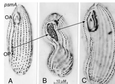
Fails psmA,psmB,psmC,psmD 10 35, 37, 57
Aborts cdaA,cdaH 11, 12 17–20, 48, 49, 52, 54, 56, 59, 60, 81, 83, 86,
87, 119–121, 156
Is displaced anteriorly cdaI,cdaK 13, 14 95
Is displaced posteriorly con1,elo1 15, 16 27, 98, 129
Circumferential positioning
Broadened domains bcd1,II8G(phgⴚ) 17B, 18 21, 23, 24, 25
Displaced landmarks hpo1 17C 55
II8G(phgⴚ) 18 88, 154
Reversed domain janA,janB,janC 17D, 19A 22, 25, 42, 43, 45, 47, 50, 53, 68, 69, 76, 141
Doublet former dbf1 19B
aMutations that are expressed only at high temperatures, or whose expression is greatly enhanced at high temperatures, are boldfaced. Gene designations are as
follows:bcd,broadened cortical domains;cda,cell division arrest;con,conical;dis,disorganized;dbf,doublet former;elo,elongated;hpo,hypoangular;jan,janus;lkn,low kinety(ciliary row)number;mp,membranellar pattern;mum,misaligned undulating membrane; NP,nonphagocytosis;phg,phagocytosis;twi,twisty;vac, (food)vacuole.
bNo defects were detectable by light microscopy.
on September 8, 2020 by guest
http://ec.asm.org/
following an order similar to that employed in the sole earlier review exclusively devoted to this topic (75): category 1, mu-tations affecting the organization of basal bodies in the ciliary rows; category 2, mutations affecting the organization of the OA; category 3, mutations affecting subdivision along the anteroposterior polar axis; category 4, mutations affecting or-ganization along the circumferential axis. Categories 3 and 4 correspond to the “body-plan” mutations of Jerka-Dziadosz and Beisson (75). Of course, some mutations fit into two or even three categories; these will be described within a single category and briefly mentioned in other categories where they also fit.
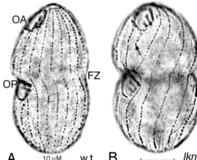
Mutations affecting the organization of basal bodies in the ciliary rows.As mentioned above, cells ofT. thermophila typ-ically have 18 to 21 longitudinal ciliary rows, and this number is preserved during vegetative growth and through conjugation (103, 104). Both the number of ciliary rows and their conser-vation during vegetative growth were affected, in a
non-tem-perature-sensitive manner, in cells homozygous for thelkn1-1
mutation. The number of ciliary rows in the mutant homozy-gotes ranged from 11 to 17 (mode, 13 to 14), whereas the cell size and the estimated total number of basal bodies remained approximately normal, so that ciliary rows were more widely
spaced in cells of homozygouslkn1-1clones than in their
wild-type sister clones but basal bodies were more numerous, and hence more crowded, within each row (Fig. 4). This is in striking agreement with Nanney’s observations of constancy in the overall number of basal bodies in cells with differing num-bers of ciliary rows (106).
Incomplete ciliary rows, which are rare in wild-type cells,
were common inlkn1-1homozygotes (Fig. 4B). In agreement
with this, the fidelity of propagation of the preexisting number
of ciliary rows in subclones (103, 104) was low inlkn1-1
ho-mozygous clones and subclones (J. Frankel, unpublished
ob-servations). The lkn1-1 mutation thus severely modifies the
normally strict cytotactic control of the propagation of ciliary rows.
A second allele,lkn1-2, expressed this phenotype to a
some-what milder degree thanlkn1-1and was not studied in detail.
In addition, theconical (con1-1, originally co) mutant clones
commonly expressed a somewhat lower mean and higher vari-ance in the ciliary row number than parallel wild-type clones (27).
Are there mutations that increase the number of ciliary rows? The majority of the mutants to be described below had a normal range of ciliary row numbers, from 18 to 21. How-ever, there were occasional exceptions, such as the original
janA-1mutant clone, which maintained a stable number of 21
to 25 ciliary rows (76) and thebig1-1mutant, which,
unsurpris-ingly, was not only larger than normal but also had more ciliary rows (57; Frankel, unpublished). This makes it probable that in
Tetrahymena, as in Euplotes (66), the “stability center” (36, 103) of ciliary row numbers is itself under genic control. The
same is likely to be true forParamecium, in which bothkin241
andcrochumutant cells are larger than wild-type cells and have increased numbers of ciliary rows (77, 80).
Thetwisty(twi1-1) mutation brings about a pronounced he-lical twist of the ciliary rows over the surface of the cell. This
FIG. 5. Silver preparations showing dividingtwi1-1(twisty) (A) and disA-1(disorganized) (B) mutant cells, both maintained at 39°C. Im-ages are focused on the ciliary rows (left) and on the OA and OP (right).
FIG. 4. Silver preparations of wild-type (w.t.) (A) andlkn1-1(low kinety number) mutant (B) cells, both maintained at 29°C, with the OAs and OPs of both cells in the final stage of development. FZ indicates the fission zone. The arrow in panel B marks an incomplete ciliary row. This figure and all subsequent figures are printed so that the viewer’s left corresponds to the cell’s right. The scale bar in this and all subsequent figures of light microscopic images indicates 10M. This and subsequent photographic images were uniformly processed in Adobe Photoshop to improve contrast and visibility.
on September 8, 2020 by guest
http://ec.asm.org/
phenotype was expressed shortly after the transfer of cells to a high temperature (39°C) and did not prevent cells from devel-oping and dividing normally (Fig. 5A). The direction of the twist generally was clockwise as viewed from the anterior end of the cell (Fig. 5A) but occasionally was counterclockwise. Interestingly, the locations of the newly developing cortical landmarks, such as the OP (Fig. 5A) and the new CVPs, were completely normal with reference to the ciliary rows. However, the fission zone, which normally bisects the ciliary rows equally
and perpendicularly at the cell equator (Fig. 4A), intwi1-1cells
bisected the ciliary rows approximately equally but not perpen-dicularly (Fig. 5A) (J. Frankel and L. M. Jenkins, unpublished observations); this provides a hint that what is being bisected is the anteroposterior axis of the cell as a whole. These pheno-typic effects were not novel, since similar observations were
reported 36 years ago for five nonallelic mutations of
Parame-cium tetraurelia(157). The best characterized of these mutated
genes was later namedscrewy1(sc1) (139)—nowscr1, with five
available alleles (http://paramecium.cgm.cnrs-gif.fr/).
The conclusion that the plane of bisection of the cell does not depend on the geometry of the ciliary row was demon-strated more clearly by cells expressing one of several
nonal-lelic disorganized mutations. The most highly expressed and
most thoroughly investigated of these is thedisA-1mutation (6,
35, 79). This mutation is located on chromosome 5 (15). It was expressed moderately at normal growth temperatures and more highly at 39°C; it brought about a disorganization of the arrangement and spatial orientation of ciliary units (Fig. 5B), without severe disruption of the internal organization of each
individual unit (6, 79). Remarkably, not only coulddisorganized
mutant cells continue to grow and divide for some time even at restrictive temperatures; they could also develop normal new OAs at their normal locations (Fig. 5B) and place the CVPs at correct intracellular latitudes and approximately correct longi-tudes (79). This, therefore, clearly demonstrates the dissocia-bility of the large-scale patterning of the major cortical land-marks and of the distinctive mechanisms that are responsible for the development of oral structures (39, 115) from the spatial order of the ciliary rows (reviewed in reference 41).
Similar mutations at four other loci bring about
disorgani-zation of ciliary rows. All of them exceptdisD-1(79) were more
weakly expressed thandisA-1, and none have been studied in
detail. disB-1, disC-1, and disD-1 were all fully temperature
sensitive, whereasdisE-1was only minimally temperature
sen-sitive.disC-1expression was weak even after prolonged
main-tenance at 39°C (Frankel, unpublished). Most of these muta-tions also express other phenotypic effects, such as oral
abnormality indisB-1and reduced numbers of ciliary rows in
disD-1anddisE-1.
In addition, the big1-1 mutation, which was selected and
characterized mainly on the basis of the unusually large size of mutant cells (57), turns out upon reexamination to bring about considerable disorganization of ciliary rows. Were it not for its
large size, it could easily have been classified as adisorganized
mutant. The same is true forcdaJ-1, a cytokinesis arrest
mu-tant. Further, a recent reexamination ofjanA-1with a sensitive
monoclonal antibody revealed previously unrecognized irreg-ularities in ciliary rows (141). These examples make it clear that (i) dramatic mutant phenotypes can lead us to overlook less prominent ones and (ii) irregularities in ciliary rows, which
are rare in wild-type cells, are probably much more common in mutants.
Mutations affecting the development of the OA. Mutations affecting oral development have been selected in three differ-ent laboratories utilizing differdiffer-ent protocols. I will review these mutations according to the ciliary structures affected, begin-ning with the one mutated gene locus for which a structural abnormality is unknown.
Suhr-Jessen and Orias isolated several allelic recessivevacA
mutations that make cells unable to form food vacuoles after growth at 39°C while retaining all of the structures of the OA that are visible under the light microscope (144). Only OAs that were newly formed after the temperature upshift were unable to form food vacuoles (145). The nature of the oral defect in these mutants is unknown. One might expect it to be a malformation in the elaborate microtubular and fibrillar sub-structure of the OA, but this normally regresses and is re-formed during cell division (see reference 40 for a brief
ac-count and references); if the vacA defect were localized in
these transient structures, one would expect old as well as new OAs to lose their capacity to form food vacuoles during culture growth in an enriched medium (123) at a restrictive tempera-ture.
A single mutation,misaligned undulating membrane(mum1
-1), affects the development of the UM selectively, with no
apparent effects on the oral membranelles or any other
struc-ture.mum1-1cells produced an excess of basal bodies in the
portion of the OP destined to become the UM, followed by the formation of overlapping UM segments where the single UM normally appears (Fig. 6B) (96). This abnormality may reduce
the efficiency of feeding, sincemum1-1cells grew at a rate 40%
lower than that of parallel wild-type controls, even under op-timal conditions (Jenkins and Frankel, unpublished).
The development of membranelles is specifically affected by mutations in five genes. Mutations at two of these gene loci (mpG-1 [Fig. 7A] and mpH-1 [data not shown]) frequently brought about the formation of OAs with two membranelles at both 28 and 39°C, whereas mutations at two other loci com-monly resulted in the formation of OAs with four
mem-branelles at both 28 and 39°C (mpC-1andmpC-2[Fig. 7B]) or
of OAs with four to five membranelles at elevated
tempera-FIG. 6. Silver preparations of the anterior ends of a wild-type (w.t.) cell (A) and amum1-1(misaligned undulating membrane) cell main-tained at 29°C (B). The UM and the three membranelles (M1, M2, and M3) are labeled in the wild-type cell, as are the lightly stained ribbed wall (RW) to the cell’s left (viewer’s right) of the UM and the darkly stained deep fiber (DF) to the cell’s left of the RW and posterior to the membranelles (these accessory structures are also visible in all of the cells in Fig. 7). Only the modified UM is labeled in themum1-1cell.
on September 8, 2020 by guest
http://ec.asm.org/
tures only (mpD-1andmpD-2[Fig. 7C]) (51). TheMPClocus
was mapped on chromosome 3 (but not 3R) (51), MPD on
chromosome 3R (57), andMPG on chromosome 4 (Jenkins
and Frankel, unpublished). These mutations had few if any additional phenotypic effects apart from the moderate changes in cell size and shape by which they were originally selected.
All of thempmutant clones exhibited normal or nearly normal
culture growth rates at temperatures at which the oral pheno-types were expressed in a large proportion of the cells. This suggests that an alteration in the number of membranelles does not by itself interfere with the ability of cells to grow and divide, at least in rich axenic media.
The extra-membranelle phenotype could also be engen-dered by mutations that have other, more-prominent effects,
such as the big1-1 mutation mentioned above, the psmD-1
“pseudomacrostome” mutation, and the cdaI-1
fission-zone-displacement mutation (see Fig. 13D), which will be described more fully below.
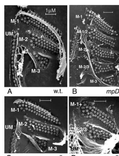
The “extra-membranelle” mutations proved useful in reveal-ing that the patternreveal-ing of the membranelles is spatially coor-dinated. In wild-type cells grown in nutrient media, the spatial “signature” of each fully formed membranelle is distinctive and remarkably uniform, so that one can easily distinguish the “M-1,” “M-2,” and “M-3” patterns of basal-body positions (159) (Fig. 8A). This might tempt one to surmise that there is a unique genically determined “blueprint” that specifies the spatial organization of each of the three membranelles. How-ever, for mutants with an increased number of membranelles—
notablympD-1—the spatial “signature” of each membranelle
depended on its relative position within the set of mem-branelles (51). Thus, in an OA with five memmem-branelles, the first membranelle had an “M-1” signature, the third had an “M-2” signature, and the fifth had an “M-3” signature. The second and fourth membranelles had intermediate structural signa-tures (“M-1/2” and “M-2/3,” respectively), such as are not seen in normal three-membranelle OAs (8). This implies the exis-tence of a spatially graded field, perhaps a field of forces responsible for displacing basal bodies as the oral cavity is sculptured late during oral development (see Fig. 28 in refer-ence 7).
Two subsequently derived mutations that reduced the
num-ber of membranelles,mpG-1andmpH-1, allowed for a clear
test of the published conclusions summarized above (51). In
mpG-1mutants, approximately one-half of the cells had three membranelles, and these had “pattern signatures” very similar to those of wild-type cells (Fig. 8, compare panels A and C). In
the other half of thempG-1cells, which expressed two
mem-branelles, both membranelles were almost invariably abnor-mal. The second membranelle had an M-2/3 pattern very
sim-FIG. 7. Silver preparations of the anterior ends ofmembranellar pattern(mp) mutant cells. All OAs of these cells have a normal UM and ribbed wall, as well as a darkly stained deep fiber located posteriorly to the membranelles. (A)mpG-1cell maintained at 39°C, with two membranelles. (B)mpC-2cell maintained at 29°C. The thin arrow indicates a small fourth membranelle, which is swept back into the oral cavity. (C)mpD-1cell maintained at 36°C, with four large membranelles lined up parallel to one another. (D)mpA-1cell, with two partially superimposed sets of membranelles: a set of four short membranelles on the cell’s anterior right side of the OA (next to the UM) and a set of three long membranelles abutting on the cell’s posterior left side of the OA. Panels B and C were modified from Fig. 3 and 2, respectively, from reference 51. Panel D is reprinted from Fig. 2c of reference 89 with permission of the author and the publisher.
FIG. 8. Scanning electron micrographs of isolated deciliated OAs from a wild-type (w.t.) cell (A), anmpD-1cell maintained at 36°C (B), andmpG-1cells maintained at 30°C (C and D), one with three mem-branelles (C) and one with two memmem-branelles (D). Anterior portions of the UMs are visible. Membranelles are labeled according to their characteristic spatial-pattern “signatures”: M-1 for the membranelle 1 pattern, M-2 for the membranelle 2 pattern, M-3 for the membranelle 3 pattern, and M 1/2 and M 2/3 for intermediate patterns. M-1⫹stands for a hypertrophied M-1 pattern. For further explanation, see the text. Panel B was modified from Fig. 12 of reference 51.
on September 8, 2020 by guest
http://ec.asm.org/
ilar to that seen in the fourth membranelle ofmpD-1cells that have five membranelles (Fig. 8, compare panels B and D). The first membranelle had a somewhat hypertrophied pattern that can be interpreted as an M-1/2 intermediate (hence it is
la-beled “M-1⫹” in Fig. 8D) (38; Frankel, unpublished). The
M-2/3 pattern is also observed in the second membranelles of severely starved wild-type cells that have only two branelles in their OAs (8). The dependence of the mem-branelle pattern signature on the number of memmem-branelles in
mpG-1cells is clearly more consistent with the “unified-mem-branellar-field” model than with the “membranellar-blueprint” alternative.
Finally, thempA-1genetic variant is the only one in
Tetra-hymenathat conforms to the first part of Beale’s proposal for finding gene-controlled structural variants, by “a search among
wild-races and varieties.” In inbred strain D ofT. thermophila,
about 10% of dividing cells and 80% of cells undergoing oral replacement expressed a unique abnormality in oral develop-ment that brought about a juxtaposition of two partial sets of membranelles (Fig. 7D), which could later fuse to form vari-able membranellar patterns (90). After outbreeding of strain D to another inbred strain (A) that did not show this abnormality, Kaczanowski (89) could demonstrate that this abnormality was based on a single recessive allelic variant that happened to be
homozygous in inbred strain D. He called itmp, and I am here
renaming itmpA-1.
A residual but highly miscellaneous category of mutations affecting oral development comprises those in which oral de-velopment is abnormal in more than the limited manner of the oral mutations considered thus far. These are illustrated in Fig. 9 in the order of severity of effect but will be described here in a different order.
I will begin with two pairs of superficially similar yet in
reality highly contrasting mutations. One of these pairs,mpB-1
andmpF-1, was initially misdiagnosed as affecting only
mem-branelles. Of these, thempB-1mutation (data not shown) had
relatively low penetrance (⬃10% abnormal cells at 29°C, rising
to 30 to 40% after prolonged maintenance at 39°C). However, it had high expressivity, with variable abnormalities affecting both the UM and the membranelles; culture growth was slow and essentially ceased after 6 h at 39°C (Frankel and Jenkins, unpublished).
In contrast, the oral phenotype of thempF-1mutation was
entirely temperature sensitive, and this mutation was both
more penetrant and more uniformly expressed at 39°C than
mpB-1. Membranelles were increased in number and reduced in size, whereas the UM was characteristically shorter than normal (compare Fig. 9A to Fig. 6A and 7). Culture growth remained normal for 7 h at 39°C (Frankel and Jenkins,
unpub-lished). In my view,mpF-1might well repay further study.
A second pair of nonallelic temperature-sensitive mutations
have the same name—defective oral apparatus—but very
dif-ferent phenotypes. Cells expressing thedoa2-1mutation (data
not shown) exhibited a rather general degeneration at 39°C, with progressive loss of oral structures, frequent oral replace-ment, rounding of the cell, signs of division blockage, and very slow growth after 2 h at 39°C (Frankel, unpublished).
Cells homozygous for thedoa1-1mutation resembleddoa2-1
cells only in that they underwent gradual dismantling of OAs at 39°C, with intermediate stages characterized by variable and disordered membranelle fragments (Fig. 9D). However,
doa1-1cells grown at 39°C underwent very little oral
replace-ment and maintained a normal shape while becoming progres-sively smaller. Culture growth continued at a rate similar to that for wild-type controls for 9 h at 39°C before dropping off afterwards. Examination of silver-stained slides strongly
sug-gested that doa1-1 cells that had completely lost their OAs
could still divide, sometimes after forming abnormal oral pri-mordia, in some cases along unexpected longitudes (Frankel
and Jenkins, unpublished). Thedoa1-1mutant deserves more
than the superficial examination it has received thus far. It remains to describe two mutations that were obtained, like
vacA, from a functional screen for the inability to form food
vacuoles but that, unlikevacA, brought about severe
morpho-logical abnormalities. One of these, NP1 (nonphagocytosis 1),
was likevacA in that it was temperature sensitive, and only
OAs formed at the restrictive temperature were unable to form food vacuoles. It differed, however, in two ways. First, although the condition was vegetatively stable, it could not be transmit-ted to sexual progeny upon conjugation, suggesting that it was caused by a mutation in the macronucleus, which is lost during conjugation (134). Second, only rudimentary, nonfunctional oral structures were produced at restrictive temperatures (124) (Fig. 9C). At these temperatures, this mutant could grow only in a rich medium that was specially devised to allow sufficient entry of critical nutrients in the absence of food vacuoles (123). The oral structures present in the NP1 macronuclear mutant were analyzed in detail by Williams and Honts (160), who
FIG. 9. Silver preparations of the anterior ends of mutant cells with general effects on the anatomy of the OA. (A)mpF-1cell maintained at 39°C, with four small membranelles and an anterior truncation of the UM (arrow); the posterior end of the UM has a direct connection to the deep fiber. (B) II8G (phg⫺) cell maintained at 37°C, with membranelles abnormally arrested in their development. (C) NP1 cell maintained at 37°C, with fragmented membranelles. (D)doa1-1cell maintained at 39°C, with rudimentary membranelles. The OAs shown in panels B through D lack UMs, ribbed walls, and deep fibers. Panel C is reprinted from Fig. 5A of reference 124 with permission of the author and the publisher.
on September 8, 2020 by guest
http://ec.asm.org/
found that the cytoskeletal elements within the OA (basal bodies and associated structures) were normal but their higher-order arrangement was abnormal. Furthermore, the an-terior OA was capable of carrying out the disassembly and reassembly of the complex microtubular and filamentous sub-structure of the anterior OA, which normally takes place dur-ing cell division. These investigators therefore concluded, “The primary lesion of NP1 may be in some early event required for the correct positioning of basal bodies within the oral appara-tus as a whole” (160).
The last mutation in this group to be considered putatively exists in a strain, known as II8G, “derived from CU399 . . . a temperature-sensitive strain forming no food vacuoles at 37°C”; the phenotype is “most probably caused by a single gene
mutation at a locus calledphg⫺” (154). This is in several ways
the most enigmatic of all the mutants described in this review. According to the original authors, “The oral structures are missing often at 37°C, but present at 28°C” (154). More re-cently, this mutant was described as being “able to proliferate [in enriched medium at 37°C] yielding small cells with very variable phenotypes including some cells without oral appara-tuses and some cells in OR [oral replacement] morphogenesis” (88). This strain was also sent to us, and we were unable to obtain progeny following crosses; we also found that the II8G cells produced silver- and protargol-stainable oral structures no matter how long the cells were kept at 39°C (Fig. 9B and 18A). Moreover, these oral structures were unlike those found in any other mutant; they consisted of a variable number of membranelles, all of which were arrested in their development at the stage of addition of a third ciliary row to two-row promembranelles (cf. reference 46), with few traces of a UM and no oral cavity. Even more remarkable is the way in which these oral structures were formed: not by the usual method of a single oral field produced next to the subequatorial region of a single ciliary row (Fig. 3; see also Fig. 12A, 13A, 14A, 15A and F, and 16A), but from several oral fields produced from adjacent ciliary rows and subsequently merged into one, with one or two promembranelles produced from each mini-field (see Fig. 18B to F) (E. M. Nelsen and J. Frankel, unpublished observations). This and other characteristics place our sample of the II8G stock within the category of alterations of circum-ferential polarity as well, so we will return to it in that section of the review.
The molecular basis of all of these defects in oral develop-ment is unknown. However, it is worth develop-mentioning that a knockout of the gene coding for one of the three major mem-brane-skeletal proteins, EpiC (162, 164), brought about a tran-sient disruption of the organization of oral structures, accom-panied by some disarray in the ciliary rows (158).
Mutations affecting subdivision along the anteroposterior axis.As indicated earlier, ciliates divide by a process of seg-mentation, generating two daughter cells in a tandem array with identical polarity (Fig. 3). An essential part of this process is the formation of a fission zone, in which the former equa-torial region of a cell becomes transformed into juxtaposed polar extremes. Mutations that affect this process of segmental subdivision can either (i) prevent it altogether, (ii) allow it to begin but abort the process before it has gone to completion, or (iii) displace its location, either anteriorly or posteriorly. I will describe below mutations that act in each of these ways.
At the outset, however, the mutations that are being left out of this account should be noted. From a topological perspec-tive, the process of cortical subdivision is already completed before division constriction begins. This distinction provides an excuse for leaving out of this account mutations that prevent constriction at some point after the formation of the fission
zone. An exemplary case iscdaC(56), which has been shown
by anatomical study (49) and temperature shift experiments (52, 146) to primarily affect the process of division furrow
constriction; other examples are alleles of cdaD, cdaE, and
cdaF (all described under earlier names in references 49 and
56), as well ascdaG andcdaJ (Jenkins and Frankel,
unpub-lished) and the recently characterizedcdaLmutation and
pos-siblecdaMmutation (E. Cole, personal communication).
The pseudomacrostome (psm) mutations, which prevent anteroposterior subdivision, all cause a switch from cell divi-sion to a peculiar form of oral replacement that brings about the formation of OAs much larger than normal, with longer UMs and membranelles (Fig. 10). The pseudomacrostome type of oral replacement differed in three respects from the typical oral replacement that normally takes place in starved cells (33, 90), especially in cells developing the “rapid
swim-mer” phenotype (110) and in exconjugants (22, 93). In psm
cells, (i) the OP extended for most of the length of the cell (Fig. 10A) rather than being confined to a small region just posterior to the old OA; (ii) oral replacement took place in nutrient medium; and (iii) it resulted in a large cell with an unusually large and prominent OA.
The pseudomacrostome phenotype has been analyzed most
extensively in the firstpsmmutation to be discovered,psmA-1,
of thePSMAgene located on chromosome 5 (57; E. Hamilton,
personal communication). It was severely temperature sensi-tive, with fairly strong expression at 25 to 28°C (22°C is “per-missive” for this allele). Penetrance and expressivity were typ-ically high (57) but were dependent on the culture medium in which the cells were grown and also differed in different
ho-FIG. 10. Silver preparations of developmental stages of psmA-1 cells maintained at 28°C. (A) Cell with an early OP, extending over most of the length of the cell, posterior to a normal-sized OA whose UM is participating in the formation of the OP. (B) Cell with an OP at an intermediate stage of development. The short arrow indicates a membranellar remnant of the old OA that is in the process of being resorbed. (C) Cell with a completed new “pseudomacrostome” OA.
on September 8, 2020 by guest
http://ec.asm.org/
mozygouspsmA-1 clones, suggesting the presence of genetic modifiers that have not yet been analyzed.
ThepsmAmutations are excellent examples of the general
rule that allelic mutations are invariably identical in qualitative
phenotype but often differ greatly in penetrance:psmA-1was
expressed at all but extremely low temperatures (57), and
psmA-2 was weakly expressed only after 2 generations of growth at a high restrictive temperature (39°C) (57), whereas
the most recently discovered allele, psmA-3, had zero
pen-etrance of the pseudomacrostome phenotype at 29°C and close to 100% penetrance of that phenotype a few hours after a shift to 39°C (Frankel, unpublished)—and therefore is the recom-mended allele for all users.
There are three additionalpseudomacrostomeloci, each
rep-resented by one allele. None of them had the degree of pen-etrance or expressivity of the pseudomacrostome phenotype
shown by favorable stocks ofpsmA-1and bypsmA-3. However,
whereaspsmC-1(also located on chromosome 5) looked and
responded much like the “weak”psmA-2, the other twopsm
mutants each had certain peculiarities: inpsmB-1(located on
chromosome 4L [15]), cells tended to have a highly tapered posterior region and commonly showed unequal division, with the resulting posterior daughter cell smaller than the anterior
one (a phenotype occasionally seen inpsmA-1as well; see Fig.
6c of reference 35).psmD-1(located on chromosome 3R) was
highly pleiotropic, since in addition to its variably expressed pseudomacrostome phenotype at 39°C, it frequently formed OAs with four membranelles (57) and also produced oral pri-mordia that shifted anteriorly and generated “hammerhead”
phenotypes similar to those expressed by thecdaI-1mutations,
to be described below (Frankel, unpublished). Such pleiotropy, observed in some (but not all) of our cortical-pattern mutants, reveals the frequent arbitrariness of the naming of these
mu-tations: psmB-2 could readily have been given new places
within themporcdaseries.
Before leaving the psmmutations, I should briefly explain
the name of this set of mutations: it refers to the macrostome
OA of the obviously dimorphicTetrahymenaspecies such as
Tetrahymena vorax, which is derived from the microstome form by a process of oral replacement (16) rather similar to that
found in pseudomacrostome T. thermophila. However, the
macrostome OA of T. vorax, with a gigantic oral pouch, is
much larger than anything ever seen inT. thermophila. Since it
is fairly clear from phylogenetic analysis that the bactivorous
microstomial form is primitive within the genusTetrahymena
and that the species capable of producing macrostome OAs are derived from the microstomial form (140), it is possible to
think of apseudomacrostomemutation as an early step in the
evolution of a macrostomial form (37).
The two mutated genes that abort anteroposterior
subdivi-sion arecdaA(five alleles) andcdaH(two alleles). The
multi-plicity of alleles probably reflects the relative ease of selecting mutants that inevitably produce chains of subcells or mis-shapen monsters at a restrictive temperature.
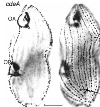
cdaA-1is perhaps the most extensively studied mutation of this set, with 20 publications (15, 17, 18, 19, 20, 48, 49, 52, 54,
56, 59, 60, 81, 83, 86, 87, 119, 120, 121, 156). cdaA-1 is a
100%-penetrant temperature-sensitive mutation, located on chromosome 4R (15; E. Hamilton, personal communication), that at 39°C permits an OP to be initiated and develop at its
normal position but prevents all other known structural (49) (Fig. 11) and molecular (81, 87, 121) processes of segmental subdivision from taking place. The temperature-sensitive
pe-riod ofcdaA-1at 36°C was an interval of about 10 min during
the middle of oral development, prior to the appearance of the fission zone (52). Micronuclear division, which accompanies
oral development (Fig. 3), occurred normally incdaA-1cells at
a restrictive temperature, but macronuclear division, which normally occurs after the fission zone forms, failed to take place under these conditions (54); periodic DNA synthesis continued within the undivided macronucleus (20, 54). The
cdaA-1cells grew, went through multiple cell cycles and addi-tional rounds of oral development (81, 86) without dividing, and eventually became gigantic, irregular monsters (49). Thus,
theCDAAgene is required for a critical step in the subdivision
of the anteroposterior axis, in the absence of which cells cannot begin to divide.
This mutation is unique among our entire collection in that it has been the subject of an extensive molecular inquiry. A protein was found that migrated differently in 2-dimensional
gels of homogenates fromcdaA-1cultures grown at a
restric-tive temperature, and this protein (called p85 because of its molecular weight) was localized near basal-body couplets that normally are situated at the anterior ends of most ciliary rows (Fig. 2) (119, 121). At a restrictive temperature, these couplets did not form at their normal sites just posterior to the fission zone, and p85 was not localized at these sites (121). These authors presumed that there is a causal connection between the localization of p85 and the capacity to form a fission zone, a line of analysis that was pursued further by the Numata group (60, 119). One problem with this analysis is that both apical basal-body couplets and p85 are absent between the fourth ciliary row to the right and the first row to the left of the OA
FIG. 11. Two focal levels of a single silver-stained cdaA-1 cell maintained at 39°C. The focus on the left side is on the OA and an OP that has completed its development. On the right side, the focus is on the cell surface. A normal set of CVPs is located near the posterior end of the cell (arrow), yet there are no CVPs, and no other indication of a fission zone, in the equatorial region. This figure was modified from Fig. 3 and 4 of reference 49 with permission of the publisher.
on September 8, 2020 by guest
http://ec.asm.org/
(100) and of the OP (60, 79), yet a fission zone develops
normally there in wild-type cells as well as incdaA-1cells at
permissive temperatures. When the cDNA that coded for p85 was cloned and sequenced, it was found that “there was no difference in the predicted amino acid sequences of wild-type andcdaA1p85” (59). Thus, the true gene product ofCDAAis
still unknown. ThisCDAAproduct was presumed by Gonda et
al. (59) to be a molecule involved in the posttranslational modification of p85. The meaning of these analyses is still unclear.
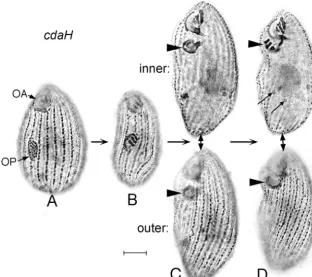
The other mutation that prevents cortical subdivision,
cdaH-1, located on chromosome 5 (15), is also 100% penetrant
at 39°C, but it has been much less studied than cdaA-1. In
cdaH-1 mutant cells grown at 39°C, oral development was initiated normally (Fig. 12A) and continued more or less nor-mally up to the stage when the fission zone would nornor-mally
appear (Fig. 12B). As withcdaA-1, no fission zone formed, but
in thecdaH-1mutant the new posterior OA sank into a
sub-surface vesicle and migrated anteriorly (Fig. 12C) to end up just posterior to the old anterior OA (Fig. 12D). At the same time, there was considerable posterior extension of the cell, perhaps exaggerating the impression of a forward migration of the OP. As can be seen in the “inner” focal planes of the topmost cells in Fig. 12C and D, the micronucleus divided whereas the macronucleus did not, consistent with similar but
far more detailed observations of cdaA-1 (Frankel,
unpub-lished).
The temperature-sensitive period forcdaH-1at 36°C (a
tem-perature at which the phenotype is still 100% penetrant but expression is less extreme than that at 39°C) was longer and
somewhat later than that ofcdaA-1: it occurred during a 25- to
30-min interval around the time when the fission zone is nor-mally formed (52).
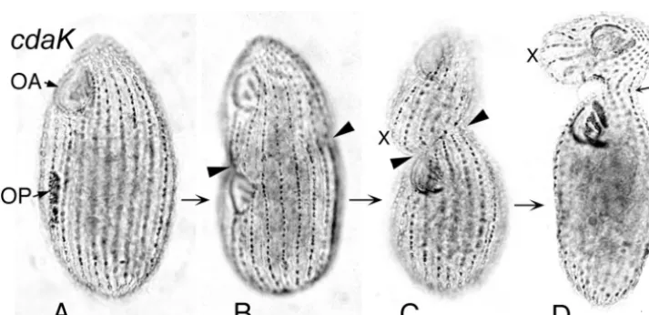
Mutations at four additional gene loci permit cortical sub-division to occur but alter its location along the
anteroposte-rior axis. Two of these,cdaI and cdaK, bring about anterior
displacement of the fission zone at restrictive temperatures.
In cells expressing all three cdaI mutations, the OP first
appeared at its normal subequatorial location (Fig. 13A), but at 39°C it then slid anteriorly on the cell surface. At the stage when the fission zone normally appears, the OP had already reached a location just posterior to the old OA, and the fission zone concomitantly was radically displaced anteriorly (Fig. 13C), while new CVPs sometimes did and sometimes did not arise anterior of it. There was then a varying amount of fur-rowing, ranging from total absence (typical of the “strongest”
allele, cdaI-3) to successful cell separation (common in the
“weakest” allele,cdaI-2) (Frankel and Jenkins, unpublished).
Most commonly, an incomplete and unilateral furrow formed, leading to a distinctive “hammerhead” phenotype (Fig. 13D). The incompleteness of the process of subdivision might be a by-product of the extreme anterior displacement of the fission zone.
In cells homozygous for thecdaK-1mutation, the OP did not
FIG. 12. Silver preparations ofcdaH-1cells maintained at 39°C, in sequential developmental stages. (A) Cell with an out-of-focus normal OA and a normal OP at an intermediate stage of development. (B) Cell with an OP at a later stage of development. (C) Two focal levels of a single cell at a stage when the nearly complete OP (arrowhead) has sunk into a vesicle underneath the surface of the cell. The upper image is focused on the oral structures, and the lower image is focused on the surface of the same cell, which shows no sign of a fission zone. (D) Two focal levels of a single cell at a stage when the OP (arrowhead) has arrived at its final location just posterior to the anterior OA. Focal levels are as in panel C. The thin arrows indicate the two divided daughter micronuclei; the undivided macronucleus is located just anterior to these.
on September 8, 2020 by guest
http://ec.asm.org/
shift, even at restrictive temperatures (36°C to 39°C). It devel-oped at its normal midbody position (Fig. 14A) and appeared to stay there. A fission zone was then formed in a highly asymmetric manner, with an anteriorly directed tilt to the left of the OP (Fig. 14C) and a sharp anterior displacement (a “cliff”) to its right (not shown here; see Fig. 1G in reference 95). Some ciliary rows remained continuous across the fission zone, preventing division from going to completion (Fig. 14D). A ventral bulge formed (Fig. 14C) and became prominent in the presumptive anterior daughter cell (Fig. 14D). A “ham-merhead” phenotype was thus generated, but in a manner
entirely different from that observed with thecdaImutant cells.
For a more detailed description and analysis of the complex phenotype of this mutant, see reference 95.
The mutations that bring about a posterior displacement of the fission zone are quite different and generate less dramatic phenotypes. In these mutant cells, displacement was not
ac-companied by any blockage of cell division. Typically, the OP began to be formed too far back, sometimes near the posterior end of the cell, followed by a partial equalization of the sizes of the two presumptive daughter cells.
The first example of this appeared in a mutation affecting cell
shape, conical (con1-1, originally co [27]) (Fig. 15). In con1-1
mutant cells, the OP appeared to form near the posterior end of the cell (Fig. 15F), followed by an apparent anterior shift in its relative position, although how much of this was attributable to genuine posterior growth and how much to a change of cell shape from conical to ovoid (Fig. 15G) is not entirely clear (27). After cell division was successfully completed, the posterior daughter cell started out smaller, and also grew more slowly and divided later, than the anterior daughter cell (27, 129).
A more recent and unequivocal example of posterior local-ization followed by partial equallocal-ization was found in the
“elon-gated”elo1-1mutant (Fig. 16), in which the OP was initiated
FIG. 13. Silver preparations ofcdaI-2cells maintained at 39°C, in sequential developmental stages. (A) Cell with an OA, mostly out of focus, and an OP that is at an early stage of development. (B) Cell with a later-stage OP, beginning to migrate anteriorly on the cell surface. (C) Cell in which the migration of the OP has been completed, and a fission zone is beginning to form anterior to it (arrowhead). The OP has four membranelles (small arrow). (D) Cell that is attempting to divide, with a unilateral furrow (arrowhead). The completely developed OP has a displaced fourth membranelle (small arrow), similar to the one in thempCcell shown in Fig. 7B.
FIG. 14. Silver preparations ofcdaK-1cells maintained at 36°C, in sequential developmental stages. (A) Cell with a normal OA, out of focus, and an OP at an early stage of development. (B) Cell with a late-stage OP, with a tilted fission zone (arrowheads) beginning to form. (C) The development of the OP is completed, and the cell is furrowing (arrowheads) along the tilted fission zone. The “X” marks a bulge along the oral meridian just anterior to the fission zone. (D) Cell attempting to divide. The “X” marks the region corresponding to the bulge in panel C, and the small arrow indicates some continuous ciliary rows that mark the region where cytokinesis is failing.