University of Pennsylvania
ScholarlyCommons
Publicly Accessible Penn Dissertations
1-1-2015
Molecular Analysis of Target Specific Peripheral
Nerve Regeneration
Jesse Isaacman-Beck
University of Pennsylvania, jisaacmanbeck@yahoo.com
Follow this and additional works at:http://repository.upenn.edu/edissertations Part of theNeuroscience and Neurobiology Commons
This paper is posted at ScholarlyCommons.http://repository.upenn.edu/edissertations/1778 For more information, please contactlibraryrepository@pobox.upenn.edu.
Recommended Citation
Isaacman-Beck, Jesse, "Molecular Analysis of Target Specific Peripheral Nerve Regeneration" (2015).Publicly Accessible Penn Dissertations. 1778.
Molecular Analysis of Target Specific Peripheral Nerve Regeneration
Abstract
Axons of the peripheral nervous system have retained the remarkable ability to regenerate. However, after peripheral nerve injury, patients often suffer from the inability to properly localize sensation and/or the loss of fine motor control, suggesting that regenerating peripheral axons reinnervate ectopic targets. Despite decades of research in myriad model systems, whether PNS axons regenerate randomly or navigate selectively to their original targets remains controversial. Moreover, while some studies suggest that regenerating axons can choose specific paths, the cell and molecular mechanisms underlying target-selective regeneration have remained elusive. Using live-cell imaging in larval zebrafish we find that after complete nerve transection regenerating motor axons exhibit a strong preference for their original muscle territory, and that axons probe both correct and incorrect trajectories extensively before selecting their original path. We demonstrate that the glycosyltransferase lysyl hydroxylase 3 is required to modify extracellular matrix collagens to provide growth and guidance to regenerating axons. Using transgenic rescue experiments, we determine that post-injury expression of lh3 and lh3 expression in Schwann cells is sufficient to restore target-selective regeneration. Moreover, we show that Schwann cells neighboring the transection site upregulate the lh3 substrate collagen4a5 and that during regeneration collagen4a5 destabilizes axons probing inappropriate trajectories to ensure target-selective regeneration. These results demonstrate that selective ECM components match subpopulations of regenerating axons with their original targets.
It has long been hypothesized that regenerating axons might reuse developmental guidance cues to reestablish synaptic connections after peripheral nerve injury. Intriguingly, Collagen4 can bind the canonical axon repellants Slit and Netrin, and we find that slit1a is upregulated with collagen4a5 in the same population of Schwann cells after injury. Additionally, we demonstrate that the slit receptors robo2 and robo3 are both required for target-selective regeneration. Together these results demonstrate that regenerating peripheral nerves reemploy developmental guidance molecules and reveal a possible mechanistic framework by which the ECM constituent collagen4a5 binds and presents the repellant slit1a to convey synaptic target selection through robo2 and robo3 repulsion of regenerating axons in vivo.
Degree Type Dissertation
Degree Name
Doctor of Philosophy (PhD)
Graduate Group Neuroscience
First Advisor Michael Granato
Subject Categories
MOLECULAR ANALYSIS OF TARGET SPECIFIC PERIPHERAL NERVE REGENERATION
Jesse Isaacman-Beck A DISSERTATION
in Neuroscience
Presented to the Faculties of the University of Pennsylvania
in Partial Fulfillment of the Requirements for the Degree of Doctor of Philosophy 2015
Supervisor of Dissertation _____________________ Michael Granato, PhD
Professor of Cell and Developmental Biology
Graduate Group Chairperson ______________________ Joshua I. Gold, PhD
Professor of Neuroscience
Dissertation Committee:
Jonathan Raper, PhD, Professor of Neuroscience
ii
Dedication
To all of my mentors - for believing in me, pushing me to and through adversity, and helping me to continually redefine success
To my parents – for showing me how to walk on when the bear beats you through the buckwheat.
iii
Acknowledgments
Graduate school is challenging. There’s nothing particularly unique to that –many
people work hard. However, I embrace the fact that my experience has been my own. I
am deeply proud of this period in my life - I have risen to adversity, overcome tumult,
managed stress, thought critically, and learned so much. I have found a meaningful new
way to engage with the world and I love it, I am a scientist. Perhaps then, it’s useful to
draw from this lexicon to describe how I got here: this journey has only been possible
through autonomous and non-autonomous growth and guidance. I could not have gotten
here without me, and I definitely would not have gotten here without the myriad mentors
who pulled me through the slog. While many who know me might jest that I could
expound on my merits until the cows come in – I hope they also know that I could write
about how great they are until this document won’t fit in the repository. For all of our
sakes then, let me try to be brief, but if I’m not, do know that I tried.
To those who know me outside of the University of Pennsylvania:
I moved into 853 N. 26th street just over 10 years ago with 5 of my closest friends:
Nick Kerr, Andy Cino, Matt Rosen, Nick Maxwell and Ben Gordon. Over the next 8
years, this group expanded to include Asher Spiller, Scott Sheppard, Nicholas Mirra, and
Kaitlin Friedman. Together we learned how to live independently from our parents and
our undergraduate institutions. In the process, we discovered that independence felt good,
iv
graduate school, and will remain a pillar for the rest of my life. Over these seven years
many of us have found partners – Jenny, Jess, Kaitlin, Sarah, Sasha, Jaci, Katrina, Andy -
and some of us have started families – Noah and Margot. In fact, I write this on the day
after Matt’s bachelor party fresh with the knowledge that with this principle of
community we will do so much good for the world.
This foundation has led me to build even more friendships – Chris and Cathy
Kerr, thank you for listening and aiding me when I needed it most. Ben Edinger, Tony
Tenisci, and Greg Nini – every morning in the gym has propelled me through this final
year. Kathy – you have helped me to grow so much as a person and a scientist. Knowing
that I can talk to you when I need to and that you will lead me to the healthiest decisions
is invaluable, thank you. I finish graduate school and leave Philadelphia knowing that I
am a better person because of all of you, and a better scientist with the values that we
have honed together.
To those who know me at the University of Pennsylvania:
I’ve had so many incredible faculty mentors in the Neuroscience Graduate Group
(NGG) including Mikey Nusbaum, Rita Balice-Gordon, Josh Gold, Kelly Jordan-Sciutto,
and Minghong Ma. Thank you all for letting me know that if I wanted to do something
for the NGG, that I should just go ahead and do it. Perhaps the most empowering lesson
that I’ve learned from you all is that I don’t need your help for most things, but when I do
v
you, Jane Hoshi, Angela Gilmore, Christine Clay, Jackie Fowlkes and so many others – I
really value every effort you’ve made on my behalf.
There are so many students within the NGG who have contributed to my success,
so I’ll thank them all up-front, and mention some specific people who have played
integral roles. Sam White – it has been a true joy to work with you. You are so diligent,
organized, and such a strong leader, thank you for showing me how to get things done
and being direct with me when it was best to let ideas go. Thanks to Natalie Rizzo,
Katherine Gribble, Shachi Doshi, and Sarah Ly who made Upward Bound Research
Fridays a reality and to all of the volunteers who made it a success. Thanks to Tanya
Weerakody, Matt Wimmer, Maria Lim, Nina Hsu, Lauren Stutzbach, and Vanessa
Troiani for all of your support and dedication to the Upward Bound summer course and
primer. Thanks to Lindsay Vass, Sam White, Adam Watson, Khaing Wang, and Drew
Jaegle for taking on and building up the Student Invited Seminar Series - so many of us
have benefitted from this incredible professional opportunity. Thanks to all of the
dedicated NGG students who helped to shape GLIA, especially those who’ve chaired –
Sam White, Kate Christisen-Lagay, Morgan Taylor, Shachee Doshi, Isaac Perron, Dave
Reiner, and Trish P. We have built a program nationally recognized for innovation in
neuroscience outreach and professional development for graduate student – I am very
proud.
Finally to the staff and students of Upward Bound Math and Science at the
University of Pennsylvania – I can’t thank you enough for all of your hard work and
vi
and you drive the UBMS ship with optimism and courage, I have been so fortunate to
work with you. Ms. Vaniece Moses, Ms. Gianna Brisbone, and Mr. David McCullough I
don’t know how you do it, but you bring a beautiful order to what could be chaos – it has
a lot to do with your grit and even more to do with your smiles. You all show me time
and again, what my mother has always said, “there’s no greater joy in the world than
improving the life of a child.”
To the 11th and 12th floor of BRB II/III, The University of Pennsylvania
It’s been so fun to go to work most days in the last seven years. For me, this is due
in big part to the incredible people with whom I science. Here again, so many have
helped me carve a path that I’ll blanket the group with my appreciation and specially
mention a dear few. First to Dave, Erin, Mona, Anthony and all of the great people who
care for our fish, I know this is often a thankless job – I can only hope I’ve thanked you
enough along the way, but let me say it again – thank you so much. To the many
technicians that have worked in the Granato and Mullins labs I echo that sentiment,
Elena, Camille, Hannah, Melissa, Amy and Sumei, thanks for making sure we have
always had what we need. There’s no way any of my work would have been possible
without everything you do.
On these floors, several graduate student and postdocs have lent their ears and
dedicated their skills to pulling me out of scientific and emotional ruts. Ben, James, and
Ed – you guys know how to dig into biochemistry with unmatched tenacity, thanks for
vii
you, I’ll never shy away from a plasmid again. Yvette, Keri, Jodi, Kristin, Josh, Lindsay,
Dina, Alison - you’ve shown me how interpersonal our profession can and should be, you
are all incredible teachers. To the members of Super Secret Journal Club – thanks for
showing me how to think critically and letting me know when I need to lay off. To the
PIs on these floors and others nearby: Shannon Fisher, Mary Mullins, Steve Scherer,
Michael Pack, Steve Dinardo, Jon Raper, and Greg Bashaw – thank you for taking the
time to mentor me and ask critical questions that challenge me to engage more deeply in
my research. Mary and Steve, both of you have gone out of your way to praise my talks –
I’ve ridden far on that positive reinforcement.
To the Granato lab:
It has been a privilege to work, learn, play and grow in the Granato lab. Each of
my labmates has contributed to my self-actualization as a scientist and each of you have
my deepest gratitude.
Owen - in our short time sitting back to back, you’ve shown me a new toolkit for making
figures and have imparted invaluable advice as I searched for a Postdoc – thank you.
Jess – you create songs about my lack of intelligence and I create detailed plans to do
ridiculous things with your pillowcases. I’m not sure of a better recipe for scientific
achievement, but I’m sure that you’ll continue to surprise me with catchy new
viii
Santanu – It has been a sincere pleasure to work with you over these many years. Thank
you for challenging me to be precise with my language, for inspiring me by attempting
the most arduous experiments, and for demonstrating how to be a postdoc and a father. I
hope all continues to go well for you in Syracuse.
Juliane – I am floored by your drive and attention to detail, thank you for pointing me to
the important controls. Even when I complained I knew you were right – my fingers are
crossed for Zurich.
Kurt – You’re too tall. I can’t even see your forehead most of the time. This is far from
the only reason I look up to you. I’m so impressed by your resilience and the care you
take with your work and I’m so thankful for your considerate and detailed feedback. It’s
great to know that I’ll get to see you and Jen on the West Coast.
Roshan – You took my job – like the only job I ever wanted. My only solace is that I am
not qualified, and you are exactly the Professor that Haverford deserves. I’ve been
mentored by Haverford Professors – they diligently push their students into rewarding
intellectual realms and they share completely in the joy of their students’ growth. You
have mentored me and you do the same. Good luck, let me know when the next position
opens up, and keep seeing all of Powerpoint’s shades of grey.
Marc – You’re too short. I can’t even see your forehead most of the time. You are
Napoleonic in your efficiency and I deeply admire the way you just get things done. You
have a brilliant sense of which experiments are the most important and you’ve seen the
big picture of all projects like a PI since I first knew you as a postdoc. Thanks for sharing
ix
Hannah – I’ve been impressed with your kindness and aptitude since you first asked me
great questions at my poster at SFN. Your skills as a copy editor will serve you so well,
thanks for applying them to my paper. Good luck!
Patti – You have the energy and enthusiasm that I wish I could still muster. Thanks for
pushing me to try. I admire your independence and I am lucky that you’re following up
with the ideas that I’ve generated. I’m sure you’ll find even better ways to do this work
than I could ever show you.
Melissa (D.) – It’s been really important to have you in the lab – us gingers have to stick
together. You face each day with optimism, enthusiasm, and kindness. Thanks for sharing
that with me – I have so appreciated your support through the many ups and downs.
Katherine – You have put up with a lot. Through it all, you’ve become an incredible
scientist. You consistently help me to think more critically about my data. Thanks for
diving in to Upward Bound Research Fridays and APICAL with me – both have
flourished under your guidance. As I enter my 8th year, I am happy to have spent 5 of
them with you.
Ali – This would not have been possible without you. That’s true scientifically – you
built the model that I succeeded with (Rosenberg et al. 2012; Rosenberg et al. 2014). It’s
also true emotionally – you gave me music and hugs to Prelim by, a front row presence to
draw strength from for my talks, and not only the push I needed to take breaks, but fun to
x
Rajiv – You, Sir, have a butt for a face. Despite this, you’re pretty much the best guy.
You know how to make a guy smile by giving him the finger; it’s quite a talent. You’ve
taught me that we must be silly to be great scientists and that by just being ourselves, we
can be very silly. I can’t wait to keep hanging out.
Michael – Look above. It’s possible that I would have made some of these connections
on my own, but to have worked with this group of people during my tenure as a graduate
student, that’s all your fault. As a Postdoc, I’ll have to find a way to cultivate another
community of enthusiastic, hard working, kind and caring scientists. Then, if I’m lucky,
I’ll have to try to build it from scratch in my own lab.
It’s also your fault that I even want to be a PI. I was a naïve graduate student,
confident in myself as a person, but unsure of my ability as a scientist. How many times
did I flounder into your office thinking I’d never crawl out from this/that nadir? Too
many to count and way more than I’d like to admit. How many times did I bounce out of
your office thinking that I was making great progress and that I’d definitely succeed in
academia? Every time. Thank you. These conversations helped me to overcome my
insecurities so that I could embrace the intellectual rigor and excitement of uncertainty
and discovery in our work. I deeply respect how much you care for us as people and
tailor our training using those principles to lead us to the joy you’ve found in academic
science. I leave the lab confident that I can do it too, but please know that I will keep
xi
To my family:
My family is the reason that I succeed and in this section alone, I could write
volumes. Thanks especially to Uncle Allen and Aunt Bobbie, Erik, Wenjing, Chloe and
Willow – we’re coming west and we know we’re in good hands. To John, Janet, John,
Carrie, Colin and Kylie, thanks for welcoming me to the Glaeser family; being a son and
brother in law and an uncle is the greatest fortune. I love you all, and I have only done
better with you in my life.
Ari – I could write a whole new senior speech about how impressed I am by you. You are
incredibly intelligent, both emotionally and intellectually. You grasp complexity with
ease and handle grey areas with clarity. I admire the courage you have to follow your
dreams in the face of adversity and know that your presence in my life means that we
both will be happy. I love you, and could not have made it to this point without you.
Mom and Dad – when I gave my first public talk, I acknowledged you as a past “funding
source”. That was cute, but it doesn’t come close to recognizing how important you have
both been in this process. You have supported me unconditionally in everything that I
have done and this has given me the confidence and freedom to achieve. I’m not sure that
it’s appropriate for a child to say that his parents did a great job, so I’ll say instead that I
hope that if Jenny and I are fortunate enough to have children that we commit as
assiduously to them and their success. I love you both and could not have come this far
without you.
Jenny – It’s been a doozie, hasn’t it? I think you know what you signed up for in dating
xii
things in perspective, your confidence in me, and your unwavering dedication to us. I
consider you a partner in everything we do and I share this success equally with you. I
know that we are better together, and that we will continue to live well and be happy
having each other. Are you ready for the next one? I know that with you by my side, I
xiii
ABSTRACT
MOLECULAR ANALYSIS OF TARGET SELECTIVE PERIPHERAL
NERVE REGENERATION
Jesse Isaacman-Beck
Michael Granato
Axons of the peripheral nervous system have retained the remarkable ability to
regenerate. However, after peripheral nerve injury, patients often suffer from the inability
to properly localize sensation and/or the loss of fine motor control, suggesting that
regenerating peripheral axons reinnervate ectopic targets. Despite decades of research in
myriad model systems, whether PNS axons regenerate randomly or navigate selectively
to their original targets remains controversial. Moreover, while some studies suggest that
regenerating axons can choose specific paths, the cell and molecular mechanisms
underlying target-selective regeneration have remained elusive. Using live-cell imaging
in larval zebrafish we find that after complete nerve transection regenerating motor axons
exhibit a strong preference for their original muscle territory, and that axons probe both
correct and incorrect trajectories extensively before selecting their original path. We
demonstrate that the glycosyltransferase lysyl hydroxylase 3 is required to modify
extracellular matrix collagens to provide growth and guidance to regenerating axons.
Using transgenic rescue experiments, we determine that post-injury expression of lh3 and
lh3 expression in Schwann cells is sufficient to restore target-selective regeneration.
Moreover, we show that Schwann cells neighboring the transection site upregulate the
lh3 substrate collagen4a5 and that during regeneration collagen4a5 destabilizes axons
xiv
demonstrate that selective ECM components match subpopulations of regenerating axons
with their original targets.
It has long been hypothesized that regenerating axons might reuse developmental
guidance cues to reestablish synaptic connections after peripheral nerve injury.
Intriguingly, Collagen4 can bind the canonical axon repellants Slit and Netrin, and we
find that slit1a is upregulated with collagen4a5 in the same population of Schwann cells
after injury. Additionally, we demonstrate that the slit receptors robo2 and robo3 are both
required for target-selective regeneration. Together these results demonstrate that
regenerating peripheral nerves reemploy developmental guidance molecules and reveal a
possible mechanistic framework by which the ECM constituent collagen4a5 binds and
presents the repellant slit1a to convey synaptic target selection through robo2 and robo3
xv
Table of Contents
Dedication………ii
Acknowledgments………...iii
Abstract………...xiii
List of Illustrations ………..xviii
General Introduction………...1
Chapter 1: Background.………..3
The path to axon regeneration: not all injuries are equal ……….3
Tissue specificity in nerve regeneration: axons preferentially regenerate to nerve…...5
Clinical observations: regenerating axons can, but do not always take the right path..7
Experimental evidence: do axons regenerate with nerve branch specificity?…………8
Preferential Motor Regeneration (PMR): A model for nerve branch selectivity……...10
Cell and molecular mechansism that govern branch selectivity in axon regeneration.12 Extracellular matrix changes after nerve transection………...13
Canonical axon guidance molecules after peripheral nerve transection………..14
xvi
Chapter 2: The lh3 glycosyltransferase directs target-selective peripheral
nerve regeneration………...20
Summary……….20
Introduction………21
Resultsand Figures………23
Discussion………..42
Experimental Procedures………...47
Author Contributions………..51
Acknowledgments………51
Supplemental Information………...52
Chapter 3: Does collagen4a5 guide peripheral nerve regeneration through canonical axon guidance cues?………....62
Introduction……….62
Resultsand Figures part 1………..65
FutureExperiments part 1……….……….66
Results and Figures part 2………..68
xvii
Discussion………...77
Experimental Procedures………...81
Author Contributions………..84
Acknowledgments………84
Chapter 4: Discussion.……….85
A zebrafish model reveals non-random pathway selection in peripheral nerve regeneration………....87
Lh3 and col4a5 reveal that Schwann cells express basal laminar cues to direct regenerating motor axons………...89
Lh3-Col4a5-Slit1A-Robo2 suggest a possible molecular framework for target selective regeneration……….91
Future studies………...92
Concluding remarks………100
Appendix I: Additional unpublished data……….101
xviii
List of Illustrations
Chapter 1
Figure 1.1 Organization of the peripheral nerve and Schwann cell-axon relationship……3
Figure 1.2 Experimental setup to search for tissue specificity in nerve regeneration…….6
Figure 1.3 Branching pattern of the Facial motor nerve and the Spinal accessory nerve…8
Figure 1.4 Experimental setup to search for branch specificity in nerve regeneration…..11
Chapter 2
Figure 2.1 Regenerating motor axons select their original trajectory with high fidelity...24
Figure 2.2 lh3 is required for pathway stabilization in early regeneration………29
Figure 2.3 lh3 is required for regenerative axonal growth and target-selective
regeneration ………32
Figure 2.4 The lh3 substrate collagen4a5 (col4a5) is upregulated after nerve transection
and directs regenerating dorsal nerve axons………....35
Figure 2.5 col4a5 destabilizes aberrant growth early in regeneration………...37
Figure 2.6 Slit1a is upregulated with collagen4a5 after nerve transection………...39
xix
Figure 2.S1 Conditional expression of Tg(hsp70:lh3myc) rescues lh3 motor axon
guidance defects……….53
Figure 2.S2 Modified Sholl analysis of target selection before and after nerve
transection………..54
Figure 2.S3 Lh3 modifies Collagens and is required for Col4a5 localization…………..55
Figure 2.S4 Basal laminae are intact in the col4a5 mutant………..….56
Chapter 3
Figure 3.1 Overexpression of col4a5 in all Schwann cells disrupts dorsal nerve
regeneration……….66
Figure 3.2 robo2 and robo3 are required for dorsal nerve regeneration………...69
Figure 3.3 robo2 is dispensable for ventral nerve regeneration………...70
Figure 3.4 col4a5 does not interact genetically with robo2 to guide dorsal nerve
regeneration………...71
Figure 3.5 A model for the molecular basis of target specific peripheral nerve
regeneration……….75
Appendix 1
xx
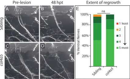
Figure A2 Semi-quantitative categories for extent of dorsal nerve regrowth…………102
Figure A3 lh3 is dispensable for ventral nerve regeneration……….103
Figure A4 Semi-quantitative categories for extent of ventral nerve regrowth …...104
Figure A5 Generation of col18a1 mutant zebrafish using TALENs………...105
Figure A6 col4a5 is dispensable for ventral nerve regeneration………...106
Figure A7 col18a1 and col19a1 are dispensable for dorsal nerve regeneration……...107
Figure A8 Zebrafish slit1a is upregulated after dorsal nerve transection………....108
Figure A9 Schwann cells are required for dorsal nerve regeneration………..………...109
Figure A10 Schwann cells are present in the correct number on the dorsal nerve in lh3
and col4a5 mutants………..…...110
General Introduction
Approximately 20 million Americans suffer from peripheral neuropathy (NINDS
2014), generating an estimated cost of 150 million dollars in treatment expenditures
annually (Pike et al. 2012; Grinsell and Keating 2014). This health care burden stems
from the broad variety of insults that cause peripheral nerve damage; chemical toxins,
including some agents used in chemotherapy, disease states, such as diabetes, and a
variety of genetic abnormalities can all lead to peripheral neuropathy (Gordois et al.
2003; Reilly et al. 2011). In these contexts, physicians are limited to prescribing palliative
care and encouraging changes in diet and activity that may stave off symptoms - the toll
on the patient and her family is extensive (Park 2014). In contrast, acute cut and crush
injuries, the most common cause of peripheral nerve damage, can be treated through
surgical nerve repair (Taylor et al. 2008). Because peripheral nerves have the intrinsic
ability to regenerate, surgeons have attempted to develop therapies to aid underlying
biological mechanisms. Unfortunately, even after surgical repair, regenerating axons
often fail to return to their targets, leaving patients in pain or chronically incapacitated
(Mackinnon and Dellon 1988). Therefore, defining the cell and molecular mechanisms
that peripheral nerves use to regenerate will shape new therapeutic responses to improve
patient recovery after peripheral nerve injury.
In over a century of research interrogating peripheral nerve regeneration, two
fundamental questions have remained elusive: do axons regenerate on target-selective
pathways? If so, what are the molecular and cellular cues that guide them? Below I will
describe how the results of these experiments have been interpreted, and suggest why
they have failed to reach definitive conclusions. Finally, I will characterize the new
model system that we have developed to model target selection in axon regeneration in
larval zebrafish and explain how we have harnessed this model to define a novel cell and
molecular framework for target-selective regeneration. My hope is that what we have
learned will ultimately aid clinicians in treating people who suffer from peripheral
Chapter 1: Background
The path to axon regeneration: not all injuries are equal.
Many studies demonstrate that the primary factor governing selectivity in nerve
regeneration is how well the integrity of the nerve tissue is maintained after injury
(reviewed in Brushart 2011). In mature peripheral nerve tissue, Schwann cells wrap
axons and secrete molecules of the extracellular matrix to confine the axon-Schwann cell
unit in basal lamina to form endoneurial tubes (Figure 1A). These tubes are grouped into
perineurial fascicles which are further wrapped in connective tissue called the epineurium
that segregate the nerve from the surrounding environment (Ushiki et al. 1990; Zochodne
compressed, but not severed, leaving axons with a defined path to grow along Schwann
cell basal lamina back to their original targets (Westerfield and Powell 1983; Ide et al.
1983; Scherer and Easter 1984; Kuffler 1986a; Nguyen et al. 2002). The consensus is that
crushed axons can and do regenerate to specific targets (Zochodne 2008; Reviewed in
Brushart 2011; Allodi et al. 2012).
In contrast, since Ramon y Cajal, researchers have debated whether transected
peripheral nerves regenerate to selective targets (Cajal 1928). Transection disrupts nerve
continuity and since the nerve is under tension, both the proximal and distal stumps
retract from the injury (Zochodne 2008). After transection, regenerating axons face the
challenge of growing through an acellular gap to reach surviving distal Schwann cells
and travel back to reinnervate their original targets. This raises a couple of questions
about how and whether regenerating axons retain different levels of target selectivity
when navigating through such a gap:
1. Can individual axons recognize and preferentially grow to distal nerve tissue?
2. Once they find this tissue, can axons locate the specific Schwann cells and basal
lamina that will guide them to the correct nerve branch?
Below, I will describe the experimental evidence arguing for and against selectivity in
regeneration at each of these levels of target specificity in the context of transection
injury and where relevant I will discuss the proposed mechanism underlying this
Tissue specificity in nerve regeneration: axons preferentially regenerate to nerve
In order to reinnervate a target and restore function an axon must first recognize
and preferentially grow to nerve tissue. Forssman provided the earliest recorded evidence
that axons regrow selectively on/to nerve tissue by extracting rat sciatic nerves and
showing that they preferentially sprouted towards brain as opposed to liver tissue in vitro (Forssman 1898). Ramon y Cajal described several experiments corroborating
Forssman’s findings in his seminal publication Degeneration and Regeneration of the Nervous System (Cajal 1928). For example, after transection of the rat sciatic nerve or various peripheral nerves in the rabbit, he noted that axons proximal to the injury
navigated through the injury scar with specificity to the distal stump and if this stump
was resected, growth and guidance diminished (Cajal 1928). Ramon y Cajal had
previously posited that target tissue released chemotactic signals to attract axons in the
development of the nervous system (Cajal 1909) and he interpreted his studies on
regeneration to indicate that target tissues provided similar neurotropic selectivity
mechanisms after injury (Cajal 1928). In the next two decades, Weiss and colleagues
performed a series of experiments to test Ramon y Cajal’s theory of neurotropism in vivo (Weiss 1937; Weiss and Taylor 1944; Weiss and Hoag 1946). Most famously, they
transected the rat sciatic nerve and placed the proximal stump into the input of a “Y-trap”
derived from fresh vascular tissue and showed that transected axons failed to regrow
preferentially to nerve tissue versus non-nerve tissue placed on either of the two outputs
dismissed the theory of neurotropism in peripheral nerve regeneration and promulgated
the belief that peripheral axons regenerate stochastically into all tissues.
This dogma was only directly challenged several decades later after a variety of
reports suggested that peripheral nerves could selectively regenerate to restore function
(Mark 1965; Grimm 1971; Stephenson 1979). In light of these findings, Lundborg et al.
directly repeated Weiss and Taylor’s experiments using an inert silicon Y trap and found
that in every case, rat sciatic axons “preferentially or exclusively” grew to nerve tissue as
opposed to tendon graft or an empty chamber (Lundborg et al. 1986; Figure 2C). Later
that year, Mackinnon et al. demonstrated that sensory neurons from the primate femoral
nerve preferentially grew to nerve tissue instead of muscle, tendon or an empty chamber,
compounding the evidence that chemotactic signals attract regenerating axons
and Taylor’s experiments may have been confounded by an immune reaction from the
fresh vascular tissue used for the “Y-trap”, but another group subsequently the exact
experiment performed by Weiss and Taylor and found that nerves preferentially regrew
to nerves through the fresh vascular Y-trap (Ochi et al. 1992). Together these
experiments support Ramon y Cajal’s prescient conclusion and reflect the current
consensus that regenerating axons can recognize and selectively grow through nerve
tissue (Zochodne 2008; Brushart 2011). While some studies (Kuffler 1986b; Abernethy et
al. 1992) suggest that Schwann cells and growth factors (Brushart et al. 2013) may guide
axons back to nerve tissue, the precise cell and molecular factors that govern the
regenerating axon’s ability to recognize nerve tissue have not been entirely elucidated.
Clinical observations: regenerating axons can, but do not always take the right path
While axons can selectively regenerate to nerve tissue, clinical insights reveal that
they do not always innervate the correct distal nerve targets. Early observations of
patients recovering from peripheral nerve injury found that these patients often suffered
from involuntary movements and inappropriate localization of sensations (Mitchell 1895;
Langley and Hashimoto 1917). More recent evidence corroborates these findings and
suggests that one important factor in functional recovery after nerve damage is the
structural complexity of the nerve (Brushart 2011). For example the human spinal
accessory nerve extends a single tributary to innervate distal targets and patients often
fully recover from damage to this nerve (Ogino et al. 1991; Nakamichi and Tachibana
the human facial nerve enters the periphery and branches in a complex pattern and reports
demonstrate that patients recovering from facial nerve damage develop involuntary
muscle contraction consistent with inappropriate target innervation (Kimura et al. 1975;
Spector et al. 1991; Figure 3B). However, even in the case of recovery from facial nerve
injury, patients recover some degree of function, suggesting that some axons regenerate
to the correct targets (Kimura et al. 1975). Thus, clinical observations suggest that axons
can select the appropriate nerve pathways in regeneration, but whether they do so through
random or target-selective methods remains unclear.
Experimental evidence: do axons regenerate with nerve branch specificity?
Researchers have addressed the question of whether nerves regenerate with nerve
conclusions. Sperry and Aurora transected the oculomotor nerve of cichlid fish, which
branches to innervate four muscles around the eye, and reported that after recovery, the
fish were able to control eye movement in the injured eye in a manner indistinguishable
from the uninjured control eye (Sperry and Arora 1965). In the same year, Mark
demonstrated that transection of the cichlid brachial plexus, which innervates the fin
through several nerve branches, led to regeneration and complete restoration of fin
movement (Mark 1965). Similar specificity was observed after nerve transection in the
axotlotl (Grimm 1971; Stephenson 1979). These authors concluded that after transection,
peripheral motor axons could regenerate with specificity to their original nerve branches.
However, Scherer repeated the oculomotor nerve transection experiments in goldfish
using HRP labeling and serial EM to determine the exact sites of axon reinnervation and
found that regenerating axons often branched and innervated inappropriate muscle targets
(Scherer 1986). Moreover, Westerfield and Powell found that after ventral root
transection in adult bullfrogs, motor axons failed to reinnervate the appropriate pathways
(Westerfield and Powell 1983). Lee and Farel confirmed this observation using
retrograde labeling of regenerated motor neurons, but demonstrated that tadpole motor
axons regenerated with specificity to their targets (Farel 1986; Lee and Farel 1988).
These experiments directly contradict the findings of Sperry and Aurora and suggest that
in adult animals, regenerating axons do not selectively regenerate to muscle targets.
Using mammalian nerve repair models, different groups have also found evidence
for and against specificity in nerve branch selection by regenerating axons. Here, several
researchers have used transection of the rat sciatic nerve as a model system. This nerve is
the nerve but branch to innervate separate areas of the leg. Politis et al. transected this
nerve prior to the branch point and attached either the proximal tibial or peroneal fascicle
to the input of a “Y Chamber” and asked whether it preferentially grew to the distal
peroneal or tibial nerve placed at the chamber outputs. Remarkably, in all cases, they
noted that these fascicles grew to their native distal targets (Politis et al. 1982; Politis
1985; Figure 4A). Evans et al. tested specificity in regeneration using electromyography
after surgical repair of the sciatic nerve repair and found that as long as the fascicles were
not intentionally misaligned, the peroneal and tibial branches selectively grew back to the
appropriate distal target (Evans et al. 1991). While these data suggest that mammalian
axons can regenerate with specificity to a given nerve branch, Abernethy et al. were not
able to reproduce Politis’ findings (Abernethy et al. 1992, Figure 4B) and Brushart et al.
called Evans’ data into question, by performing experiments that suggested that any
selectivity that Evans et al. found could be the result of confounding technical factors
(Brushart et al. 1995). Therefore, the question of whether regenerating axons selectively
choose specific nerve branches after transection has remained controversial.
Preferential Motor Regeneration (PMR): A model for nerve branch selectivity
In contrast to the contentious debate surrounding whether axons regenerate
selectively to specific nerve branches, there is a great deal of evidence that motor axons
preferentially grow to motor nerves. In their seminal studies, Brushart and colleagues
took advantage of the rat femoral nerve – a mixed nerve that consists of sensory and
targets (Brushart 1988; Brushart 1993). They transected the nerve prior to branching,
sutured the proximal and distal nerves together with a silicone tube and used retrograde
tracing from the segregated branches to show that motor axons preferentially
reinnervated the motor branch – a process they termed “Preferential Motor Regeneration”
(Brushart 1988). Further experiments demonstrated that PMR occurs in primates, can
occur independently of the target end organ and is more apparent in juvenile animals
(Brushart 1993; Le et al. 2001). PMR has been independently verified in several
laboratories (Al-Majed et al. 2000; Franz 2005), demonstrating that regenerating motor
axons somehow recognize and selectively reinnervate motor nerves.
Because PMR is widely accepted as selective regeneration to specific nerve
branches, several studies have attempted to identify the mechanism underlying this
(Franz 2005; Franz et al. 2008). However, studies looking for PMR at different time
points after injury found that motor axons initially innervate both sensory and motor
nerve branch, but were detected less frequently in the sensory nerve branch over time
(Brushart 1993; Ghalib et al. 2001; Höke et al. 2006; Figure 4C), suggesting that the
initial choice of pathway is random, but motor nerves selectively maintain motor axons
(Höke et al. 2006; Brushart et al. 2013). Subsequently, many studies have shown that the
cells in denervated motor and sensory nerves respond to injury with different expression
patterns of trophic factors, extracellular matrix components, and even canonical guidance
molecules (Siironen et al. 1992; Martini 1994; Höke et al. 2006; Reviewed in Zochodne
2008; Brushart 2011; Brushart et al. 2013). Together these studies indicate that both
peripheral motor axons and cells in the distal motor nerve respond to transection by
changing their expression phenotype and some of these regulated genes may contribute to
selectivity in motor nerve regeneration. However, to date, the molecular pathways that
are required to direct axons to particular nerve branches have remained elusive. Below I
will outline molecular-genetic changes that occur after nerve transection and highlight
potential mechanisms that could underlie nerve branch selectivity.
Cell and molecular mechanisms that govern branch selectivity in axon regeneration
In response to transection, changes in intracellular calcium and the loss of
homeostatic retrograde trophic signals from the dorsal nerve, peripheral neurons undergo
well-characterized transcriptional changes (Raivich et al. 1991; Mandolesi 2004; Patodia
proximal axons to regenerate into the acellular gap towards distal nerve targets.
Concomitantly, several nerve-associated cells, including fibroblasts, macrophages and
Schwann cells enter the gap to establish an environment supportive to axonal
regeneration (Richardson et al. 1980; Schröder et al. 1993; Paíno et al. 1994; Xu et al.
1997; Parrinello et al. 2010; Rosenberg et al. 2014). Schwann cells are particularly
important in this process as axons cross the acellular gap to reestablish connections with
distal nerve Schwann cells and their basal lamina (Ide 1983; Westerfield and Powell
1983; Sketelj et al. 1989). Schwann cells also alter their transcriptional profiles in
response to nerve transection to adopt a pro-regenerative repair state (Fu and Gordon
1997; Jessen et al. 2008; Patodia and Raivich 2012; Arthur-Farraj et al. 2012). While
growth and survival of both neurons and Schwann cells is critical for successful axon
regeneration (Rosenberg et al. 2014) here I will focus on transcriptional changes that are
critical for intracellular communication between regrowing axons and their distal
Schwann cell targets that may provide target selectivity in regeneration.
Extracellular matrix changes after nerve transection.
Several pieces of evidence strongly suggest that molecules of the Extracellular
matrix (ECM) facilitate nerve regeneration (Nathaniel and Pease 1963; Pollard and
Fitzpatrick 1973; Forman and Berenberg 1978; Scherer and Easter 1984; Kuffler 1986a;
Martini 1994). First, after mouse sciatic nerve transection, ECM molecules are the second
most upregulated class of genes in the distal nerve stump (Kubo et al. 2002). These
proteoglycans, and Fibronectin (Lefcort et al. 1992; Siironen et al. 1992; Tona et al.
1993; Masaki et al. 2000; Wallquist et al. 2002). Furthermore, regenerating peripheral
axons grow along Schwann cell basal lamina (Haftek and Thomas 1968; Ide et al. 1983;
Sketelj et al. 1989). In fact, mouse sciatic axons can regenerate through basal lamina
devoid of other cells (Ide et al. 1983). Together these studies have led to the belief that
constituents of the basal lamina are simply permissive to regenerating axons. However, in
the developing organism, cues from the ECM provide critical guidance to axons as they
navigate to their targets (Ackley et al. 2001; Bülow and Hobert 2006; Xiao et al. 2011;
Poulain and Chien 2013). This raises the possibility that Schwann cells upregulate
molecules of the basal lamina to direct regenerating axons to specific distal nerve targets.
These basal lamina molecules can interact directly with growth cone receptors, including
Integrins, which are upregulated by motor neurons after peripheral nerve injury and are
required for nerve regeneration in vivo (Werner et al. 2000; Hammarberg et al. 2000). Moreover, they can bind to and present canonical axon guidance molecules to migrating
growth cones (Xiao et al. 2011). However, because ECM molecules are often critical for
developmental growth processes (Löhler et al. 1984; Guo et al. 1991; George et al. 1993;
Smyth et al. 1999; Myllyharju 2004; Poschl 2004; Ruotsalainen et al. 2006), their roles in
peripheral nerve regeneration have only been characterized in vivo in a few instances (Chen 2003; Edwards and Hammarlund 2014) and not in the context of nerve branch
selectivity.
In development, axons navigate to their target in response to contact dependent
and chemotactic guidance ligands expressed by cells along their path (Tessier-Lavigne
and Goodman 1996; Huber et al. 2003). Growth cones respond to these ligands if they
express the cognate receptors, and through combinatorial mechanisms, functionally
diverse sets of axons travel through the same environment to select different targets.
After peripheral nerve transection, several canonical axon guidance ligands and receptors
are upregulated (Pasterkamp et al. 1998; reviewed in Zochodne 2008; Brushart 2011).
For example, Schwann cells in the distal nerve upregulate Slit-2 and Netrin-1 after rat
sciatic nerve transection (Madison et al. 2000; Tanno et al. 2005). Moreover
downregulation of Unc5H in rats, knockout of Neuropilin-2 in mice and loss of Slit-Robo
signaling in C. elegans leads to diminished growth in peripheral axon regeneration suggesting that these canonical axon guidance molecules can facilitate axon regeneration
(Bannerman et al. 2008; Gabel et al. 2008; Webber et al. 2011; Rosenberg et al. 2014).
However, whether these molecules are re-employed to guide peripheral nerves to distinct
nerve branches in regeneration remains unclear.
A zebrafish model for nerve branch selection in axon regeneration.
Together, the data addressing selective regeneration at nerve branches suggest
that some regenerating axons are able to select the correct branch pathway while others
veer onto aberrant nerve branch trajectories. All of these studies were conducted in the
context of injuries that required invasive surgery, and many involved nerve repair with
order to address whether and how axons regenerate to a specific branch we sought to
define a system where we could follow the tenets of Goran Lundborg:
“To study the behavior of the regenerating nerve elements in a situation where all external factors, including surgery and foreign material, were minimized, and where possible regeneration-promoting factors mobilized by the nerve tissue itself should be free to act” (Lundborg and Hansson 1979).
Moreover, we believe that the best way to characterize how a regenerating axon chooses
a specific path is to watch the dynamics of this process in real time in vivo. Historically, researchers have analyzed specificity in regeneration using fixed samples at different
time points after nerve injury. If axons do not receive the appropriate trophic support they
often degenerate (reviewed in Zochodne 2008; Brushart 2011). Without the ability to
track regenerating axons in vivo, these experiments could not definitively determine
whether axons regenerate to specific targets or regenerate randomly, but selectively
degenerate from inappropriate targets. While several researchers have successfully
characterized the dynamics of axon regeneration in live invertebrate models
(Hammarlund et al. 2009; Chen et al. 2011; Fang and Bonini 2012; Klinedinst et al.
2013), there are few vertebrate examples (Speidel 1932; Speidel 1948; Fangboner et al.
1980; Rieger and Sagasti 2011; Rosenberg et al. 2012; Villegas et al. 2012; Pan et al.
2013; Lewis and Kucenas 2014). Screens in these invertebrate models have already
revealed invaluable information regarding the axon-intrinsic mechanisms that drive axon
regeneration (Chen et al. 2011; Nix et al. 2014). However, as previously described the
intercellular interactions that occur within a vertebrate nerve are critical for axon
regeneration (Scherer and Easter 1984; Hall 1986; Kuffler 1986b; Parrinello et al. 2010;
whether vertebrate nerves regenerate to selective nerve branches and if so what
environmental cues drive this selectivity.
We therefore chose the peripheral motor nervous system of the 5 day
post-fertilization (dpf) larval zebrafish motor to test the environmental and intercellular
interactions required for branch pathway selection in regeneration. In the larval zebrafish,
motor nerves consist of ~100 axons that exit spinal cord and initially fasciculate along a
common path before diverging along two branches to innervate targets in the ventral
myotome (~60-80 axons) and in the dorsal myotome respectively (Myers et al. 1986;
Westerfield et al. 1986; Westerfield 1987). We and others have previously shown that
these motor nerves consist of the same cell types as mammals, are myelinated and form
stable neuromuscular junctions by 5dpf, and undergo all of the hallmarks of degeneration
after nerve transection (Kucenas et al. 2008; Rosenberg et al. 2012; Binari et al. 2013;
Lewis and Kucenas 2014; Rosenberg et al. 2014). Moreover, using non-invasive
laser-transection and transgenic markers, we have used this model to observe the intercellular
interactions between Schwann cells, Macrophages, and axons in regeneration in real time
in vivo. Using this system, we have demonstrated that Schwann cells and the axon guidance receptor dcc provide critical directional information to ventrally directed axons as they regenerate across the injury gap (Rosenberg et al. 2012; Rosenberg et al. 2014).
Here we have adopted this model to define whether and how axons regenerate
selectively to their original target areas. We first transected both nerve branches proximal
(Uemura et al. 2005). In Chapter 2, we demonstrate that the majority of fascicles that
innervated the dorsal myotome in development regenerate to this target region (80%) and
conclude that vertebrate axons can regrow with selectivity to their original nerve branch.
Using real time in vivo imaging we show that regenerating axons search the transection gap extensively before stabilizing growth to the correct nerve branch and destabilizing
growth to inappropriate targets. We take advantage of the genetic tractability of zebrafish
to show that collagen modifications provided by the glycosyltransferase lysyl hydroxylase 3 (lh3) are required for growth and guidance of regenerating dorsal axons. We next demonstrate that after injury, lh3’s substrate collagen4a5 – a resident of the basal lamina - is upregulated in a specific subset of Schwann cells ventral and ventrolateral to the
transection gap and is required to destabilize regenerating axons that search in these
inappropriate target regions. Finally, we show that lh3 acts post injury in Schwann cells to guide regenerating axons. Collectively, these data demonstrate that after nerve
transection, local Schwann cells modify the basal lamina to direct axons on target
selective pathways in regeneration (Chapter 2).
We next asked how these basal lamina cues might destabilize regenerating axons.
Collagen4 is known to bind the axon guidance repellents Netrin and Slit (Yebra et al.
2003; Xiao et al. 2011) and in rodent models, both netrin and slit are upregulated in Schwann cells after peripheral nerve transection (Madison et al. 2000; Tanno et al. 2005).
Together these results suggest that axons regenerating to different branches require
different guidance cues. Furthermore they provide a possible mechanistic framework by
which Schwann cells modify the basal lamina to aggregate canonical guidance cues to
direct regenerating axons (Isaacman-Beck et al., in press).
In Chapter 3, we follow up on these findings with several unpublished
discoveries. We demonstrate that overexpression of col4a5 in all Schwann cells disrupts target-selectivity in regeneration suggesting that col4a5 may be required in specific locations and at specific levels to direct regenerating dorsal axons. Furthermore we show
that the slit receptors robo2 and robo3 are required for dorsal nerve regeneration suggesting that slit-robo repulsion might guide regenerating axons. Finally, we outline future experiments to determine the cell type requirements for robo2 and robo3 in peripheral nerve regeneration and ultimately to define whether col4a5 presents slits to regenerating motor axons to direct them on target selective pathways.
Chapter 2:
The
lh3
glycosyltransferase directs target-selective peripheral nerve
regeneration
The data in Chapter 2 are in press at Neuron.
Jesse Isaacman-Beck, Valerie Schneider, Clara Franzini-Armstrong and Michael Granato
Summary
Functional PNS regeneration requires injured axons to return to their original
synaptic targets, yet the mechanisms underlying target-selective regeneration have
remained elusive. Using live-cell imaging in zebrafish we find that regenerating motor
axons exhibit a strong preference for their original muscle territory, and that axons probe
both correct and incorrect trajectories extensively before selecting their original path. We
show that this process requires the glycosyltransferase lh3 and that post-injury expression
of lh3 in Schwann cells is sufficient to restore target-selective regeneration. Moreover,
we demonstrate that Schwann cells neighboring the transection site express the lh3
substrate collagen4a5 and that during regeneration collagen4a5 destabilizes axons
probing inappropriate trajectories to ensure target-selective regeneration, possibly
through the axonal repellant slit1a. Our results demonstrate that selective ECM
components match subpopulations of regenerating axons with their original targets, and
reveal a previously unappreciated mechanism that conveys synaptic target selection to
Introduction
Axons of the peripheral nervous system have the remarkable ability to regenerate
following injury and form functional connections with their original targets. Damage to
peripheral nerves such as acute mechanical trauma, disease or chemical insult triggers the
well-characterized program of Wallerian degeneration that results in axonal
fragmentation and debris clearance involving immune and Schwann cells (Waller 1849;
Vargas and Barres 2007). Concomitantly, denervated Schwann cells de-differentiate to a
more stem cell-like phenotype with critical roles in supporting axonal regrowth from the
proximal nerve stump (Zochodne 2008; Rosenberg et al. 2014). There, intrinsic and
extrinsic factors promote sprouting of axonal growth cones, which then begin their
regenerative journey to re-establish functional connections with their original synaptic
targets (reviewed in Zochodne 2008; Brushart 2011).
Not surprisingly, the degree of functional regeneration depends largely on the type of
injury (Kruspe et al. 2014). For example, crush injuries leave the nerve-ensheathing basal
lamina intact, thereby providing an uninterrupted tube-like permissive substrate that can
lead regenerating axons back to their appropriate targets (Haftek and Thomas 1968;
Westerfield and Powell 1983; Scherer and Easter 1984; Sketelj et al. 1989). In contrast,
nerve transections disrupt the continuity of the nerve and nerve basal lamina, forcing
regenerating axons to navigate across the injury gap through an acellular environment
(Forman and Berenberg 1978; Forman et al. 1979). This challenge is even greater in
cases when regenerating axons encounter a nerve branch choice point distal to the injury
original targets, thereby decreasing the degree of functional regeneration (reviewed in
Brushart 2011). Moreover, misguided axons can innervate inappropriate targets, leading
to involuntary muscle contractions such as those observed in facial palsy (Kimura et al.
1975; Spector et al. 1991). Several studies argue that this sparse and/or ectopic axonal
reinnervation is the result of regenerating axons selecting their path at branch points in a
stochastic manner (Westerfield and Powell 1983; Scherer 1986; Westerfield 1987;
English 2005), while others conclude that regenerating axons somehow ‘recognize’ their
original trajectory (Mark 1965; Sperry and Arora 1965; Grimm 1971; Stephenson 1979;
Kuffler 1986b; Brushart 1988; Lee and Farel 1988). However, the mechanisms and
molecules that enable regenerating axons to select their original trajectory as they
encounter branch choice points in vivo have remained elusive.
Extracellular matrix (ECM) components and their modifying enzymes are known to
provide critical guidance to developing axons, and while several ECM components are
transcriptionally upregulated following peripheral nerve injury, their roles in axonal
regeneration have not been well defined in vivo (Kubo et al. 2002; Chen et al. 2011; Nix
et al. 2014). In regenerating peripheral nerves, ECM components are the second most
upregulated class of genes, and while regenerating axons are known to associate with the
ECM as they to return to their targets (Chernousov and Carey 2000; Kubo et al. 2002;
Chen et al. 2011; Nix et al. 2014), the role of ECM components and their modifying
enzymes has not been fully elucidated in genetic loss of function studies, mainly due to
their frequent essential requirement during developmental processes (Löhler et al. 1984;
Ruotsalainen et al. 2006). Here, we take advantage of the optical transparency and
stereotyped peripheral motor nerve architecture in larval zebrafish to determine the role
of ECM components in target specific regeneration of spinal motor axons. We find that
the collagen-modifying glycosyltransferase lysyl hydroxylase 3 (lh3) is critical for
regenerative growth and guidance of axons of the dorsal but not ventral nerve branch, and
that lh3 expression during regeneration and in Schwann cells is sufficient to restore
dorsal regeneration. Furthermore we show that in vivo lh3 exerts its function at least in
part through its well-established substrate collagen4a5 (Wang et al. 2000; Ruotsalainen et
al. 2006), and that following nerve transection collagen4a5 mRNA is selectively
upregulated in Schwann cells at the lesion site. Combined our results revise the widely
held assumption that during regeneration ECM components serve primarily as permissive
substrates, and reveal an underappreciated yet specific role in directing regenerating
axons towards their original targets.
Results
Regenerating motor axons select their original trajectory with high fidelity
Following complete nerve transection, peripheral axons can successfully traverse a
short, acellular injury gap, yet whether axons randomly extend towards their original
targets when confronted with a path choice or whether mechanisms for target-selective
innervation exist has long been a point of contention (Westerfield and Powell 1983;
Scherer 1986; Kuffler 1986b; Brushart 1993; English 2005). As a first step to distinguish
between these possibilities in a live vertebrate system, we took advantage of the simple
approximately 100 fasciculated axons, which separate into two main nerve branches
shortly after exiting from the spinal cord: a ventral nerve branch consisting of 60-80
axons with synaptic targets in the ventral myotome, and a dorsal nerve branch consisting
of 20-30 axons innervating the dorsal myotome (Myers et al. 1986; Westerfield et al.
1986 and Figure 1A; Westerfield 1987).
To test whether regenerating motor axons preferentially select their original
branch-specific nerve path, we laser-transected the entire motor nerve proximal to where the
trajectories of the dorsal and ventral branches diverge, creating a ~9µm gap between the
proximal and distal nerve stumps (Rosenberg et al. 2012; Binari et al. 2013; Lewis and
Kucenas 2014; Rosenberg et al. 2014). We labeled both ventral and dorsal nerve axons
using the Tg(Xla.Tubb:DsRed) transgene (Peri and Nüsslein-Volhard 2008), and
Dorsal
branch
(~20 axons)
Ventral
branch
(~60 axons)
A Prelesion Dorsal branch ~25 axons Ventral branch ~70 axons Choice point Transection Site Tg(isl1:GFP) Tg(Xla.Tubb:DsRed) ROI B 48 hpt C 80% 5 dpf; PrelesionFigure 1: Regenerating motor axons select their original trajectory with high fidelity. (A,B)
selectively labeled the dorsal branch with the Tg(isl1:GFP) transgene (Uemura et al.
2005), thereby enabling us to monitor target-selective regeneration in vivo (Figure 1A,B).
Prior to nerve transection (pre-lesion), the majority of Tg(isl1:GFP) labeled fascicles
extend within a very narrow area that spans 20º of the dorsal myotome (Figure 1A, B).
Forty-eight hours post nerve transection, 80% of these fascicles regenerated to this
original area (Figure 1C; see Supplementary Experimental Procedures for more details on
quantification). This is a significantly higher fraction than the 50% expected for a
‘random’ mechanism given a binary choice between the 20º dorsal target area and regions
outside, demonstrating that following complete nerve transection, regenerating axons of
the dorsal nerve branch retain the ability to select their original branch-specific trajectory.
Furthermore, transection of only the dorsal nerve branch resulted in the same degree of
branch-specific regrowth of Tg(isl1:GFP) positive fascicles (data not shown), indicating
that target-selective regeneration of dorsal nerve axons occurs independently of injury to
ventral nerve axons. Together these results reveal that when confronted with a choice
point, regenerating zebrafish motor axons select their original path with high fidelity,
consistent with the existence of non-random genetic mechanisms that promote
target-selective regeneration.
Regenerating axons probe the transection gap extensively before selecting their
original path
We next used live-cell imaging to examine the behavior of regenerating axons as they
encounter a branch choice point. One possibility is that regenerating axons exclusively