Iancu et al.BMC Medical Imaging 2010, 10:14 http://www.biomedcentral.com/1471-2342/10/14
Open Access
C A S E R E P O R T
© 2010 Iancu et al; licensee BioMed Central Ltd. This is an Open Access article distributed under the terms of the Creative Commons Attribution License (http://creativecommons.org/licenses/by/2.0), which permits unrestricted use, distribution, and reproduction in any medium, provided the original work is properly cited.
Case report
Brainstem infarction in a patient with internal
carotid dissection and persistent trigeminal artery:
a case report
Daniela Iancu*
1,2, Rene Anxionnat
1and Serge Bracard
1Abstract
Background: The primitive trigeminal artery (PTA) is the most commonly described fetal anastomosis between the carotid and vertebrobasilar circulations.
Case presentation: We report a 42-year-old patient presenting with internal carotid dissection, and imaging features of brainstem infarction.
Conclusion: Based on the imaging studies we presume occlusive carotid dissection with extensive thrombosis within a persistent trigeminal artery as the cause of this brainstem ischemia.
Background
Several fetal anastomoses have been described between the carotid and vertebrobasilar circulations. These anas-tomoses regress while the P1 segments develop, but they can occasionally persist in adult age [1]. The primitive trigeminal artery (PTA) is the most common of them rep-resenting about 85% of cases with prevalence between 0.1% and 0.76% [2].
We report a patient with brainstem infarction caused by a persistent PTA thrombosis secondary to occlusive dissection of the homolateral internal carotid artery (ICA).
Case presentation
A 42-year-old woman presented with right-side motor deficit and dysarthria. She experienced diffuse head-aches, regressive episodes of ill-defined visual distur-bance and right-side numbness the previous day. She reported osteopathic cervical manipulations in the previ-ous week.
Neurologic examination revealed right-sided hemipa-resis, hypoesthesia, central facial palsy and dysarthria. No Horner's sign, cranial nerve palsy, abnormal cardiac or carotid bruit were found.
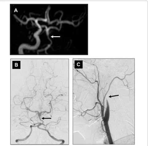
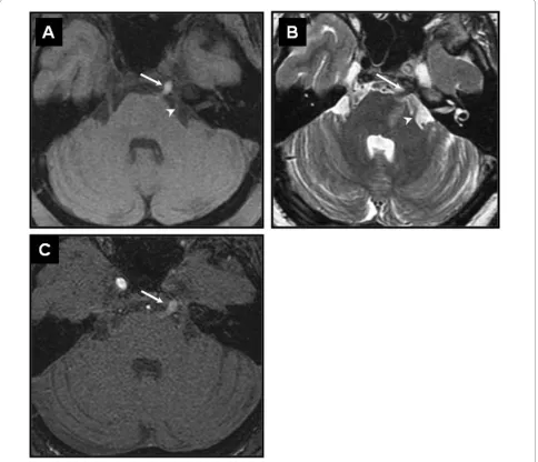
Magnetic resonance imaging (MRI) revealed left anter-olateral pontine infarction (Figures 1A, B, and 1C). No acute infarction was seen in the left-ICA territory. Three-dimensional time-of-flight MRA (3D-TOF MRA) showed occlusion of the left ICA and hypoplastic vertebral (VA) and proximal basilar (BA) arteries (Figure 2A). The left hemispheric supply was provided by the right-ICA via anterior communicating artery. Additional thin axial sec-tions T2 and T1WI [3] were unable to demonstrate dis-sections on vertebrobasilar system. However, a well defined 5mm structure, hyperintense on T1WI with fat-saturation and hypointense on T2WI was seen within the prepontine cistern via Meckel's cave with a similar course to the trigeminal nerve, which was identified separately. In view of its location and orientation we presumed that this structure corresponded to a thrombosed persistent PTA and concluded that the brainstem stroke was due to an extensive thrombosis caused by occlusive ICA dissec-tion via the PTA (Figure 3).
A digital subtraction angiography (DSA) showed irreg-ular localized filling defect within the distal hypoplastic BA (Figure 2B) and a flame-shaped occlusion of the left ICA which is characteristic of occlusive dissection (Fig-ure 2C).
Further investigation did not reveal concomitant car-diac or coagulation disorders. Intravenous Heparin was initiated and the patient was discharged 3 weeks later
* Correspondence: [email protected]
1 Department of Neuroradiology, CHU Nancy, Nancy, 54035, France
with residual motor deficit. MRA follow-up showed per-sistent ICA and PTA occlusion.
Discussion
Persistent PTA is often associated with intracranial aneu-rysms, arteriovenous malformations, carotid cavernous fistulas, Moyamoya and cerebellar hemangioblastoma [4-7]. Its clinical significance is usually uncertain but presen-tation may include cranial nerve dysfunction or suba-rachnoid haemorrhage [4-7].
Very few cases of brainstem or occipital infarction due to embolism from the ICA stenosis via persistent PTA have been reported [8,9]. To our knowledge, this is the only case report documenting a persistent PTA thrombo-sis responsible for a brainstem infarction. The diagnothrombo-sis was difficult since flow was completely absent in the PTA
even on DSA. One clue was the diminutive aspect of both VA and proximal BA, usually found in persistent PTA. Moreover, concomitant dissection of the ICA and BA would have been unlikely.
Iancu et al.BMC Medical Imaging 2010, 10:14 http://www.biomedcentral.com/1471-2342/10/14
Page 3 of 5
edge [1]. The pontine artery could give an accessory branch to the trigeminal ganglion but its main territory is the protuberance [1]. In this case the anterolateral pon-tine infarction corresponds to this territory.
Stroke secondary to cervical ICA dissection generally involves embolic mechanisms instead of hypoperfusion. Since dissection rarely extends beyond the petrous
seg-ment of the ICA [10], we propose that extensive throm-bosis, due to the dissection, is the mechanism that occluded the intracavernous carotid segment and the PTA. The DSA aspect showing small VA and proximal BA with localized endovascular filling defect is suggestive of pre-existent PTA pattern with a distal extension of the thrombosis into the PTA and BA.
Conclusion
This is a very rare case of MRI documented persistent PTA thrombosis responsible for brainstem infarction. In patients presenting with brainstem ischemia associated with occlusion or stenosis of the homolateral ICA, persis-tent PTA should be considered.
Consent
Written informed consent was obtained from the patient for publication of this case report and any accompanying images. A copy of the written consent was provided to the editorial office of this journal.
Competing interests
The authors declare that they have no competing interests.
Authors' contributions
DI performed the literature search and compiled data presented in this report. RA provided the expertise for selective imaging and contributed to the diag-nosis. SB provided intellectual input and critically revised the manuscript. All authors read and approved the final manuscript.
Acknowledgements
We acknowledge Dr. Marshall Wilkinson, Dr. Thomas Mammen and Dr. Andreea Nistor from the University of Manitoba, for reviewing and helping with editing this manuscript.
Author Details
1Department of Neuroradiology, CHU Nancy, Nancy, 54035, France and 2Department of Radiology, Section of Neuroradiology, University of Manitoba,
Health Sciences Centre, 820 Sherbrook St, Winnipeg, Manitoba R3A 1R9, Canada
Received: 22 March 2010 Accepted: 2 July 2010 Published: 2 July 2010
This article is available from: http://www.biomedcentral.com/1471-2342/10/14 © 2010 Iancu et al; licensee BioMed Central Ltd.
This is an Open Access article distributed under the terms of the Creative Commons Attribution License (http://creativecommons.org/licenses/by/2.0), which permits unrestricted use, distribution, and reproduction in any medium, provided the original work is properly cited.
BMC Medical Imaging 2010, 10:14
Iancu et al.BMC Medical Imaging 2010, 10:14 http://www.biomedcentral.com/1471-2342/10/14
Page 5 of 5
References
1. Lasjaunias P, Berenstein A: Surgical neuroangiography: functional anatomy
of craniofacial arteriesVolume 1. Berlin, Springer-Verlag; 1987.
2. Piotin M, Miralbes S, Cattin F, Marchal H, Amor-Sahli M, Moulin T, Bonneville JF: MRI and MR angiography of persistent trigeminal artery.
Neuroradiology 1996, 38:730-733.
3. Leclerc X, Lucas C, Godefroy O, Nicol L, Moretti A, Leys D, Pruvo JP:
Preliminary experience using contrast-enhanced MR angiography to assess vertebral artery structure for the follow-up of suspected dissection. Am J Neuroradiol 1999, 20:1482-1490.
4. Redekop GJ: Extracranial carotid and vertebral artery dissection: a review. Can J Neurol Sci 2008, 35:146-152.
5. Hurst RW, Howard RS, Zager E: Carotid cavernous fistula associated with persistent trigeminal artery: endovascular treatment using coil embolization. Skull Base Surg 1998, 8:225-228.
6. Komiyama M, Nakajima H, Nishikawa M, Yasui T, Kitano S, Sakamoto H, Fu Y: High incidence of persistent primitive arteries in moyamoya and quasi-moyamoya diseases. Neurol Med Chir 1999, 39:416-420. 7. Murai Y, Kobayashi S, Tateyama K, Teramoto A: Persistent primitive
trigeminal artery aneurysm associated with cerebellar hemangioblastoma. Case report. Neurol Med Chir 2006, 46:143-146. 8. Gasecki AP, Fox AJ, Lebrun LH, Daneault N: Bilateral occipital infarctions
associated with carotid stenosis in a patient with persistent trigeminal artery. The Collaborators of the North American Carotid
Endarterectomy Trial (NASCET). Stroke 1994, 25:1520-1523. 9. Foerch C, Berkefeld J, Halbsguth A, Ziemann U, Neumann-Haefelin T:
Brain stem infarction caused by proximal internal carotid artery stenosis in a patient with a persisting primitive trigeminal artery.
Cerebrovasc Dis 2006, 22:200-202.
10. Abe T, Matsumoto K, Aruga T: Primitive trigeminal artery variant associated with intracranial ruptured aneurysm and cerebral arteriovenous malformation - case report. Neurol Med Chir 1994,
34:104-107.
Pre-publication history
The pre-publication history for this paper can be accessed here: http://www.biomedcentral.com/1471-2342/10/14/prepub
doi: 10.1186/1471-2342-10-14
Cite this article as: Iancu et al., Brainstem infarction in a patient with internal carotid dissection and persistent trigeminal artery: a case report BMC Medical