C A S E R E P O R T
Open Access
Newly recognized cerebral infarctions on
postmortem imaging: a report of three
cases with systemic infectious disease
Sakon Noriki
1,4*, Kazuyuki Kinoshita
2,4, Kunihiro Inai
3,4, Toyohiko Sakai
2,4, Hirohiko Kimura
2,4, Takahiro Yamauchi
5,
Masayuki Iwano
6and Hironobu Naiki
3,4Abstract
Background:Postmortem imaging (PMI) refers to the imaging of cadavers by computed tomography (CT) and/or magnetic resonance imaging (MRI). Three cases of cerebral infarctions that were not found during life but were newly recognized on PMI and were associated with severe systemic infections are presented.
Case presentations:An 81-year-old woman with a pacemaker and slightly impaired liver function presented with fever. Imaging suggested interstitial pneumonia and an iliopsoas abscess, and blood tests showed liver dysfunction and disseminated intravascular coagulation (DIC). Despite three-agent combined therapy for tuberculosis, she died 32 days after hospitalization. PMI showed multiple fresh cerebral and cerebellar infarctions and diffuse ground-glass shadows in bilateral lungs. On autopsy, the diagnosis of miliary tuberculosis was made, and non-bacterial
thrombotic endocarditis that involved the aortic valve may have caused the cerebral infarctions.
A 74-year-old man on steroid therapy for systemic lupus erythematosus presented with severe anemia, melena with no obvious source, and DIC. Imaging suggested intestinal perforation. The patient was treated with antibiotics and drainage of ascites. However, he developed adult respiratory distress syndrome, worsening DIC, and renal
dysfunction and died 2 months after admission. PMI showed infiltrative lung shadow, ascites, an abdominal aortic aneurysm, a wide infarction in the right parietal lobe, and multiple new cerebral infarctions. Autopsy examination showed purulent ascites, diffuse peritonitis, invasive bronchopulmonary aspergillosis, and non-bacterial thrombotic endocarditis that likely caused the cerebral infarctions.
A 65-year-old man with an old pontine infarction presented with a fever and neutropenia. Despite appropriate treatment, his fever persisted. CT showed bilateral upper lobe pneumonia, pain appeared in both femoral regions, and intramuscular abscesses of both shoulders developed. His pneumonia worsened, his level of consciousness decreased, right hemiplegia developed, and he died. PMI showed a newly diagnosed cerebral infarction in the left parietal lobe. The autopsy revealed bilateral bronchopneumonia, right-sided pleuritis with effusion, an intramuscular abscess in the right thigh, and fresh multiple organ infarctions. Systemic fibrin thrombosis and DIC were also found. Postmortem cultures showedE. coliandBurkholderia cepacia.
Conclusion:Cerebral infarction that is newly recognized on PMI might suggest the presence of severe systemic infection.
Keywords:Case report, Postmortem imaging, Cerebral infarction, Infection, Cause of death, Autopsy, Pathology
* Correspondence:[email protected]
1Division of Tumor Pathology, Department of Pathological Sciences, School
of Medical Sciences, University of Fukui, 23-3 Shimoaizuki, Matsuoka Eiheiji-cho, Yoshida-gun, 910-1193 Fukui, Japan
4Autopsy Imaging Center, School of Medical Sciences, University of Fukui,
Fukui, Japan
Full list of author information is available at the end of the article
Background
Cadavers can be evaluated using diagnostic imaging, which comprises one aspect of medical assessment at the time of death. Imaging of cadavers has also been re-ferred to as postmortem imaging (PMI). However, this procedure is variously described as virtopsy in Switzerland [1], virtual autopsy in France [2], radio-autopsy in Germany [3], and radio-autopsy imaging (Ai) in Japan [4]. Although the descriptions and concept of PMI in these countries differ somewhat, all involve analysis of a cadaver by computed tomography (CT) and/or mag-netic resonance imaging (MRI) to acquire postmortem medical information.
PMI is a useful diagnostic tool for a forensic case [5] that has no antemortem medical information. However, PMI is often performed in hospital deaths. On the other hand, the rate of the hospital autopsies has been de-creasing worldwide in recent years, because the hospital autopsy requires consent in most countries. Further-more, the brain examination rate of the hospital autopsy is only 20% at our hospital, because another consent is required for the brain examination. Therefore, PMI that can examine the intracerebral state of the cadaver is use-ful. The findings of PMI are interpreted by taking into consideration the postmortem changes based on the findings of imaging of the living body. Characteristic in-terpretations of the findings of PMI have yet to be developed.
In this paper, three cases of cerebral infarctions that were not found during life but were newly recognized on PMI are reported. The autopsies revealed severe sys-temic infectious diseases in all three cases. The aim of this paper is to suggest the possibility of the presence of a severe systemic infection when cerebral infarction is newly recognized on PMI.
Case presentations
Case 1
An 81-year-old woman visited a hospital for a pacemaker check. At that time, slightly impaired liver function was noted. She then developed a fever of 38 °C. Although care-ful examinations to identify the cause of the fever were performed, the source could not be identified. Various cul-tures were also negative. Although antibiotic treatment was given, her fever and general status did not improve, and she was admitted to our hospital. On physical exam-ination at admission, her temperature was 38 °C, blood pressure was 140/87 mmHg, and her pulse was 76/min. On blood tests, hemoglobin (Hb) was 11.8 g/dl, C-reactive protein (CRP) was 5.54 mg/dl, soluble interleukin-2 recep-tor (sIL-2R) was 3732 U/ml (standard 144–518 U/ml), and a tendency to disseminated intravascular coagulation (DIC) (PLT 5.4 x 10⁴/μl, FDP 156μg/ml, D-dimer 80μg/ ml) was seen. Slightly impaired liver function (aspartate
aminotransferase (AST) 120 IU/l, alanine aminotransfer-ase (ALT) 81 IU/l) was also found.
The chest CT on the second day after hospitalization showed a diffuse ground glass shadow and suspected interstitial pneumonia. On the fifth day after admission, contrast-enhanced CT showed a low-density area (LDA) in the left iliopsoas muscle, suggesting an iliopsoas muscle abscess. Staphylococcus aureus, Escherichia coli (E. coli), or tuberculosis was considered as the causative organism of the iliopsoas muscle abscess. Various dys-functions, such as interstitial pneumonitis, liver damage, and DIC, were also present simultaneously. We assumed that there was a solitary underlying disease that could explain her clinical picture, for example, hematological disease, and treatment was started.
Since intense accumulation of fluoro-deoxy-glucose (FDG) was observed in bilateral lung fields on FDG-positron-emission tomography (PET), an inflammatory disorder, especially tuberculosis was suspected, and three-agent combined therapy was started on the 8th day after admission. However, neither the fever nor her general status improved, and the ground glass appear-ance had deteriorated further on the chest CT on the 16th day after admission.
On the 22nd day after admission (9 days before death), the patient’s respiratory condition deteriorated suddenly, and methylprednisolone pulse therapy resulted in no im-provement. She died on the 32nd day after hospitalization. PMI and an autopsy (only thoracoabdominal) were performed 14 h after death (Additional file 1).
PMI findings
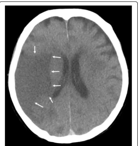
Multiple LDAs were recognized in the right middle cere-bral artery (MCA) region (Fig. 1), right cerebellum, and left basal ganglia on PMI. They seemed to be infarctions. Since no atrophy was found in the brain, the infarctions seemed to be relatively fresh lesions. There was neither a mass effect nor hemorrhage in the brain. The ground glass shadow was widespread in bilateral lungs, and part of the lungs showed infiltrative shadow and the crazy paving pattern. As the cause of the interstitial shadow, adult respiratory distress syndrome (ARDS), acute inter-stitial pneumonia, or an infectious disease such as Pneumocystis jirovecii pneumonia was considered. No airway obstruction was found. A cardiac pacemaker was confirmed. Neither brain CT nor brain MRI was done during the patient’s lifetime.
Pathological findings
On autopsy, white viscous liquid flowed when an inci-sion was made into the left iliopsoas muscle (Fig. 2a). The wall of the abscess was composed of lymphocytes and fibrous tissue, and the contents of the abscess con-sisted of necrotic material (Fig. 2b). A few neutrophils
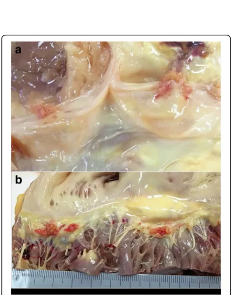
infiltrated into the abscess. A few acid fast bacilli were noted in the abscess with Ziehl-Neelsen staining. These histological findings and polymerase chain reaction (PCR) testing showed that the lesion was tuberculous. In addition, tuberculous nodules were found microscopic-ally in bilateral lungs (left 730 g, right 842 g), liver (1118 g), spleen (94 g), left kidney (182 g), bone marrow, and lymph nodes surrounding the pancreas, and miliary tuberculosis was diagnosed. Moreover, vegetations (4 mm and 5 mm in diameter) were noted on the aortic valve (Fig. 3a). The vegetations consisted of fibrin thrombus without bacterial colonies (Fig. 3b), and they were diagnosed as non-bacterial thrombotic endocardi-tis. This thrombus might have detached from the valve and become the emboli that resulted in the cerebral infarctions.
Case 2
A 74-year-old man was taking a steroid (30 mg/day of predonin) for systemic lupus erythematosus (SLE) and was being followed in the outpatient department. A blood test showed severe anemia (Hb 3.8 g/dl), and he was hospitalized 2 months before death. Melena was found, but no bleeding source was identified even on gastroscopy and colonoscopy. Then, 14 days after admis-sion, free air was found at the subphrenic region on chest X-ray and CT, and intestinal perforation was sus-pected. The patient was given a course of antibiotic treatment and drainage of ascites because of his general status.
The patient’s manifestations were relieved, but the inflam-matory response increased again, and his respiratory condi-tion suddenly worsened, requiring intensive care unit (ICU) admission. The onset of ARDS was suspected. He had DIC on admission, and it was exacerbated with progression of the infection. His inflammatory response, renal failure, and respiratory condition deteriorated, and he died 2 months after admission. PMI and an autopsy (only thoracoabdom-inal) were performed 2 h after death (Additional file 2).
PMI findings
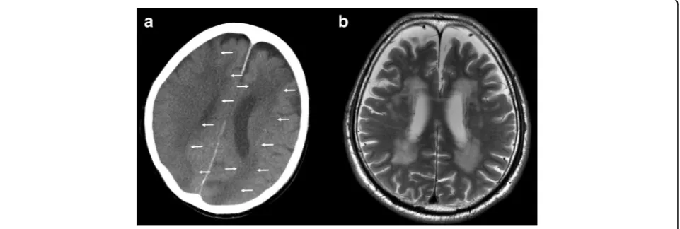
Ascites and an abdominal aortic aneurysm were found on abdominal CT, but the free air had disappeared. On chest CT, infiltrative shadow was found, and ARDS, pneumonia, and interstitial pneumonia were considered. Brain CT showed a wide LDA area in the right parietal lobe, and cerebral infarction was diagnosed (Fig. 4a) [6]. Multiple new cerebral infarctions were seen on PMI. Brain MRI was done 1 year 5 months before death. The
Fig. 1Brain postmortem CT image 14 h after death (Case 1). An LDA was found in the middle cerebral artery area (arrows)
T2-weighted image of the MRI corresponding to the CT image is shown (Fig. 4b). At that time, no cerebral in-farction was found.
Pathological findings
Yellowish white purulent ascites of 1800 ml was found in the abdominal cavity at autopsy, and it showed diffuse peritonitis. However, the perforation site of the intestine was not confirmed. Moreover, invasive bronchopulmon-ary aspergillosis was present in the lungs (Fig. 5a,b) [6], and the background lung showed diffuse alveolar dam-age. Aspergillus species were also found in the periton-eum. There were a few vegetations, up to 10 mm in diameter, on the aortic valve in the heart (Fig. 6a) [6]. Three vegetations, 12 mm, 10 mm, and 7 mm in diam-eter, were also found on the mitral valve (Fig. 6b) [6]. They were regarded as the cause of the cerebral infarc-tion; these thrombotic vegetations had separated from the valves. In addition, cholesterin crystal embolism was
found in the kidney, heart, liver, spleen, and it was thought that this had caused the progressive renal dysfunction.
Case 3
A 65-year-old man developed a left pontine infarction 2 months before hospitalization. He had a fever of about 39 °C. Blood tests showed white blood cells of 300/μl (3% neutrophils; neutropenia), and he was hospitalized in the Department of Hematology. Drug-induced neutropenia was suspected, and granulocyte colony-stimulating factor and antibiotics were administered with stopping of oral medicine. However, his fever remained, and pneumonia of both upper lobes was diagnosed by CT on the fourth day after admission, and an antifungal drug was added. Pain in both femoral regions then appeared. His pneumonia got worse, and intramuscular abscesses of both shoulders were noted 30 days before death. Though an antimicrobial drug was added, the pneumonia worsened, and an
Fig. 3Vegetations of the aortic valve at autopsy (Case 1).aThe macroscopic appearance of the aortic valve. The aortic valve has two vegetations of 4 mm and 5 mm in diameter.bLoupe image of the aortic valve. Vegetations consist of fibrin without bacterial colonies, and non-bacterial thrombotic endocarditis was diagnosed (HE stain. Original magnification × 1)
Fig. 4The postmortem CT and antemortem MRI (Case 2).aThe postmortem CT image of the brain 2 h after death. LDAs were found widely, resulting in a diagnosis of cerebral infarction (arrows). (with permission [6])bAntemortem MRI, T2-weighted image showing no cerebral infarc-tion. The brain MRI was taken 1 year 5 months before death
inflammatory pleural effusion developed. The patient’s level of consciousness decreased, and complete paralysis of the right side arm and leg appeared. He died 3 days after his consciousness level decreased and the right hemi-plegia developed. PMI and autopsy were performed 2 h after death (Additional file 3).
PMI findings
An LDA was found in the left parietal lobe, and it was a newly diagnosed cerebral infarction on PMI (Fig. 7a). Brain MRI was done 22 days before death. The T2-weighted image of the MRI corresponding to the CT image is shown (Fig. 7b). At that time, no cerebral in-farction was found.
Pathological findings
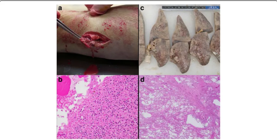
The autopsy revealed a severe systemic infection: bilat-eral bronchopneumonia (left 954 g: right 857 g), right-sided pleuritis with effusion, and an intramuscular ab-scess in the right thigh (Fig. 8). Moreover, systemic fibrin thrombosis and a bleeding tendency due to DIC were seen. The thrombosis resulted in fresh multiple organ infarctions, such as myocardial infarction, left renal in-farction, splenic inin-farction, and cerebral infarctions in the left frontal and parietal lobes. It was also confirmed that the old pontine infarction formed a cyst inside the left pons. The pus of the right thigh abscess was cultured at autopsy, andE. coliwas detected.E. coliand Burkhol-deria cepacia were also detected by blood culture from the right atrium.
Discussion
As described above, both cases 1 and 2 did not show neurological manifestations of the cerebral infarctions during their lifetime, and there were no findings on
Fig. 5The aortic valve and mitral valve at autopsy (Case 2).aThe macroscopic appearance of the aortic valve. The aortic valve has two vegetations of 4 mm and 5 mm.bThe macroscopic appearance of the mitral valve. The mitral valve has some vegetations diagnosed as non-bacterial thrombotic endocarditis histologically. (with permission [6])
brain CT at the final antemortem imaging. However, in both cases, cerebral infarctions were recognized on the postmortem brain CT. In case 3, neurologic symptoms appeared in the agonal stage, and cerebral infarction was newly recognized on postmortem brain CT. The autopsy revealed severe infectious diseases in all three cases.
From October 2010 to January 2014, there were 106 cases that underwent both PMI and autopsy at the Univer-sity of Fukui Hospital. PMI was performed at the Ai center
of the University of Fukui with an 8-slice multi-detector CT scanner (Hitachi Medico, Tokyo, Japan) used exclu-sively for autopsies, as described previously [7]. The corpus was placed in the supine position, and a full-body scan from the vertex to the toes was performed. The scanning conditions were 120 kV, 250 mA, 8 × 2.5 collimation, 1.125 pitch, 0.8-s rotation time, 5-mm slice thickness, and 5-mm increments [7, 8]. No contrast reagent was used in these cases.
Fig. 7The postmortem CT and antemortem MRI (Case 3).aBrain postmortem CT image 7 h after death. The left parietal lobe has an LDA that was diagnosed as cerebral infarction (arrows).bAntemortem MRI, T2-weighted showing no cerebral infarction. The brain MRI was taken 22 days before death
Fig. 8The left thigh and left lung at autopsy (Case 3).aThe macroscopic appearance of the thigh. After incision into the abscess of the left thigh, leakage of pus is noted.bThe microscopic appearance of the abscess. Numerous necrotic cells and neutrophils are noted. The pus was cultured, andE. coliwas detected (HE stain. Original magnification × 20).cThe cut surface of the left lung after fixation. The lung was diffusely firm, boggy, and heavy. Whitish lesions were found.dThe microscopic appearance of the lung. The alveoli were filled with eosinophilic fluid and neutrophils (HE stain. Original magnification × 4)
In three of 106 cases (2.8%), cerebral infarction was newly recognized on PMI, and severe infection was diag-nosed on the subsequent autopsy. However, this rate is limited to our hospital, which is a limitation of this re-port. Many of the autopsied subjects may have had an increased frequency of severe infectious diseases such as ARDS, sepsis, and pneumonia. However, our recent autopsy-based study of histiocytic hyperplasia with hemophagocytosis (polyhemophagocytosis) [9] showed that the incidence of polyhemophagocytosis was equal to that shown in the German, US, and Japanese literature [9–11], suggesting that the characteristics of our autop-sied subjects were not biased. Therefore, there might be such cases in other hospitals. However, the exact fre-quency of cerebral infarction being newly recognized on PMI and severe infection being diagnosed on autopsy is not known.
Some comparison studies of PMI findings and patho-logical diagnosis by autopsy have been done. In two UK centers in Manchester and Oxford, 182 unselected cases were assessed by radiology (CT and MRI) and autopsy for the cause of death [12]. In Aachen University Hos-pital, 29 cases were analyzed by CT and autopsy [13], and in the ICUs of Hamburg, 47 cases were analyzed by autopsy and PMI [14]. However, none of these reported cases similar to the ones presented here.
The reported case of a woman in her 40s with familial hypercholesterolemia is similar [15]. She underwent aor-tic valve replacement and coronary bypass surgery for aortic stenosis and stenosis of the left main coronary ar-tery. However, she developed acute myocardial infarction and died of postoperative mediastinitis and multiple organ failure. PMI and autopsy were done. An LDA was found in the right parietal lobe on PMI. The autopsy re-vealed that she had disseminated cryptococcosis and de-veloped multiple organ failure due to sepsis.
In the present three cases and the one case in the lit-erature, the systemic infectious disease had already been known before death. However, if the cerebral infarction is newly found on PMI even if infection is not suspected or adequate testing is not possible before the death, sys-temic infectious diseases might be suspected.
The cause of cerebral infarction in all cases was con-sidered to be a thrombus of thrombotic endocarditis or DIC due to the severe infection. In fact, some studies re-ported the association of cerebral infarction with prior infection and inflammatory processes [16–20]. However, no article has referred to the relationship between cere-bral infarction in PMI and systemic infection. Thus, we would like to emphasize that findings of brain infarc-tions on PMI might imply systemic infection.
The rate of hospital autopsies has been decreasing worldwide in recent years. For example, autopsies are performed in less than 10% of all U.S. deaths [21]. In the
United Kingdom, the mean hospital autopsy rate in 2013 was 0.69% of hospital deaths [22]. In Japan, according to the Japan Council for Quality Health Care, the autopsy rate in 2012 was 4.0%. To make matters worse, another consent is required for the brain examination, so the rate of brain examination at autopsy is still lower; it is only about 20% at our hospital. Therefore, the finding of brain infarction on PMI of the cadaver is important be-cause it suggests the presence of systemic infection.
Systemic infection such as miliary tuberculosis is regarded as a disease for which diagnosis is difficult on PMI at present. However, there is a possibility that sys-temic infection might become a disease for which a diag-nosis can be suspected on PMI based on an accumulation of cases similar to those presented here. Thus, we believe that we can improve the certainty of PMI by comparative examinations between PMI and autopsy findings.
Conclusion
Three cases with systemic infectious disease were pre-sented in this report. All of them showed newly recog-nized cerebral infarctions on PMI. Cerebral infarction that is newly recognized on PMI might suggest the pres-ence of severe systemic infection.
Additional files
Additional file 1:Timeline of Case 1. (DOCX 20 kb)
Additional file 2:Timeline of Case 2. (DOCX 19 kb)
Additional file 3:Timeline of Case 3. (DOCX 19 kb)
Abbreviations
Ai:Autopsy imaging; ALT: Alanine aminotransferase; ARDS: Adult respiratory distress syndrome; AST: Aspartate aminotransferase; CRP: C-reactive protein; CT: Computed tomography; DIC: Disseminated intravascular coagulation;E. coli:Escherichia coli; FDG: Fluoro-deoxy-glucose; Hb: Hemoglobin; HE: Hematoxylin-eosin.; ICU: Intensive-care unit; LDA: Low density area; MCA: Middle cerebral artery; MRI: Magnetic resonance imaging; PET: Positron-emission tomography; PMI: Postmortem imaging; sIL-2R: Soluble interleukin-2 receptor; SLE: Systemic lupus erythematosus
Acknowledgement
None.
Funding
This report was partially supported by Grants-in-Aid from the Ministry of Edu-cation, Culture, Sports, Science and Technology of Japan to S. Noriki (MEXT/ JSPS KAKENHI Grant Number 26108009 and 26670348).
Availability of date and materials
All relevant data are within the paper. To protect the privacy of our patients, their full transcripts are not openly available. Information about the data and conditions for access are available from the corresponding author (Dr. Sakon Noriki, [email protected]).
Authors’contributions
management of the patient of Case 2. All authors read and approved the final manuscript.
Authors’information
SN: MD, PhD, Associate Professor, Division of Tumor Pathology, Department of Pathological Sciences, School of Medical Sciences, University of Fukui, Fukui, Japan 23-3 Shimoaizuki, Matsuoka, Eiheiji, Fukui 910-1193, Japan. KK: MBBS, Junior Associate Professor of Radiology, School of Medical Sciences, University of Fukui. TS: MD, PhD, Associate Professor of Radiology, School of Medical Sciences, University of Fukui. KI: MD, PhD, Junior Associate Professor of Molecular Pathology, School of Medical Sciences, University of Fukui. HK: MD, PhD, Professor of Radiology, School of Medical Sciences, University of Fukui. TY: MD, PhD, Professor of Hematology and Oncology, School of Medical Sciences, University of Fukui. MI: MD, PhD, Professor of Nephrology, School of Medical Sciences, University of Fukui. HN: MD, PhD, Professor of Molecular Pathology, School of Medical Sciences, University of Fukui.
Competing interests
The author(s) declare that they have no competing interests.
Consent for publication
Written, informed consent for publication of this case report and any accompanying images was obtained from the family of each deceased patient prior to autopsy.
Ethics approval and consent to participate
All research protocols were approved by the ethics review board of our institute and conformed to the provisions of the Declaration of Helsinki. The full name of the Ethics Committee that approved the study is“Research Ethics Committee, University of Fukui.”
Author details
1
Division of Tumor Pathology, Department of Pathological Sciences, School of Medical Sciences, University of Fukui, 23-3 Shimoaizuki, Matsuoka Eiheiji-cho, Yoshida-gun, 910-1193 Fukui, Japan.2Division of Radiology, Department of Radiology and Laboratory Medicine, School of Medical Sciences, University of Fukui, 23-3 Shimoaizuki, Matsuoka Eiheiji-cho, Yoshida-gun, 910-1193 Fukui, Japan.3Division of Molecular Pathology,
Department of Pathological Sciences, School of Medical Sciences, University of Fukui, Fukui, Japan.4Autopsy Imaging Center, School of Medical Sciences,
University of Fukui, Fukui, Japan.5Division of Hematology and Oncology, Faculty of Medical Sciences, University of Fukui, 23-3 Shimoaizuki, Matsuoka Eiheiji-cho, Yoshida-gun, 910-1193 Fukui, Japan.6Division of Nephrology, Department of General Medicine, University of Fukui, 23-3 Shimoaizuki, Matsuoka Eiheiji-cho, Yoshida-gun, 910-1193 Fukui, Japan.
Received: 9 May 2016 Accepted: 22 December 2016
References
1. Thali MJ, Yen K, Schweitzer W, Vock P, Boesch C, Ozdoba C, et al. Virtopsy, a new imaging horizon in forensic pathology: virtual autopsy by postmortem multislice computed tomography (MSCT) and magnetic resonance imaging (MRI)–a feasibility study. J Forensic Sci. 2003;48:386–403.
2. Clarot F, Proust B, Eurin D, Vaz E, Le Dosseur P. Sudden infant death syndrome and virtual autopsy: scalpel or mouse? Arch Pediatr. 2007;14:636– 9. Epub 2007 Apr 17. [in French].
3. Oesterhelweg L, Lorenzen M, Braun C, Rohwedder D, Adam G, Püschel K. Radiosektion - computertomographie-assistierte rekonstruktion eines erweiterten suizids. Rechtsmedizin. 2006;17:44–7 [in Germany]. 4. Ezawa H, Yoneyama R, Kandatsu S, Yoshikawa K, Tsujii H, Harigaya K.
Introduction of autopsy imaging redefines the concept of autopsy: 37 cases of clinical experience. Pathol Int. 2003;53:865–73.
5. Ruder TD, Thali MJ, Hatch GM. Essentials of forensic post-mortem MR imaging in adults. Br J Radiol. 2014;87:20130567.
6. Noriki S, Kinoshita K. New findings on pathological autopsy - comparison studies between Ai and autopsy. Innervision. 2015;7:44–6 [in Japanese]. 7. Inai K, Noriki S, Kinoshita K, Nishijima A, Sakai T, Kimura H, et al. Feasibility of
liver weight estimation by postmortem computed tomography images: an autopsy study. Pathol Int. 2014;64:315–24.
8. Inai K, Noriki S, Kinoshita K, Sakai T, Kimura H, Nishijima A, et al. Postmortem CT is more accurate than clinical diagnosis for identifying the immediate cause of death in hospitalized patients: a prospective autopsy-based study. Virchow Arch. 2016;[in press]
9. Inai K, Noriki S, Iwasaki H, Naiki H. Risk factor analysis for bone marrow histiocytic hyperplasia with hemophagocytosis: an autopsy study. Virchows Arch. 2014;465:109–18.
10. Strauss R, Neureiter D, Westenburger B, Wehler M, Kirchner T, Hahn EG. Multifactorial risk analysis of bone marrow histiocytic hyperplasia with hemophagocytosis in critically ill medical patients–a postmortem clinicopathologic analysis. Crit Care Med. 2004;32:1316–21.
11. Suster S, Hilsenbeck S, Rywlin AM. Reactive histiocytic hyperplasia with hemophagocytosis in hematopoietic organs: a reevaluation of the benign hemophagocytic proliferations. Hum Pathol. 1988;19:705–12.
12. Roberts IS, Benamore RE, Benbow EW, Lee SH, Harris JN, Jackson A, et al. Post-mortem imaging as an alternative to autopsy in the diagnosis of adult deaths: a validation study. Lancet. 2012;379:136–42.
13. Wichmann D, Heinemann A, Weinberg C, Vogel H, Hoepker WW, Grabherr S, et al. Virtual autopsy with multiphase postmortem computed tomographic angiography versus traditional medical autopsy to investigate unexpected deaths of hospitalized patients: a cohort study. Ann Intern Med. 2014;160:534–41.
14. Westphal SE, Apitzsch J, Penzkofer T, Mahnken AH, Knuchel R. Virtual CT autopsy in clinical pathology: feasibility in clinical autopsies. Virchows Arch. 2012;461(2):211–9.
15. Tanei Z, Takazawa Y, Miura Y, Saitou A, Fukayama M. A case report of familial hypercholesterolemia with right coronary artery obstruction with the artificial valve seat at aortic valve replacement and coronary bypass surgery for aortic stenosis and ischemic heart disease. Pathol Clin Med. 2012;30:993–1000 [in Japanese].
16. Macko RF, Ameriso SF, Barndt R, Clough W, Weiner JM, Fisher M. Precipitants of brain infarction. Roles of preceding infection/inflammation and recent psychological stress. Stroke. 1996;27:1999–2004.
17. Ameriso SF, Wong VL, Quismorio Jr FP, Fisher M. Immunohematologic characteristics of infection-associated cerebral infarction. Stroke. 1991;22:1004–9. 18. Das S, Ghosh KC, Pulai S, Pulai D, Bhar D, Gangopadhyay PK. Systemic
infection and inflammation as trigger factors of ischemic stroke. Ann Neurosci. 2011;18:17–20.
19. Grau AJ, Buggle F, Becher H, Zimmermann E, Spiel M, Fent T, et al. Recent bacterial and viral infection is a risk factor for cerebrovascular ischemia: clinical and biochemical studies. Neurology. 1998;50:196–203.
20. Dalager-Pedersen M, Sogaard M, Schonheyder HC, Nielsen H, Thomsen RW. Risk for myocardial infarction and stroke after community-acquired bacteremia: a 20-year population-based cohort study. Circulation. 2014;129:1387–96. 21. Nemetz PN, Tanglos E, Sands LP, Fisher Jr WP, Newman 3rd WP, Burton EC.
Attitudes toward the autopsy–an 8-state survey. MedGenMed. 2006;8:80. 22. Turnbull A, Osborn M, Nicholas NJ. Hospital autopsy: endangered or extinct?
Clin Pathol. 2015;68:601–14.
• We accept pre-submission inquiries
• Our selector tool helps you to find the most relevant journal
• We provide round the clock customer support
• Convenient online submission
• Thorough peer review
• Inclusion in PubMed and all major indexing services
• Maximum visibility for your research
Submit your manuscript at www.biomedcentral.com/submit