C A S E R E P O R T
Open Access
Concurrent isolated retroperitoneal HGSC
and STIC defined by somatic mutation
analysis: a case report
Kazuaki Suda
1, Hirofumi Nakaoka
2,3, Chihiro Hata
2,3, Natsumi Yahata
4, Masanori Isobe
1, Hitoshi Kameyama
5,
Toshifumi Wakai
5, Teiichi Motoyama
6, Ituro Inoue
2,3, Kosuke Yoshihara
1*and Takayuki Enomoto
1Abstract
Background:Retroperitoneal high-grade serous carcinoma (HGSC) is extremely rare and the origin remains unclear. We present a case of retroperitoneal HGSC and coexisting serous tubal intraepithelial carcinoma (STIC), which is considered as the main origin of ovarian HGSC. We reviewed the available literature and discussed about the origin of this rare disease.
Case presentation:A 58-year-old female with a 93 × 65 × 62 mm-solid tumor with a cystic part was located immediately dorsal to the rectum underwent bilateral salpingo-oophorectomy, total abdominal hysterectomy, and en bloc resection of the retroperitoneal tumor together with lower anterior resection of the rectum. Histological diagnosis was retroperitoneal HGSC and STIC at the right fallopian tube. Two deleterious somatic mutations inTP53 andBRCA2genes were shared between retroperitoneal HGSC and STIC.
Conclusions:In addition to clinical features in the previous reports, our genetic findings suggest the origin of retroperitoneal HGSC might be STIC.
Keywords:Retroperitoneal high-grade serous carcinoma, Serous tubal intraepithelial carcinoma, Somatic mutation, Case report
Background
High-grade serous carcinoma (HGSC) is the most com-mon histological type of ovarian cancer. Since genetic relationships between intraepithelial carcinoma of the fimbria and pelvic HGSC were clarified in 2007 [1, 2], a paradigm shift in the origin of ovarian cancer has been occurring. At present, serous tubal intraepithelial carcin-oma (STIC) is recognized as the precursor lesion of HGSC [3–5], and a recent genomic study also endorses the theory that HGSC originates in fallopian tube [6]. In fact, the knowledge that STIC cells migrate onto the peri-toneum corresponds to the clinical feature of HGSC char-acterized by peritoneal dissemination and massive ascites.
On the other hand, retroperitoneal HGSC is very rare case [7–13] and the precursor of retroperitoneal HGSC
remains unclear. In this case report, we present a case of HGSC entirely existing in the retroperitoneal space with-out the evidence of lymphovascular invasion. Further-more, we demonstrate that retroperitoneal HGSC and coexisting STIC shared the same somatic mutations using next-generation sequencing.
Case presentation
A 58-year-old (gravida 2, para 2) woman presented the nearby hospital complaining of persistent defecation dis-order and vomiting. Although her family history was notable for pancreatic cancer in her father, there was no other familial history of cancer, including breast and ovarian cancer. Her past medical history is unremark-able. Her past surgical history includes right ovarian cystectomy for a dermoid cyst at the age of 30. A com-puted tomography (CT) scan showed a large pelvic tumor adjacent to the rectum. Laboratory findings showed that her serum level of cancer antigen (CA) 125 * Correspondence:[email protected]
1Department of Obstetrics and Gynecology, Niigata University Graduate
School of Medical and Dental Sciences, 1-757 Asahimachi-dori, Niigata 951-8510, Japan
Full list of author information is available at the end of the article
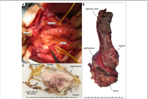
increased to 315.2 IU/ml. Magnetic resonance imaging (MRI) demonstrated that a 93 × 65 × 62 mm-solid tumor with cystic parts was located immediately dorsal to the rectum (Fig.1). CT and MRI showed no evidence of dis-semination, lymph node metastasis, nor distant metasta-sis. Colonoscopy showed strong extrinsic compression at the rectum with intact mucosa; however, biopsy of the rectum and the tumor site was not performed during colonoscopy. Based on the MRI finding that a perirectal cystic tumor was present without peritoneal dissemin-ation, stage IA ovarian cancer was suspected, and she was referred to our hospital for treatment. At laparot-omy, the tumor was located dorsal to the rectum and existed entirely in the retroperitoneal space (Fig. 2a). There were no apparent lesions in the peritoneal cavity including bilateral adnexa, uterus, and peritoneum. Peri-toneal washing cytology was negative. After bilateral salpingo-oophorectomy and total abdominal hysterec-tomy, en bloc resection of the retroperitoneal tumor to-gether with lower anterior resection of the rectum was performed (Fig.2b). Whereas the tumor was adhered to the rectal wall, the tumor itself was relatively well-capsulated and easily separated from surrounding fat tissues. Based on pathological diagnosis of the retro-peritoneal tumor: high-grade serous carcinoma, she re-ceived 6 cycles of adjuvant chemotherapy with carboplatin, paclitaxel and bevacizumab according to the standard treatment strategy for ovarian cancer. After the combination therapy, bevacizumab was administered for 3 cycles of tri-weekly maintenance therapy but was dis-continued because of general fatigue. She has been alive without evidence of recurrence for 20 months since her initial surgery.
Pathological examination
Macroscopically, the retroperitoneal tumor measured 80 × 55 × 35 mm in size and was divided into solid and
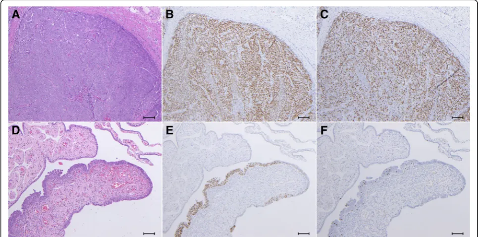
cystic parts. The rectum and the peritoneum separated the tumor from the peritoneal cavity (Fig. 2c). The cyst part covered with a thick wall included bloody serous fluid. Removed genital organs (e.g. uterus, fallopian tubes, and ovaries) presented no abnormal gross findings except for uterine fibroids. Microscopically, a cyst wall which was composed of fibrous tissue contained hemosiderin-laden macrophages. A solid part of the tumor, characterized by extensive atypical nuclei and lace-like pattern by coalescence of papillae, revealed high-grade serous carcinoma (Fig. 3a). Although the tumor was invasive into rectal mascularis propria and adjacent fat tissues, the surgical margin, peritoneal inva-sion, and lymphovascular involvement were negative. In addition, neither cancer metastasis nor endosalpingiosis were identified in the lymph nodes in the adjacent fat tissue. There were no invasive lesions in genital organs but STIC lesion was detected at the right fallopian tube (Fig.3d).
The cells of the retroperitoneal tumor and STIC were immunohistochemically positive for p53 (Fig. 3b & e). Ki-67 was diffusely and partially positive in the tumor and STIC lesions, respectively (Fig.3c and f ).
Target-gene sequencing of microdissected cancer cells from the retroperitoneal tumor
The retroperitoneal tumor sample was immediately sep-arated from surgical specimen, embedded in Tissue-Tek O.C.T. compound (Sakura Finetek) in a Tissue-Tek Cryomold (Sakura Finetek), and frozen in liquid nitro-gen. Serial 8-μm-thick frozen sections were mounted on MMI Membrane Slides (Molecular Machines & In-dustries), fixed with 100% methanol, and stained with toluidine blue. We performed laser-microdissection (LMD) using the MMI CellCut system (Molecular Ma-chines & Industries) to isolate tumor cells [14], followed by DNA extraction.
Fig. 1Preoperative T2-weighted magnetic resonance imaging showed the pelvic mass composed of cystic and solid parts inaaxial andb
Somatic mutations were investigated by target gene se-quencing in the retroperitoneal tumor. We selected 120 genes that were frequently mutated in ovarian and endo-metrial cancers from the TumorPortal website [14, 15] and involved in the homologous recombination repair pathway. We performed target sequencing of the 120 genes with a pool and capture method described in our previous study [14, 16]. The DNA libraries from the retroperitoneal tumor and the peripheral blood samples were sequenced via Illumina HiSeq 2500 platform in a rapid run mode with a 2 × 100 bp paired-end module. Referring to sequencing data from peripheral blood as a control, we called somatic mutations using Strelka soft-ware [17].
Somatic mutations detected in retroperitoneal tumor were listed in Table 1. Among six non-silent somatic mutations, a mutation ofTP53 (c.536A > G, p.179H > R) showed the highest mutant allele frequency (MAF) of 0.94. A nonsense mutation of BRCA2 (c.6385G > T, p.2129E > X) also showed high MAF of 0.84. The MAFs of these two mutations were significantly higher than 0.5 (binomial test, P < 0.0001), suggesting the presence of
either loss-of-heterozygosity events or homozygous mu-tations at these sites. We detected no obviously patho-genic mutations in the patient’s germline data obtained by sequencing for her blood cells.
Deep sequencing of variant sites
To explore the genetic relevance between the retroperi-toneal tumor and STIC lesion, deep sequencing was per-formed focusing on the two variant sites (TP53: c.536A andBRCA2: c.6385G) which were the most dominant in the retroperitoneal tumor. To obtain the STIC lesion, LMD was also performed using formalin-fixed paraffin-embedded (FFPE) tissue sections of 10-μm thickness. For LMD experiment, one per ten serial slides (n= 1, n= 11, etc.) was immunostained for P53. DNA samples derived from the retroperitoneal tumor and STIC were amplified by PCR reactions using the follow-ing oligonucleotide primers. Forward and reverse primers for TP53 c.536A > G [p.179H > R] were
5’-CTGCTCACCATCGCTATCTG-3′ and 5’-CACA
TGACGGAGGTTGTGAG-3′, respectively. For BRCA2 c.6385G > T [p.2129E > X], forward and reverse primers
were 5′- CTGCTCACCATCGCTATCTG-3′ and
5’-CACATGACGGAGGTTGTGAG-3′, respectively.
The PCR products were subjected to a deep sequencing via Illumina MiSeq v2 platform with a 2 × 150 bp paired-end module. The tag counts of paired-end se-quences supporting the reference and mutant alleles were measured by using only high confidence base calls (base quality > 20) at the mutation sites.
As a result, the somatic mutations of TP53 and BRCA2 were shared between the retroperitoneal tumor and STIC lesion (Table 2). The MAFs of these two mu-tations were lower in STIC lesion. There is a possibility that STIC lesion was inadequately isolated from sur-rounding normal tubal epithelium, stroma and infiltrat-ing immune cells through LMD, because the area of STIC lesion was limited.
Discussion
In this report, we presented HGSC existing entirely in retroperitoneal space without intraperitoneal malignant
lesion except STIC in the fallopian tube, and demon-strated the genetic relationship between retroperitoneal HGSC and STIC lesion. In addition, extremely high MAF of the two mutations suggested that mutations might occur at the early stage of tumorigenesis, and it seemed reasonable to interpret that the origin of the retroperitoneal HGSC might be STIC.
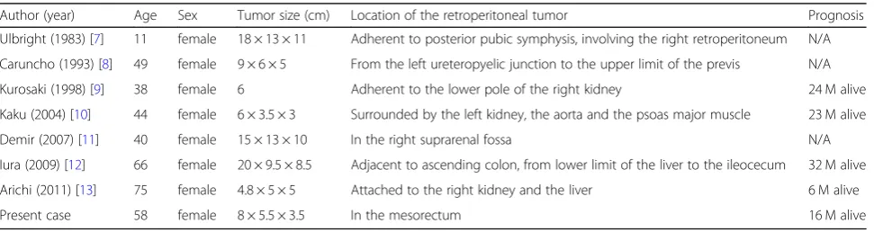
To date, seven case reports of retroperitoneal HGSC are retrieved by searching NCBI PubMed service with the keywords of “retroperitoneal serous carcinoma” and “serous carcinoma & retroperitoneum” (Table3) [7–13]. All patients were female, and onset age ranged from 11 to 75. In these reports, locations of the tumors varied from pelvis to upper abdomen in the retroperitoneal space. To explain how HGSC arose in retroperitoneal space, some theories represented by metaplasia of coel-omic mesothelium and supernumerary ovary were dis-cussed [7–12]. Unfortunately, no detailed analysis was performed in these reports to identify the pathogenesis
of retroperitoneal HGSC and the origin of
Fig. 3Histological images ofa,b,cthe retroperitoneal HGSC andd,e,fSTIC examined bya,dhematoxylin and eosin staining, immunohistochemical staining forb,ep53 andc,fKi-67. All panels are shown at power field of × 100. Scale bars show 100μm
Table 1Mutations detected by target-gene sequencing of the retroperitoneal high-grade serous carcinoma
Gene Amino acid (DNA) substitution No. wild-type reads No. mutant reads MAF
TP53 179H > R (536A > G) 3 50 0.94
BRCA2 2129E > X (6385G > T) 17 89 0.84
MLL4 2009A > V (6026C > T) 21 39 0.65
SIGLEC9 Y117H (349T > C) 33 33 0.50
MUC6 1758H > L (5273A > T) 179 15 0.08
retroperitoneal HGSC remains unclear. In the two re-ports, bilateral salpingooophorectomy (BSO) was per-formed but there was no description about coexisting STIC. As shown in the article focused on the pathogen-esis of epithelial ovarian cancer [18], a small STIC is fre-quently missed. In this case, STIC was diagnosed by a skilled gynecological pathologist (TM) based on mor-phological appearance and immunostaining for p53. For more accurate diagnosis of STIC, p16 was shown to be a useful biomarker in identifying STIC, separate from morphologically normal fallopian tube epithelium or HGSC [19, 20]. Based on the results in earlier studies, we attempted immunostaining for p16. However, all sec-tions having the STIC lesion were used for performing LMD, which eliminated the opportunity of immuno-staining for p16 or any further examination.
There are some theories about how STIC cells were transferred into the retroperitoneal space without peri-toneal spread. Firstly, STIC cells could have migrated by lymphovascular metastasis in spite of“intraepithelial car-cinoma”as Schneider et al. have described. They claimed that STIC should be regarded as a malignant lesion with metastatic potential by presenting that STIC cases ac-companied lymph node metastases without any other intra-abdominal/peritoneal spread [21]. In this case, there were no evidence of the lympovascular invasion pathologically in surgical specimens including fallopian tubes. The patient was incompletely staged for periton-eal disease (or ovarian cancer), lacking pelvic and para-aortic lymphadenectomy, omentectomy, and peri-toneal biopsy. Some articles have demonstrated micro-scopic metastasis at components for FIGO staging in
early stage ovarian cancer. In the study of occult metas-tasis, lymphatic and omental/peritoneal involvement of serous carcinoma was detected at the rate of 17% (12/ 69) and 4% (3/69), respectively [22]. Another study re-ported that microscopic metastasis was found at pelvic peritoneum or omentum in two (9%) of 23 patients with apparent early serous ovarian cancer [23]. However, in apparent early stage epithelial ovarian cancer, random peritoneal biopsy and omentectomy beyond careful in-spection of peritoneum was considered to have little sig-nificance [24]. However, we could not further assess the metastasis of STIC cells.
Secondly, tubal epithelial cells could have implanted directly into the mesentery, which resulted in endosal-pingiosis. Kurman et al. have described the association between proliferative tubal epithelium and low-grade serous tumor and proposed the possibility that endosal-pingiosis results from implantation of tubal epithelial cells [25]. Besides, it was reported that endosalpingiosis could be the origin of serous carcinoma in the mesen-trium. McCoubrey et al. pathologically demonstrated the transformation from benign ciliated serous-type epithe-lium to well-differentiated serous carcinoma in a cystic endosalpingiosis in the mesosigmoid [26]. Combining these studies, we reasoned thatTP53-mutated tubal epi-thelial cells developed into HGSC in the mesocolon via endosalpingiosis. In addition, our inference could be the-oretically supported by the concept of “precursor es-cape,”which was recently raised by Crum [27]. Based on this novel concept, TP53-mutated tubal epithelial cells already have the potential to be the precursor of HGSC at other than tubal epithelium since they were p53 Table 2Deep sequencing information of the tumor and STIC
Targeted gene Amino acid (DNA) substitution Sample No. wild-type reads No. mutant reads MAF
TP53 179H > R (536A > G) Tumor 1685 7383 0.814
STIC 152,901 110,704 0.420
BRCA2 2129E > X (6385G > T) Tumor 6162 39,716 0.866
STIC 96,243 86,018 0.472
MAFdenotes mutant allele frequency
Table 3Previous case reports of retroperitoneal HGSC
Author (year) Age Sex Tumor size (cm) Location of the retroperitoneal tumor Prognosis
Ulbright (1983) [7] 11 female 18 × 13 × 11 Adherent to posterior pubic symphysis, involving the right retroperitoneum N/A Caruncho (1993) [8] 49 female 9 × 6 × 5 From the left ureteropyelic junction to the upper limit of the previs N/A Kurosaki (1998) [9] 38 female 6 Adherent to the lower pole of the right kidney 24 M alive Kaku (2004) [10] 44 female 6 × 3.5 × 3 Surrounded by the left kidney, the aorta and the psoas major muscle 23 M alive Demir (2007) [11] 40 female 15 × 13 × 10 In the right suprarenal fossa N/A Iura (2009) [12] 66 female 20 × 9.5 × 8.5 Adjacent to ascending colon, from lower limit of the liver to the ileocecum 32 M alive Arichi (2011) [13] 75 female 4.8 × 5 × 5 Attached to the right kidney and the liver 6 M alive
Present case 58 female 8 × 5.5 × 3.5 In the mesorectum 16 M alive
signature or early serous proliferations. In this case, one possibility is that p53 signature might have migrated into the mesentery and resulted in HGSC, while p53 signa-ture in the fallopian tube grew into STIC.
Conclusions
We presented a case of retroperitoneal HGSC possessing genetic relationship with STIC, suggesting that STIC is the precursor of retroperitoneal HGSC. When the tumor lying in the retroperitoneal space was diagnosed with HGSC pathologically, complete resection of the tumor with BSO and elaborated pathological examination of fallopian tubes should be recommended.
Abbreviations
BSO:Bilateral salpingo-oophorectomy; CA125: Cancer antigen 125; CT: Computed tomography; HGSC: High-grade serous carcinoma; MRI: Magnetic resonance imaging; PCR: Polymerase Chain Reaction; STIC: Serous tubal intraepithelial carcinoma
Acknowledgements
We are grateful to Junko Kajiwara, Junko Kitayama, Yumiko Sato, and Anna Ishida for their technical assistance.
Funding
This work was supported in part by Japan Society for the Promotion of Science (JSPS) JP16H06267 (Grant-in-Aid for Young Scientists A for K. Yoshihara).
Availability of data and materials
The datasets used and/or analyzed during the current study are available from the corresponding author on reasonable request.
Authors’contributions
KS and HN performed and analyzed experiments. KS and KY interpreted all data, prepared the figures, and drafted the manuscript. All authors read and approved the final manuscript.
Ethics approval and consent to participate
Institutional ethics review board at Niigata University approved this study. The patient gave written informed consent.
Consent for publication
Written informed consent was obtained from the patient.
Competing interests
The authors declare that they have no competing interests.
Publisher’s Note
Springer Nature remains neutral with regard to jurisdictional claims in published maps and institutional affiliations.
Author details
1Department of Obstetrics and Gynecology, Niigata University Graduate
School of Medical and Dental Sciences, 1-757 Asahimachi-dori, Niigata 951-8510, Japan.2Department of Genetics, School of Life Sciences, Graduate University for Advanced Studies (SOKENDAI), Hayama, Japan.3Division of
Human Genetics, National Institute of Genetics, Mishima, Japan.4Department
of Obstetrics and Gynecology, Niigata Prefectural Shibata Hospital, Shibata, Japan.5Division of Digestive and General Surgery, Niigata University Graduate School of Medical and Dental Sciences, Niigata, Japan.
6Department of Molecular and Diagnostic Pathology, Niigata University
Graduate School of Medical and Dental Sciences, Niigata, Japan.
Received: 26 November 2018 Accepted: 1 February 2019
References
1. Lee Y, Miron A, Drapkin R, Nucci MR, Medeiros F, Saleemuddin A, Garber J, Birch C, Mou H, Gordon RW, et al. A candidate precursor to serous carcinoma that originates in the distal fallopian tube. J Pathol. 2007;211(1): 26–35.
2. Kindelberger DW, Lee Y, Miron A, Hirsch MS, Feltmate C, Medeiros F, Callahan MJ, Garner EO, Gordon RW, Birch C, et al. Intraepithelial carcinoma of the fimbria and pelvic serous carcinoma: evidence for a causal relationship. Am J Surg Pathol. 2007;31(2):161–9.
3. Kuhn E, Kurman RJ, Vang R, Sehdev AS, Han G, Soslow R, Wang TL, Shih Ie M. TP53 mutations in serous tubal intraepithelial carcinoma and concurrent pelvic high-grade serous carcinoma--evidence supporting the clonal relationship of the two lesions. J Pathol. 2012;226(3):421–6.
4. McDaniel AS, Stall JN, Hovelson DH, Cani AK, Liu CJ, Tomlins SA, Cho KR. Next-generation sequencing of tubal intraepithelial carcinomas. JAMA Oncol. 2015;1(8):1128–32.
5. Bashashati A, Ha G, Tone A, Ding J, Prentice LM, Roth A, Rosner J, Shumansky K, Kalloger S, Senz J, et al. Distinct evolutionary trajectories of primary high-grade serous ovarian cancers revealed through spatial mutational profiling. J Pathol. 2013;231(1):21–34.
6. Labidi-Galy SI, Papp E, Hallberg D, Niknafs N, Adleff V, Noe M, Bhattacharya R, Novak M, Jones S, Phallen J, et al. High grade serous ovarian carcinomas originate in the fallopian tube. Nat Commun. 2017;8(1):1093.
7. Ulbright TM, Morley DJ, Roth LM, Berkow RL. Papillary serous carcinoma of the retroperitoneum. Am J Clin Pathol. 1983;79(5):633–7.
8. Caruncho M, Pombo F, Arnal-Monreal F. Primary retroperitoneal serous cystadenocarcinoma of‘ovarian-type’: US and CT findings. Eur J Radiol. 1993;17(2):115–6.
9. Kurosaki Y, Kuramoto K. Case report: serous cystadenocarcinoma of the retroperitoneum: CT and sonographic appearance. Clin Radiol. 1998;53(12): 916–8.
10. Kaku M, Ohara N, Seima Y, Imanishi K, Tomura N, Kobayashi A, Yamasaki M, Hirata Y, Murao S. A primary retroperitoneal serous cystadenocarcinoma with clinically aggressive behavior. Arch Gynecol Obstet. 2004;270(4):302–6. 11. Demir MK, Unlu E, Genchellac H, Temizoz O, Ozdemir H: Primary serous
papillary carcinoma of the retroperitoneum: magnetic resonance imaging findings with pathologic correlation. Australas Radiol 2007, 51 Spec No.: B71–B73.
12. Iura A, Sasajima Y, Katsumata N, Kasamatsu T. Serous adenocarcinoma of the retroperitoneum, as a type of multifocal mullerian carcinoma. Int J Clin Oncol. 2009;14(3):254–7.
13. Arichi N, Yasumoto H, Mitsui Y, Hiraoka T, Honda S, Shiina H, Igawa M. A case of primary retroperitoneal serous adenocarcinoma. Int J Urol. 2011; 18(12):844–6.
14. Suda K, Nakaoka H, Yoshihara K, Ishiguro T, Tamura R, Mori Y, Yamawaki K, Adachi S, Takahashi T, Kase H, et al. Clonal expansion and diversification of Cancer-associated mutations in endometriosis and Normal endometrium. Cell Rep. 2018;24(7):1777–89.
15. Lawrence MS, Stojanov P, Mermel CH, Robinson JT, Garraway LA, Golub TR, Meyerson M, Gabriel SB, Lander ES, Getz G. Discovery and saturation analysis of cancer genes across 21 tumour types. Nature. 2014;505(7484): 495–501.
16. Ahmadloo S, Nakaoka H, Hayano T, Hosomichi K, You H, Utsuno E, Sangai T, Nishimura M, Matsushita K, Hata A, et al. Rapid and cost-effective high-throughput sequencing for identification of germline mutations of BRCA1 and BRCA2. J Hum Genet. 2017;62(5):561–7.
17. Saunders CT, Wong WS, Swamy S, Becq J, Murray LJ, Cheetham RK. Strelka: accurate somatic small-variant calling from sequenced tumor-normal sample pairs. Bioinformatics (Oxford England). 2012;28(14):1811–7. 18. Kurman RJ, Shih Ie M. The origin and pathogenesis of epithelial ovarian
cancer: a proposed unifying theory. Am J Surg Pathol. 2010;34(3):433–43. 19. Novak M, Lester J, Karst AM, Parkash V, Hirsch MS, Crum CP, Karlan BY,
Drapkin R. Stathmin 1 and p16(INK4A) are sensitive adjunct biomarkers for serous tubal intraepithelial carcinoma. Gynecol Oncol. 2015;139(1):104–11. 20. Sehdev AS, Kurman RJ, Kuhn E, Shih Ie M. Serous tubal intraepithelial
21. Schneider S, Heikaus S, Harter P, Heitz F, Grimm C, Ataseven B, Prader S, Kurzeder C, Ebel T, Traut A, et al. Serous tubal intraepithelial carcinoma associated with Extraovarian metastases. International journal of gynecological cancer : official journal of the International Gynecological Cancer Society. 2017;27(3):444–51.
22. Ayhan A, Gultekin M, Celik NY, Dursun P, Taskiran C, Aksan G, Yuce K: Occult metastasis in early ovarian cancers: risk factors and associated prognosis. Am J Obstet Gynecol 2007, 196(1):81.e81–86.
23. Shroff R, Brooks RA, Zighelboim I, Powell MA, Thaker PH, Mutch DG, Massad LS. The utility of peritoneal biopsy and omentectomy in the upstaging of apparent early ovarian cancer. International journal of gynecological cancer : official journal of the International Gynecological Cancer Society. 2011; 21(7):1208–12.
24. Powless CA, Bakkum-Gamez JN, Aletti GD, Cliby WA. Random peritoneal biopsies have limited value in staging of apparent early stage epithelial ovarian cancer after thorough exploration. Gynecol Oncol. 2009;115(1):86–9. 25. Kurman RJ, Vang R, Junge J, Hannibal CG, Kjaer SK, Shih Ie M. Papillary tubal
hyperplasia: the putative precursor of ovarian atypical proliferative (borderline) serous tumors, noninvasive implants, and endosalpingiosis. Am J Surg Pathol. 2011;35(11):1605–14.
26. McCoubrey A, Houghton O, McCallion K, McCluggage WG. Serous adenocarcinoma of the sigmoid mesentery arising in cystic endosalpingiosis. J Clin Pathol. 2005;58(11):1221–3.