R E S E A R C H A R T I C L E
Open Access
Aspergillus fumigatus during COPD
exacerbation: a pair-matched retrospective
study
Xunliang Tong
1†, Anqi Cheng
2†, Hongtao Xu
3, Jin Jin
4, Yimeng Yang
4, Sainan Zhu
5and Yanming Li
4*Abstract
Background:Recently awareness of the importance of Aspergillus colonization in the airway of patients with chronic obstructive pulmonary disease (COPD) was rising. The aim of this study was to investigate the clinical features and short-term outcomes of COPD patients with Aspergillus colonization during acute exacerbation.
Methods:A pair-matched retrospective study on patients presenting with COPD exacerbation was conducted from January 2014 to March 2016 in Beijing Hospital, China.
Results:Twenty-three patients with Aspergillus colonization and 69 patients as controls, diagnosed of COPD exacerbation, were included in this study at a pair-matched ratio of 1:3. In stable stage, the percentage of patients with high-dose corticosteroids inhalation in the Aspergillus colonization group is higher than that of in control group (65.5% vs 33.3%,p= 0.048). Multivariate analysis showed that corticosteroids use was the risk factor for isolation of Aspergillus. In acute exacerbation stage, patients in Aspergillus colonization group received higher dose of inhaled corticosteroids and more types of antibiotics than control group. The short-time outcome hinted that the remission time and the duration of hospitalization were longer in the Aspergillus colonization group than in the control group (remission time: 11 ± 4 days vs 7 ± 4 days,p= 0.001; duration: 15 ± 5 days vs 12 ± 4 days,p= 0.011).
Conclusions:Aspergillus colonization in the lower respiratory tract of COPD patients showed typical clinical manifestations, affected their short time outcome and provided a dilemma of clinical treatment strategy.
Keywords:COPD, Aspergillus fumigatus, Fungal colonization
Background
The prevalence of chronic obstructive pulmonary disease (COPD) is rapidly growing and is associated with signifi-cant morbidity and mortality, which increases economic
health burden [1–3]. COPD is characterized by
irrevers-ible airflow obstruction with underlying emphysema and small airway obliteration, which commonly co-exist. Pathogenic microorganisms, such as bacteria, are com-monly colonized in the airways of COPD patients, possibly contributing to increased airway inflammation, and have
been implicated in COPD exacerbations [4–6]. However,
fungal colonization and its potential role in acute exacer-bations of COPD (AECOPD) are poorly understood.
Aspergillus spp. is a ubiquitous fungus in the environ-ment with high sporulation capacity [7,8]. After Aspergillus sporulates, conidia with a diameter of 2–3μm are released into the air, enter the airway, and reach alveoli [9]. There-fore, the lung is the main organ affected by Aspergillus. Iso-lation of Aspergillus spp. from lower respiratory tract (LRT) samples (e.g. sputum, bronchial aspirate, or broncho-alveolar lavage) provides important etiologic evidence for its identification. Aspergillus spp. causes various diseases in lungs, such as Aspergillus colonization, Aspergillus
infec-tion and allergic bronchopulmonary aspergillus [10]. In
COPD patients, impairment of the defense mechanisms of airways facilitates the binding of conidia to epithelial cells, which may cause Aspergillus colonization in the airway [11]. Positive isolation of Aspergillus spp. in LRT samples from COPD patients is common, and a previous study re-ported that the positive identification rate was nearly 29% * Correspondence:lymyl@263.net
†Equal contributors
4Department of Respiratory and Critical Care Medicine, Beijing Hospital,
Beijing 100730, People’s Republic of China
Full list of author information is available at the end of the article
[12]. However, clinical manifestations of COPD patients with Aspergillus spp. colonization from LRT have rarely been summarized and are difficult to distinguish, often leading to debate in clinical practice. Consequently, it is dif-ficult for clinicians to make treatment strategies for Asper-gillus colonization. The aims of this study were to investigate the clinical features of COPD patients with As-pergillus colonization in LRT, to analyze the risk factors that could predict the possibility of Aspergillus spp. isola-tion and to summarize the clinical treatment choices and outcomes of COPD patients with Aspergillus colonization.
Methods Study design
This pair-matched study was conducted from January 2014 to March 2016 in the Department of Respiratory and Critical Care Medicine in Beijing Hospital, China. Patient data were collected from the electronic medical records system of our hospital and by additional chart review. The
data used was part of our project“Study on Aspergillus of
COPD patients”, which was approved by the ethics
com-mittee of Beijing Hospital (Approval notice number 2013BJYYEC-024-01). The informed consent was ob-tained from all participants in written form.
Mentioned conditions
Conditions
A Age between 18 and 90 years old.
B The diagnosis of COPD was based on the Global Initiative for GOLD guidelines.
COPD exacerbations are defined as an acute worsening of respiratory symptoms that result in additional therapy.
C Positive isolation of Aspergillus spp. in an LRT sample by microbiologic examination. LRT samples included sputum, bronchial aspirate, and bronchoalveolar lavage fluid (BALF).
D Patients without any chest CT findings of chronic pulmonary aspergillosis (CPA), invasive pulmonary aspergillosis (IPA), allergic bronchopulomnary aspergillosis (ABPA), pneumonia.
Inclusion criteria
Definition
Aspergillus colonization
fulfilled above condition C and D above.
Aspergillus colonization group
fulfilled all conditions above, including A, B, C and D.
Control group COPD patients who were admitted a day before or after the Aspergillus colonization group patients were recruited to the control group. At the same time, patients fulfilled above conditions A, B and D.
Exclusion criteria
(1)Immunocompromised patients, including those with allogeneic or autologous hematopoietic stem cell transplantation, neutropenia, hematologic malignant disease and solid tumor, hematologic stem cell or solid-organ transplantation, and AIDS, as were patients receiving high-dose immunosuppressive agents (e.g. for connective tissue disease
and vasculitis).
(2)Patients with chest CT findings of CPA, IPA, ABPA, pneumonia.
(3)Patients with other underlying lung diseases.
Microbiological examination
When patients admitted into hospital, their LRT samples were delivered for microbiological examination the next
day. Microbiological examination included direct
microscopy and bacterial and fungal pathogen
cultvation. All samples were cultured on conventional media, including blood agar, chocolate agar, MacConkey
agar and Sabouraud’s dextrose agar. Bacterial infection
was defined as a colony count≥105cfu/mL. Aspergillus
and other fungal isolates were identified using
microculture and standard morphological procedures.
Data collection
Following information was collected from the electronic
medical records system of our hospital: patient
characteristics (sex, age, etc.), lung function, administration of corticosteroids and nutritional status before admission, clinical symptoms and signs, chest imaging and CT scan data, laboratory test results (IgE, eosinophil counts, etc.), microbiologic findings of LRT samples, treatment during hospitalization, remission time (defined as the duration of stabilization of clinical symptoms and disappearance of signs, like the typical symptoms of cough, sputum, wheeze, and the typical signs of wheezing rale), length of hospitalization or ICU stay, and mortality.
Statistical analysis
Normally distributed continuous variables were
expressed as the mean ± standard deviation and were
compared with a t-test. Non-normally distributed
con-tinuous variables were expressed as medians and
quar-tiles and were compared with the Mann-WhitneyU-test.
Categorical variables were compared with chi-square test
or Fisher’s exact test. Logistic regression was used to
identify independent risk factors for Aspergillus
colonization. The first logistic regression analysis uni-variately considered the explanatory variables in stable
stage. Only variables with p value < 0.10 in univariate
the entry of p= 0.10 and removal of p= 0.05. Kaplan-Meier method was used to estimate the time from
ad-mission to read-mission of symptoms. Pvalues < 0.05 were
considered statistically significant. The statistical analysis was performed using SPSS 19.0.0 (IBM Corporation, Armonk, NY, USA).
Results
Study performance
A total of 504 patients diagnosed with AECOPD were admitted to hospital from January 2014 to March 2016. Aspergillus spp. was identified in the LRT samples of 42 patients (all from qualified sputum specimen), resulting in a detection rate of 8.33%. According to the exclusion criteria, 19 patients with Aspergillus spp. in their sputum samples were excluded from this study, including 9 patients with IPA infection, 5 patients with other pulmonary diseases (asthma: 4 cases, bronchiectasis: 1 case), 2 patients with cancer (lung cancer: 1 case, prostate cancer: 1 case), 1 patient being treated with immunosuppressive agents for connective tissue disease, 1 patient with agranulocytosis and 1 patient who was unable to participate lung function test. Finally, 23 patients with Aspergillus colonization were included in the study group. After performing matching at a ratio of 1:3 (one study group patient to three control group patients), a total of 92 patients were enrolled in this study shown in Fig.1.
Demographic characteristics and treatment during stable stage
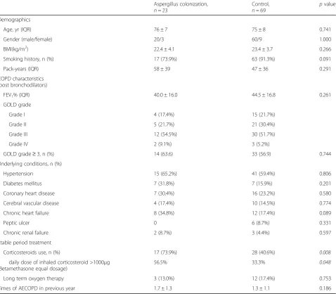
Patients in Aspergillus colonization group consisted of 20 men and 3 women with a median age of 76 ± 7 years. Most of these patients were current smokers with a smoking duration of more than 40 pack-years. All patients
were classified by GOLD stage severity. The forced
expira-tory volume 1 (FEV1) of patients in Aspergillus
colonization group was worse according to GOLD severity classification than that of patients in control group
(FEV1% predicted: 40.0% ± 16.0% vs 44.5% ± 16.8%).
Pa-tients in Aspergillus colonization group also had more underlying diseases, including hypertension, diabetes mel-litus and coronary heart disease, than patients in control group. The percentage of inhaled corticosteroid (ICS) use during the stable stage was higher in Aspergillus colonization group than that of control group (73.9% vs
40.6%, p= 0.008). Besides, more patients received a daily
dose of beclomethasone greater than 1000 μg (30.4% vs
11.6%, p= 0.048), which was significantly different from
patients in control group. The demographic characteristics of patients in Aspergillus colonization and control groups were listed in Table1.
Univariable baseline analysis showed that smoking history, chronic heart failure and corticosteroids use were risk factors for Aspergillus colonization in COPD
patients’ LRT. After multivariable adjustment, only
corticosteroids use was independently associated with
Aspergillus colonization in COPD patients’ LRT (OR
4.685, 95% CI 1.529–14.355), shown in Table2.
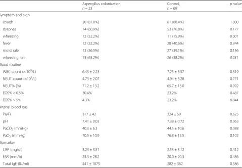
Clinical manifestations in the exacerbation stage
The clinical characteristics and selected laboratory
abnormalities of included patients were shown in Table3.
Most patients exhibited fever, cough, dyspnea and wheezing. Wheezing and wheezing rales were the most specific symptom and sign, which were significantly common in Aspergillus colonization group (wheezing:
52.2% vs 15.9%,p= 0.001; wheezing rales: 65.2% vs 38.2%,
p= 0.031). Laboratory results showed that the number
and percentage of eosinophils (EOS) were significantly decreased in patients in Aspergillus colonization group compared to those in control group (EOS% > 5%: 4.3% vs
23.2%, p= 0.044). Arterial blood gas was measured in all
patients, but no significant differences were observed between two groups. Some inflammatory markers, such as C-reactive protein (CRP) and erythrocyte sedimentation rate (ESR), and total IgE levels, were not significantly different between two groups.
Detection of pathogenic bacteria and other fungi
To investigate whether Aspergillus colonization was associated with combined identification of other specific microbial pathogens, we compared the cultivation results of LRT samples from the Aspergillus colonization group and control group. Particular attention was paid to
pathogenic bacteria identification, including Acinetobacter
baumannii, methicillin-resistant Staphylococcus aureus
(MRSA), Pseudomonas aeruginosa, Klebsiella pneumonia, Stenotrophomonas maltophilia and Enterococcus faecium. The percentage of pathogens identification in two groups
showed no statistical difference (Additional file 1: Table
S1). Besides, some of other fungi were also detected in our microbiologic cultivation, especially Saccharomycetes.
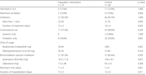
Patient treatment during hospitalization and short-term outcomes
The Aspergillus colonization group included a higher percentage of patients who received more than one class
of antibiotics (52.2% vs 20.3%, p= 0.006) and had a
longer duration of antibiotic usage (13 ± 5 vs 10 ± 4 days,
p= 0.003) than that of the control group. In addition to
Table 1Basic information of COPD patients in stable stage
Aspergillus colonization,
n= 23
Control,
n= 69 p
value
Demographics
Age, yr (IQR) 76 ± 7 75 ± 8 0.741
Gender (male/female) 20/3 60/9 1.000
BMI(kg/m2) 22.4 ± 4.1 23.4 ± 3.7 0.266
Smoking history, n (%) 17 (73.9%) 63 (91.3%) 0.091
Pack-years (IQR) 58 ± 39 47 ± 36 0.291
COPD characteristics (post bronchodilators)
FEV1% (IQR) 40.0 ± 16.0 44.5 ± 16.8 0.261
GOLD grade
Grade I 4 (17.4%) 15 (21.7%)
Grade II 5 (21.7%) 21 (30.4%)
Grade III 12 (54.5%) 30 (51.7%)
Grade IV 2 (9.1%) 3 (5.2%)
GOLD grade≥3, n (%) 14 (63.6) 33 (56.9) 0.744
Underlying conditions, n (%)
Hypertension 15 (65.2%) 41 (59.4%) 0.806
Diabetes mellitus 7 (31.8%) 7 (15.9%) 0.201
Coronary heart disease 7 (30.4%) 16 (23.2%) 0.580
Cerebral vascular disease 4 (17.4%) 10 (14.5%) 0.774
Chronic heart failure 8 (34.8%) 12 (17.4%) 0.089
Peptic ulcer 0 6 (8.7%) 0.331
Chronic renal failure 2 (8.7%) 3 (4.4%) 0.597
Stable period treatment
Corticosteroids use, n (%) 17 (73.9%) 28 (40.6%) 0.008
daily dose of inhaled corticosteroid >1000μg (Betamethasone equal dosage)
56.5% 33.3% 0.048
Long term oxygen therapy 3 (13.0%) 12 (17.4%) 0.753
Times of AECOPD in previous year 1.7 ± 1.3 1.3 ± 1.1 0.186
Table 2Univariate and multivariate logistic analysis for Aspergillus colonization
Characteristics Univariate analysis Multivariate analysis
OR (95% CI) pvalue OR (95% CI) pvalue
BMI 0.929 (0.817–1.057) 0.264
Smoking history 0.270 (0.077–0.944) 0.040 0.275 (0.073–1.036) 0.056
Pack-years 1.008 (0.994–1.022) 0.291
FEV1% pre (post bronchodilators) 0.982 (0.951–1.014) 0.259
GOLD grade 1.968 (0.638–6.075) 0.239
Hypertension 1.280 (0.479–3.424) 0.622
Diabetes mellitus 1.719 (0.593–4.983) 0.319
Coronary heart disease 1.449 (0.507–4.139) 0.488
Cerebral vascular disease 1.242 (0.349–4.421) 0.738
Chronic heart failure 2.533 (0.878–7.313) 0.086 3.147 (0.978–10.130) 0.055
Chronic renal failure 2.095 (0.328–13.397) 0.435
Corticosteroids use 4.149 (1.456–11.825) 0.008 4.685 (1.529–14.355) 0.007
Long term oxygen therapy 0.713 (0.182–2.787) 0.626
Times of AECOPD in previous year 1.299 (0.880–1.918) 0.188
Italicizedp-values are statistically significant, ie.p< 0.05
Table 3Characteristics and examination results of COPD patients in acute exacerbation stage
Aspergillus colonization,
n= 23
Control,
n= 69 p
value
Symptom and sign
cough 20 (87.0%) 61 (88.4%) 1.000
dyspnea 14 (60.9%) 53 (76.8%) 0.177
wheezing 12 (52.2%) 11 (15.9%) 0.001
fever 12 (52.2%) 28 (40.6%) 0.344
moist rale 13 (56.5%) 27 (39.1%) 0.156
wheezing rale 15 (65.2%) 26 (38.2%) 0.031
Blood routine
WBC count (× 109/L) 6.45 ± 2.23 7.25 ± 3.57 0.319
NEUT count (×109/L) 4.73 ± 2.07 4.94 ± 3.28 0.771
NEUT% (%) 71.2 ± 13.2 65.7 ± 13.0 0.092
EOS% < 0.5% 30.4% 23.2% 0.487
EOS% > 5% 4.3% 23.2% 0.044
Arterial blood gas
Pa/Fi 317 ± 42 324 ± 59 0.625
pH 7.41 ± 0.03 7.38 ± 0.72 0.063
PaCO2(mmHg) 40.3 ± 6.3 44.5 ± 10.6 0.088
PaO2(mmHg) 70.5 ± 10.9 76.8 ± 15.5 0.102
Biomarker
CRP (mg/dl) 3.23 ± 3.51 2.53 ± 3.12 0.412
ESR (mm/h) 25.3 ± 28.2 20.0 ± 20.3 0.436
Total IgE (IU/ml) 441 ± 1075 282 ± 362 0.386
antibiotic treatment, systemic corticosteroids and/or ICS were used to control wheezing symptom. The percentage of patients in Aspergillus colonization group who use high dose of inhaled corticosteroids (accumulated budesonide dose: no less than 40 mg) were higher than that of control
group (30.4% vs 5.8%,p= 0.002). Remission time curve was
estimated by Kaplan-Meier analysis. Median remission time was 10 days in the Aspergillus colonization group and 6 days in the control group (log-rank testp= 0.004) (Fig.2). The duration of hospitalization were longer in the Aspergil-lus colonization group than it is in the control group (15 ± 5 days vs 12 ± 4 days,p= 0.011), shown in Table4.
Discussion
AECOPD is often associated with infectious agents, including bacteria, virus and fungi. A previous study was conducted in critically ill patients with isolation of
Aspergillus spp. from the respiratory tract, with
mortality rates of 50% in the colonization group and 80% in the invasive infection group after 9 months of
follow-up [13]. Therefore, clinicians usually focus on
in-fection when a positive Aspergillus spp. culture is ob-tained from the LRT. However, the significance of a more frequent clinical phenomenon, Aspergillus spp. colonization, has yet to be clarified. In this research, a pair-matched observational study was conducted to in-vestigate the differences in the clinical manifestations and short-term outcomes between COPD patients with and without Aspergillus colonization in LRT.
Cigarette smoking, one of the major risk factors for the development of COPD, induces structural and functional changes in airway epithelium in vitro and in
vivo [14–16]. In our study, the number of patients with
a history of smoking was higher in the Aspergillus colonization group than in the control group, which indicated that smoking is a potential risk for Aspergillus colonization. Cigarette smoking and repeated airway inflammation could alter the structure and function of lung and injure a profound effect on the host defense
against invading pathogens and particulates, thus
impairing the airway epithelium [17,18] and mad COPD
patients more susceptible to Aspergillus colonization.
Meanwhile, most patients in the Aspergillus
colonization group received higher doses of ICS during stable stage treatment, in contrast to the control group. Our findings were consistent with a previous study that suggested that high-dose corticosteroids use was a risk for Aspergillus colonization or positive Aspergillus culture [19,20].
In our study, Aspergillus-colonized patients presented with wheezing and wheezing rales in the acute exacerba-tion period. More than half of the patients received systemic corticosteroids and/or ICS. The percentage of corticosteroid usage in the two groups was similar, but in the Aspergillus colonization group, patients received higher doses of ICS. This finding suggested that Aspergillus colonization contributed to an increased severity of exacerbations in COPD patients. These phe-nomena suggested that high-dose ICS treatment was re-lated to Aspergillus colonization and induced similar clinical manifestations to allergic reactions due to Asper-gillus colonization. A previous study on the mechanism involved in Aspergillus-related allergic reactions was based on the Aspergillus hyphae and involved antigen-triggered mast cell degranulation and release of
hista-mine and inflammatory factors [21–24]. These data
showed that Aspergillus colonization may aggravate air-way hyper-responsiveness and worsen airair-way inflamma-tion and bronchoconstricinflamma-tion. But no cohort study of patients with repeated cultures of Aspergillus have been done in COPD patients, it is unclear whether fungal colonization contributes to lower lung function or is a marker of more severe lung disease and aggressive ther-apy. In our study, patients with Aspergillus colonization had a longer time to be stable and a longer duration of
hospitalization, which indicated that Aspergillus
colonization was related with clinical manifestations and short-term outcomes of COPD patients.
After demonstrating the significance of Aspergillus colonization in the airways of COPD patients, we highlighted an essential clinical treatment dilemma: whether to eliminate colonization with anti-fungal ther-apy or to stabilize wheezing with continuous ICS. Be-cause Aspergillus colonization is clinically significant,
the treatment strategy should aim to eliminate
has not been determined. Aspergillus colonization in-duces sustainable inflammation in the airway, which leads to worsening lung function. Meanwhile, poorer lung function is significantly associated with Aspergillus colonization. However, in clinical practice, ICS are commonly used to control wheezing and airway inflam-mation, which could also enhance the risk of Aspergillus colonization in the airway. It is concerning that once As-pergillus colonization in the airways of COPD patients is identified, a vicious cycle is established. Unfortunately, no accurate timing, biomarker, or scoring system exists that could determine the optimal antifungal therapy.
There were some limitations to this study: (1) we could not determine the timing of Aspergillus colonization: the acute exacerbation or stable stage; (2) we only conducted this retrospective study without a long-term observation from the beginning of Aspergillus colonization to its caus-ing symptomatic clinical manifestation. Thus, whether Aspergillus colonization could affect the clinical process was still unknown, including lung function decline, the frequency of acute exacerbation, and daily symptoms; and (3) we performed this research in a single center and recruited a small sample of patients.
Conclusions
Our findings show that Aspergillus colonization in the
airway could cause significant change in clinical
manifestations and treatment outcome of COPD patients. This study provides new insight for clinicians when managing patients with fungal colonization and also
indicates the directions for future comprehensive studies, including the development of a reliable and rapid method to accurately identify infection and colonization and to develop a full-scale investigation and treatment strategy of COPD patients with Aspergillus colonization.
Additional file
Additional file 1:Table S1.Detection of pathogenic bacteria and other fungi. (DOCX 14 kb)
Abbreviations
ABPA:Allergic bronchopulomnary aspergillosis; AECOPD: Acute exacerbation of chronic obstructive pulmonary disease; BALF: Bronchoalveolar lavage fluid; COPD: Chronic obstructive pulmonary disease; CPA: Chronic pulmonary aspergillosis; CRP: C-reactive protein; CT: Computed tomography;
EOS: Eosinophils; ESR: Erythrocyte sedimentation rate; FEV1: Forced expiratory
volume 1; GOLD: Global initiative for chronic obstructive lung disease; ICS: Inhaled corticosteroids; ICU: Intensive care unit; IPA: Invasive pulmonary aspergillosis; LRT: Lower respiratory tract
Acknowledgments
The authors thank Academician Chen Wang for his guidance and assistance with this work. We acknowledge the China National Center for Protein Sciences Beijing for providing facility support.
Funding
This project was supported by project grant 2012AA02A511 from the National High Technology Research and Development Program, China; grant 2012BAI05B02 from the National Key Technology Research and Development Program, China; grant 81400037 from the National Natural Science Foundation of China; and grant 201302017 from the Public Health Special Research of the Ministry of Health of the People’s Republic of China.
Table 4Clinical treatment and short-term outcomes in AECOPD patients
Aspergillus colonization,
n= 23
Control,
n= 69 p
value
Admitted in ICU 4 (17.4%) 11 (15.9%) 1.000
Machined ventilation 3 (13.0%) 9 (13.0%) 0.906
Antibiotics 21 (95.5%) 66 (95.7%) 1.000
More than 1 class 52.2% 21.7% 0.006
Duration of treatment (days) 13 ± 5 10 ± 4 0.003
Corticosteroid use 17 (77.3%) 42 (60.9%) 0.204
Systemic only 0 2 (2.90%) 1.000
Inhalation only 8 (34.8%) 20 (29.0%) 0.601
Dose of usage
Budesonide (inhaled,≥40 mg) 30.4% 5.8% 0.002
Methylprednisolone (iv,≥120 mg) 39.1% 31.9% 0.524
Bronchodilators (aerosol inhalation) 21 (91.3%) 57 (82.6%) 0.505
Ipratropium Bromide (mg) 16.3 ± 11.6 14.8 ± 8.2 0.631
Salbutamol (mg) 115 ± 98 83 ± 53 0.308
Remission time (days) 11 ± 4 7 ± 4 0.001
Duration of hospitalization (days) 15 ± 5 12 ± 4 0.011
Availability of data and materials
The data that support the findings of this study are available from Beijing Hospital Clinical Database Centre, but restrictions apply to the availability of these data, which were used under license for the current study, and so are not publicly available. Data are however available from the authors upon reasonable request and with permission of Beijing Hospital Clinical Database Centre.
Authors’contributions
YL and XT contributed to the conception and design of the study, acquisition of the data, and interpretation of the results and drafted the manuscript. AC contributed to the acquisition of the mycological data and revision of the manuscript for important intellectual content. HX contributed to the acquisition of the bacteriological data and revision of the manuscript for important intellectual content. SZ performed the statistical analysis and revised the manuscript for important intellectual content. All authors read and approved the final manuscript.
Ethics approval and consent to participate
The data used in this study was part of our project“Study on Aspergillus of COPD patients”, which was approved by the ethics committee of Beijing Hospital (Approval notice number 2013BJYYEC-024-01). The informed consent to participate was obtained from all participants in written form.
Consent for publication
Not applicable.
Competing interests
The authors declare that they have no competing interests.
Publisher’s Note
Springer Nature remains neutral with regard to jurisdictional claims in published maps and institutional affiliations.
Author details
1
Department of Geriatrics, Beijing Hospital, National Center of Gerontology, Beijing 100730, People’s Republic of China.2Tobacco Medicine and Tobacco
Cessation Centre, Center of Respiratory Medicine, China-Japan Friendship Hospital; WHO Collaborating Centre for Tobacco Cessation and Respiratory Diseases Prevention; National Clinical Research Center for Respiratory Diseases, Beijing 100029, People’s Republic of China.3Department of
Laboratory Medicine, Beijing Hospital, Beijing 100730, People’s Republic of China.4Department of Respiratory and Critical Care Medicine, Beijing
Hospital, Beijing 100730, People’s Republic of China.5Statistics Department, First Hospital of Peking University, Beijing 100034, People’s Republic of China.
Received: 16 June 2017 Accepted: 9 March 2018
References
1. WHO. The top 10 causes of death.http://www.who.int/mediacentre/ factsheets/fs310/en/. Date last updated: 27 Oct 2016.
2. WHO. Burden of COPD.http://www.who.int/respiratory/copd/burden/en/. Date last updated: 15 Mar 2017.
3. Mathers CD, Loncar D. Projections of global mortality and burden of disease from 2002 to 2030. PLoS Med. 2006;3(11):e442.
4. Banerjee D, Khair OA, Honeybourne D. Impact of sputum bacteria on airway inflammation and health status in clinical stable COPD. Eur Respir J. 2004; 23(5):685–91.
5. Patel IS, Seemungal TAR, Wilks M, Lloyd-Owen SJ, Donaldson GC, Wedzicha JA. Relationship between bacterial colonisation and the frequency, character, and severity of COPD exacerbations. Thorax. 2002;57(9):759–64. 6. Wedzicha JA, Donaldson GC. Exacerbations of chronic obstructive
pulmonary disease. Respir Care. 2003;48(12):1204–13.
7. Streifel AJ, Lauer JL, Vesley D, Juni B, Rhame FS. Aspergillus fumigatus and other thermotolerant fungi generated by hospital building demolition. Appl Environ Microbiol. 1983;46(2):375–8.
8. Latgé JP. The pathobiology of aspergillus fumigatus. Trends Microbiol. 2001; 9(8):382–9.
9. Bulpa P, Dive A, Sibille Y. Invasive pulmonary aspergillosis in patients with chronic obstructive pulmonary disease. Eur Respir J. 2007;30(4):782–800.
10. Soubani AO, Chandrasekar PH. The clinical spectrum of pulmonary aspergillosis. Chest. 2002;121(6):1988–99.
11. Latgé JP. Aspergillus fumigatus and aspergillosis. Clin Microbiol Rev. 1999; 12(2):310–50.
12. Pashley CH, Fairs A, Morley JP, et al. Routine processing procedures for isolating filamentous fungi from respiratory sputum samples may underestimate fungal prevalence. Med Mycol. 2012;50(4):433–8. 13. Garnacho-Montero J, Amaya-Villar R, Ortiz-Leyba C, et al. Isolation of
Aspergillus spp. from the respiratory tract in critically ill patients: risk factors, clinical presentation and outcome. Crit Care. 2005;9(3):R191–9.
14. Kohansal R, Martinez-Camblor P, Agustí A, Buist AS, Mannino DM, Soriano JB. The natural history of chronic airflow obstruction revisited: an analysis of the Framingham offspring cohort. Am J Respir Crit Care Med. 2009;180(1):3–10. 15. Nyunoya T, Mebratu Y, Contreras A, Delgado M, Chand HS, Tesfaigzi Y.
Molecular processes that drive cigarette smoke-induced epithelial cell fate of the lung. Am J Respir Cell Mol Biol. 2014;50(3):471–82.
16. Amitani R, Murayama T, Nawada R, et al. Aspergillus culture filtrates and sputum sols from patients with pulmonary aspergillosis cause damage to human respiratory ciliated epithelium in vitro. Eur Respir J. 1995;8(10):1681–7. 17. Jones JG, Minty BD, Lawler P, Hulands G, Crawley JC, Veall N. Increased alveolar
epithelial permeability in cigarette smokers. Lancet. 1980;1(8159):66–8. 18. Hulbert WC, Walker DC, Jackson A, Hogg JC. Airway permeability to horseradish peroxidase in guinea pigs: the repair phase after injury by cigarette smoke. Am Rev Respir Dis. 1981;123(3):320–6.
19. Muquim A, Dial S, Menzies D. Invasive aspergillosis in patients with chronic obstructive pulmonary diseases. Can Respir J. 2005;12(4):199–204. 20. Bafadhel M, McKenna S, Agbetile J, et al. Aspergillus fumigatus during stable
state and exacerbations of COPD. Eur Respir J. 2014;43(1):64–71.
21. Urb M, Snarr BD, Wojewodka G, et al. Evolution of the immune response to chronic airway colonization with Aspergillus fumigatus hyphae. Infect Immun. 2015;83(9):3590–600.
22. Saluja R, Metz M, Maurer M. Role and relevance of mast cells in fungal infections. Front Immunol. 2012;3:146.
23. Urb M, Pouliot P, Gravelat FN, Olivier M, Sheppard DC. Aspergillus fumigatus induces immunoglobulin E-independent mast cell degranulation. J Infect Dis. 2009;200(3):464–72.
24. Chaudhary N, Marr KA. Impact of Aspergillus fumigatus in allergic airway diseases. Clin Transl Allergy. 2011;1(1):4.
• We accept pre-submission inquiries
• Our selector tool helps you to find the most relevant journal
• We provide round the clock customer support
• Convenient online submission
• Thorough peer review
• Inclusion in PubMed and all major indexing services
• Maximum visibility for your research
Submit your manuscript at www.biomedcentral.com/submit