Original Research Article.
A Comparative Study to Find Gender Wise Differences in Lumbo-Pelvic
Alignment in Patient with Chronic Low Backache
Pawan Kumar Mahato
1, Sunil Kumar Srivastava
2*, Amit Kumar Saxena
3, Prachi Saffar Aneja
41Ph.D Scholar, 3Professor & Head, 4Professor, Department of Anatomy,
SGT Medical College Hospital & Research Institute, Budhera, Gurugram, Haryana, India.
2*Professor & Head, Department of Anatomy, G S Medical College & Hospital, Pilkhuwa, UP, India. ABSTRACT
Introduction: In patients with low back pain, radiographic parameters that most highly correlate with patient-reported outcomes are focal i.e, rotatory subluxation, regional i.e, loss of lordosis and global i.e, sagittal malalignment. Recently, the literature has also confirmed the impact of spinopelvic alignment on patient reported outcomes. For the further measurement of lumbo-pelvic alignment following parameters are assessed i.e. lumbar lordosis, pelvic incidence, sacral slope and pelvic tilt. The present study aims evaluate gender wise differences in lumbo-pelvic alignment in patient with chronic low backache.
Material and Methods: The present study was performed on patients with clinical symptoms of chronic low back pain. Parameters studied are Lumbar Lordosis, Pelvic Incidence angle, Pelvic Tilt and Sacral Slope. Data obtained was arranged as mean or percentage as required.
Results: In present study, In Group I, the mean value of lumbar lordosis amongst males was 29.64 ± 9.69 and that amongst females was 30.71 ± 9.06 with the p value of 0.639. In Group II, the mean value of lumbar lordosis amongst males was 32.79 ± 7.86 and that amongst females was 35.04 ± 7.22 with the p value of 0.256. In Group I, the mean value of pelvic incidence amongst males and females was 52.69 ± 11.86 and 52.52 ± 11.17 respectively. In Group II, the mean value of pelvic incidence amongst males and females was 54.00 ± 11.13 and 55.72 ± 10.45 respectively. In Group I, the mean value of pelvic tilt amongst males and females was
13.74 ± 8.78 and 13.81 ± 7.95 respectively. In Group II, the mean value of pelvic tilt amongst males and females was 14.42 ±4.75 and 12.44 ± 4.69 respectively.
Conclusion: The present study found no significant difference in the lumbar lordosis, pelvic tilt and sacral slope between males and females with chronic low back pain. Parameters of the sagittal plane were extracted as being associated with low back pain too. But with respect to the literature and inconsistency of different multivariate analysis approaches, those parameters also might to some extent be affected by aging and degeneration.
Keywords: Low Back Pain; Lumbo-Pelvic Alignment; Lumbar
Lordosis.
*Correspondence to:
Dr. Sunil Kumar Srivastava,
Professor & Head, Department of Anatomy, G S Medical College & Hospital,
Pilkhuwa, Uttar Pradesh, India.
Article History:
Received: 05-12-2017, Revised: 03-01-2018, Accepted: 22-01-2018
Access this article online
Website:
www.ijmrp.com
Quick Response code
DOI:
10.21276/ijmrp.2018.4.1.138
INTRODUCTION
Low Back pain is usually described by the length of time for which symptoms persist: Acute low back pain lasts less than 6 weeks. Sub-acute low back pain lasts between 6 to 12 weeks and chronic low back pain persists for more than 12 weeks.1 For those, whose
conditions have transitioned from acute to chronic pain (pain persisting) for 3 months or longer, there are often few physical abnormalities.2 These patients adjust the sagittal alignment not
only by spinal inclination, but also by pelvic rotation or even hip and knee flexion to maintain a static horizontal gaze with the least expenditure of energy.3-5 Pain can arise from the intervertebral
disc in which case, greatest pain provocation will be associated with movements and functions in the sagittal plane. Lumbar pain
can also arise from afflictions within the zygapophyseal joint mechanism, which will produce the greatest pain provocation during three-dimensional movements due to maximal stress to either the synovium or joint cartilage. Finally, patients can experience pain associated with irritation to the dural sleeve dorsal root ganglion or chemically irritated lumbar nerve root. Pain can also arise from muscle.6
Radiographic parameters that most highly correlate with patient-reported outcomes are focal i.e, rotatory subluxation, regional i.e, loss of lordosis and global i.e, sagittal malalignment.7 These
can lead to pain and radiculopathy.8 Recently, the literature has
also confirmed the impact of spinopelvic alignment on patient reported outcomes.7,9 For the further measurement of
lumbo-pelvic alignment following parameters are assessed i.e. Lumbar Lordosis, Pelvic Incidence, Sacral Slope and Pelvic Tilt.
Lumbar lordosis is an important factor in decreasing longitudinal stiffness, improvement of muscle movement and control of different mechanical tensions in the lumbar spine. The normal curvature of the lumbar spine is visible in the sagittal plane. The average Lumbar Lordosis angle in male 59.30 and in female 60.01
by using Cobb’s angle technique.10
Pelvic tilt is defined as angle between the vertical line and the line joining the midpoint of sacral plate and the axis of the femoral head. Pelvic incidence is defined as the angle between the perpendicular to the sacral plate and the line joining the mid-point of the sacral plate and the axis of the femoral neck. Sacral slope is defined as the angle between the horizontal line and the sacral plate.11 Spinopelvic harmony12 describes the relationship between
spinal and pelvic parameters and introduces the more generalised concept of balance. It has been reported that chronic low back pain and lumbar degenerative diseases are associated with a reduction in sacral slope and increase in pelvic tilt.123 So, the
Lumbo-pelvic parameters including lumbar lordosis, pelvic incidence, pelvic tilt and sacral slope is useful to find out the reasons of low back pain on standing radiographs.14 The present
study aims evaluate gender wise differences in lumbo-pelvic alignment in patient with chronic low backache.
MATERIALS AND METHODS
The present study was performed on 64 patients with clinical symptoms of chronic low back pain and 64 X-rays of normal populations already present in department of radiology of SGT Hospital having age between 19–70 years. The patients were divided into two groups Group I involved patients with chronic low back ache and Group II enrolled Normal Subjects. The present study was designed to find any gender differences in lumbo-pelvic alignment in patients with chronic low backache. Pelvic incidence is the angle between superior endplate of S1 and line joining hip axis to center of superior endplate of S1. Pelvic tilt is the angle between vertical line and line joining hip axis to center of superior endplate of S1. Sacral slope is defined as the angle between superior endplate of S1 and horizontal line. The controls were included if there was no diagnosis dealing with back pain complaints, no serious back pain history for 2 years, and no back pain at all in the last 6 months. Patients who were fulfilling the inclusion criteria were selected and designated as sample. All subjects had a standing left lateral radiograph including the spine and pelvis from which sagittal spinopelvic alignment was assessed. All films were obtained with the subject standing with arms crossed and knees fully extended with adequate lateral view of overlapping femoral heads and visualization from above the C7 vertebral body to the sacral endplate. Parameters studied are Lumbar Lordosis, Pelvic Incidence angle, Pelvic Tilt and Sacral Slope. Data obtained was arranged as mean or percentage as required.
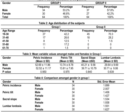
Table 1: Gender distribution of the patients
Gender GROUP I GROUP II
Frequency Percentage Frequency Percentage
Male 34 53.2% 37 57.8%
Female 30 46.8% 27 42.2%
Total 64 100% 64 100%
Table 2: Age distribution of the subjects
Group I Group II
Age Range Frequency Percentage Frequency Percentage
19-30 27 42.2 45 70.3
31-40 17 26.6 18 28.2
41-50 8 12.5 1 1.5
51-60 11 17.2 0 0
61-70 1 1.5 0 0
Table 3: Mean variable values amongst males and females in Group I
Gender Pelvic Incidence
(Mean ± SD) (Mean ± SD) Pelvic Tilt Sacral Slope (Mean ±SD) Lumbar Lordosis (Mean ± SD)
Male 52.69 ± 11.86 13.74 ± 8.78 40.21 ± 9.58 29.64 ± 9.69
Female 52.52 ± 11.17 13.81 ± 7.95 39.71 ± 10.79 30.71 ± 9.06
P value 0.950 0.975 0.840 0.639
Table 4: Comparison amongst gender in group I
Gender N Std. Error Mean
Pelvic incidence Male 34 1.899
Female 30 2.007
Pelvic tilt Male 34 1.406
Female 30 1.427
Sacral slope Male 34 1.534
Female 30 1.938
Lumbar lordosis Male 34 1.551
RESULTS
From the table1, it is inferred that there were a total of 34 (53.2%) males in Group I and 37 (57.8%) males in Group II. In Group I and Group II, there were 30 (46.8%) and 27 (42.2%) females respectively.
From table 2, in Group I, there were 27 subjects (42.2%) who were aged between 19-30 years. There were 26.6% (n=17) subjects between 31-40 years. There were 8 subjects (12.5%) subjects between 41-50 years of age. There were 11 subjects between 51-60 years of age. Least number of subjects were between 61-70 years i.e. 1.5%. In Group II, there were maximum number of subjects that were aged between 19-30 years. There were 45 subjects (70.3%) in this age group. There was only one subject who was aged between 41-50 years of age. There were 18 subjects (28.2%) who were aged between 31-40 years of age. The table 3 shows the mean value of variables amongst subjects in Group I. The mean value of pelvic incidence amongst males and females was 52.69 ± 11.86 and 52.52 ± 11.17 respectively. On applying t test, the p value was 0.950. There was no significant difference in the pelvic incidence between males and females. The mean value of pelvic tilt amongst males and females was 13.74 ± 8.78 and 13.81 ± 7.95 respectively. On applying t test, the p value was 0.975. There was no significant difference in the pelvic tilt between males and females. The mean value of sacral slope amongst males was 40.21 ± 9.58 and that amongst females was 39.71 ± 10.79 with the p value of 0.840. Hence there was no significant difference in the sacral slope between males and females. The mean value of lumbar lordosis amongst males was 29.64 ± 9.69 and that amongst females was 30.71 ± 9.06 with the p value of 0.639. Hence there was no significant difference in the lumbar lordosis between males and females.
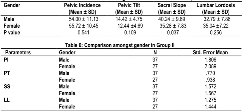
Table 4 shows the standard error amongst males and females in Group I. The standard error in pelvic incidence amongst males was 1.899 and amongst females were 2.007. The standard error in pelvic tilt amongst males was 1.406 and females were 1.427. The standard error in sacral slope amongst males and females was 1.534 and 1.938 respectively. The standard error in lumbar lordoisis amongst males and females was 1.551 and 1.627 respectively.Table 5 shows the mean value of variables amongst subjects in Group II. The mean value of pelvic incidence amongst males and females was 54.00 ± 11.13 and 55.72 ±10.45 respectively. On applying t test, the p value was 0.541. There was no significant difference in the pelvic incidence between males and females. The mean value of pelvic tilt amongst males and females was 14.42 ± 4.75 and 12.44 ±4.69 respectively. On applying t test, the p value was 0.109. There was no significant difference in the pelvic tilt between males and females. The mean value of sacral slope amongst males was 40.24 ± 9.69 and that amongst females was 35.28 ± 7.83 with the p value of 0.037. Hence there was a significant difference in the sacral slope between males and females. The mean value of lumbar lordosis amongst males was 32.79 ± 7.86 and that amongst females was 35.04 ± 7.22 with the p value of 0.256. Hence there was no significant difference in the lumbar lordoisis between males and females. Table 6 shows the standard error amongst males and females in Group II. The standard error in pelvic incidence amongst males was 1.806 and amongst females were 2.089. The standard error in pelvic tilt amongst males was 0.770 and females were 0.938. The standard error in sacral slope amongst males and females was 1.572 and 1.567 respectively. The standard error in lumbar lordosis amongst males and females was 1.275 and 1.444 respectively.
Table 5: Mean variable values amongst males and females in Group II
Gender Pelvic Incidence
(Mean ± SD)
Pelvic Tilt (Mean ± SD)
Sacral Slope (Mean ± SD)
Lumbar Lordosis (Mean ± SD)
Male 54.00 ± 11.13 14.42 ± 4.75 40.24 ± 9.69 32.79 ± 7.86
Female 55.72 ± 10.45 12.44 ±4.69 35.28 ± 7.83 35.04 ±7.22
P value 0.541 0.109 0.037 0.256
Table 6: Comparison amongst gender in Group II
Parameters Gender N Std. Error Mean
PI Male 37 1.806
Female 27 2.089
PT Male 37 .770
Female 27 .938
SS Male 37 1.572
Female 27 1.567
LL Male 37 1.275
Female 27 1.444
DISCUSSION
LBP is a work-related musculoskeletal disorder that causes substantial economic losses to individuals as well as to the community.15
Chronic low back pain (CLBP) has a high morbidity with high social and economic effects. Pathology in any segment of the trunk or lower leg can disturb the global postural equilibrium, resulting in compensatory changes in other segments. Low back pain (LBP) is an important clinical, social, economic and public health problem affecting the population indiscriminately. The
studies done all over the world show that the prevalence of chronic lower back pain is increasing. This increase in the prevalence is a concern for worry because it is a condition responsible for substantial social impact and an important source of demand for health services.16 In Indian context the
epidemiological study on back pain in various occupational groups are not widely available. Current treatments are inadequate for many patients. With current therapies many patients fail to achieve adequate relief for chronic pain.17 The present study was
in patient with chronic low backache and to find any differences in lumbo-pelvic alignment. The age distribution of patients in this study was, majority of the subjects were between 19- 30 years of age. The least number of subjects were between 61-70 years of age. In Group I, there were 26.6% (n=17) subjects between 31-40 years. There were 8 subjects (12.5%) subjects between 41-50 years of age. There were 11 subjects between 51-60 years of age. In Group II, there was only one subject who was aged between 41-50 years of age. There were 18 subjects (28.2%) who were aged between 31-40 years of age. In a study conducted by J. Schroeder et al (2013)92 the age of the subjects with chronic lower
back pain was similar to that of this study. They also enrolled subjects more than 19 years of age. They divided the subjects into two groups, one was more than 40 years of age and the other less than 40 years of age. The age range was also similar to a study conducted by Emmanuelle Chaleat-Valayer et al,18 in which
subjects between 18-60 years were included in the study. In present study, there were a total of 34 (53.2%) males in Group I and 37 (57.8%) males in Group II. In Group I and Group II, there were 30 (46.8%) and 27 (42.2%) females respectively. There were majority of males subjects in our study. This was in accordance with the study conducted by Emmanuelle Chaleat-Valayer et al.18
There were 111 males and 87 females in their study.
In present study, In Group I, the mean value of lumbar lordosis amongst males was 29.64 ± 9.69 and that amongst females was 30.71 ± 9.06 with the p value of 0.639. In Group II, the mean value of lumbar lordosis amongst males was 32.79 ± 7.86 and that amongst females was 35.04 ± 7.22 with the p value of 0.256. Hence there was no significant difference in the lumbar lordosis between males and females. The results of this study were comparable to the study conducted by Schroeder et al19 who
found that the spine was significantly flatter for the older male patients compared to the male controls with the p value of 0.013. Gelb D20 and Takeda N21reported an age related decrease in
lumbar angle which means with age, there is flatter lordosis. In this study, in Group I, the mean value of pelvic incidence amongst males and females was 52.69 ± 11.86 and 52.52 ± 11.17 respectively. In Group II, the mean value of pelvic incidence amongst males and females was 54.00 ± 11.13 and 55.72 ± 10.45 respectively. There was no significant difference between males and females in both the groups. In present study, in Group I, the mean value of pelvic tilt amongst males and females was 13.74 ± 8.78 and 13.81 ± 7.95 respectively. In Group II, the mean value of pelvic tilt amongst males and females was 14.42 ±4.75 and 12.44 ± 4.69 respectively. There was no significant difference in the pelvic tilt amongst males and females in both the groups. But studies conducted by Jackson RP et al,22 Rajnics et al23 and
Barrey et al24 show a significantly larger pelvic tilt in subjects of
low back pain.
In our study, in Group I, The mean value of sacral slope amongst males was 40.21 ± 9.58 and that amongst females was 39.71 ± 10.79. In Group II, The mean value of sacral slope amongst males was 40.24 ± 9.69 and that amongst females was 35.28 ± 7.83. There was no significant difference between the males and females in both the groups as the p value was more than 0.05. Based on a previous literature review25,it was found that there are
three main risk factors for recurrent and chronic LBP: (1) history of LBP with associated limitations and treatments, (2) dissatisfaction at work and (3) poor general medical condition. Other risk factors
such as socioeconomic and employment status, psychological status and physically demanding work are also suggested. Although psychosocial and environmental factors seem important in predicting recurrence and chronicity in LBP, morphological and postural factors can also potentially influence the occurrence of LBP.
It is recommended for patients with non-specific chronic low back pain to remain physically active as long periods of inactivity will adversely affect recovery.26 A variety of different types of exercise
have been explored to treat chronic low back pain, including low-to-moderate intensity aerobic exercise,27,28 high intensity aerobic
exercise,29,30 core stabilization and muscular strength exercises 31-36 and flexibility programmes.37-39
However, the most effective form of exercise as a method of rehabilitation for non-specific chronic low back pain is unknown40,41
reflecting its complexity162and more research is required.42
Physical activity (PA) to increase aerobic capacity and muscular strength, especially of the lumbar extensor muscles, is important for patients with CLBP in assisting them to complete activities of daily living.43 However, different exercises have been found to
result in varying levels of effectiveness in reducing lower back pain.44 In addition, too much or too little PA can be associated with
low back pain,45, suggesting that PA as an intervention for low
back pain is complex.
Sagittal spino-pelvic alignment plays a very important role in spinal biomechanics. Differences in spinal curvatures in erect posture require changes in the ventro-dorsal position of the spine (i.e., the position of the spine in the mid-sagittal plane).46 When
the spine is situated closer to the line of gravity (more ventral position), a smaller PI and spinal curvatures is needed to keep an economic upright posture. When the spine is situated further back from the line of gravity (more dorsal position), a higher PI and spinal curvatures is needed to maintain an economic upright balance.46 As biomechanical overloading of the spine is known to
cause and worsens several spinal disorders, sagittal spino-pelvic alignment has been studied extensively in the past two decades and referential values are described in both asymptomatic adults and children.
CONCLUSION
The present study found no significant difference in the lumbar lordosis, pelvic tilt and sacral slope between males and females with chronic low back pain. Sagittal spinopelvic balance in modern humans is achieved when spinal curvatures and pelvic/sacral orientation are aligned in the same manner. In a well-aligned spine, the line of gravity falls close to the acetabulum. Pelvic incidence significantly influences the sagittal spinal geometry, specifically lumbar lordosis in healthy modern humans. Parameters of the sagittal plane were extracted as being associated with low back pain too. But with respect to the literature and inconsistency of different multivariate analysis approaches, those parameters also might to some extent be affected by aging and degeneration.
REFERENCES
2. Classification of chronic pain. Descriptions of chronic pain syndromes and definitions of pain terms. Prepared by the International Association for the Study of Pain, Subcommittee on Taxonomy. Pain Suppl 1986; 3: S1-226.
3. Klineberg E, Schwab F, Smith JS, Gupta MC, Lafage V, Bess S. Sagittal spinal pelvic alignment. Neurosurgery Clinics 2013; 24(2): 157-62.
4. Barrey C, Roussouly P, Perrin G, Le Huec JC.Sagittal balance disorders in severe degenerative spine. Can we identify the compensatory mechanisms? Eur Spine J 2011; 20(Suppl 5): 626-33.
5. Watelain E, Dujardin F, Babier F, Dubois D, Allard P. Pelvic and lower limb compensatory actions of subjects in an early stage of hip osteoarthritis. Arch Phys Med Rehabil 2001; 82: 1705-11. 6. Van Tulder M, Koes B, Bombardier C. Low back pain. Best Pract Res Clin Rkheumatol 2002; 16: 761-75.
7. Schwab F, Farcy J, Bridwell K. A clinical impact classification of scoliosis in the adult. Spine (Phila Pa 1976). 2006; 31: 2109-14. 8. Liu W, Chen XS, Jia LS, Song DW. The clinical features and surgical treatment of degenerative lumbar scoliosis: a review of 112 patients. Orthopaedic surgery 2009; 1(3): 176-83.
9. Glassman SD, Hamill CL, Bridwell KH, Schwab FJ, Dimar JR, Lowe TG. The impact of perioperative complications on clinical outcome in adult deformity surgery. Spine 2007; 32(24): 2764-70. 10. Ashraf A, Farahngiaz S, Johran BP, Setayeshpour N, Naseri M, Nasseri A. Correlation between radiologic sign of lumbar lordosis and functional status in patients with chronic mechanical low back pain. Asian Spine 2014; 8(5): 565-70.
11. Guo J, Liu Z, Lv F, Zhu Z, Qian B, Zhang X, Lin X, Sun X, Qiu Y. Pelvic tilt and trunk inclination: new predictive factors in curve progression during the Milwaukee bracing for adolescent idiopathic scoliosis. European Spine Journal. 2012 Oct 1;21(10):2050-8.
12. Patel A, Schwab F, Ungar B, Farcy JP, Lafage V. Analysis of sagittal plane deformity and correction. Current Orthopaedic Practice 2010; 21(4): 356-63.
13. Barrey C, Roussouly P, Perrin G, Le Huec JC.Sagittal balance disorders in severe degenerative spine. Can we identify the compensatory mechanisms? Eur Spine J 2011; 20(Suppl 5): 626-33.
14. Shin MH, Ryu KS, Hur JW, Kim JS, Park CK. Comparative study of lumbo-pelvic sagittal alignment between patients with and without sacroiliac joint pain after lumbar interbody fusion. Eurspine 2013; 38(21): E1334-41.
15. Alperovitch-Najenson D, Santo Y, Masharawi Y, Katz- Leurer M, Ushaev D, Kalichman L. Low back pain among professional bus drivers: Ergonomic and Occupational- Psychosocial Risk factors. IMAJ. 2010; 12: 26-31.
16. Meucci RD, Fassa AG, Paniz VM, Silva MC, Wegman DH. Increase of chronic low back pain prevalence in a medium-sized city of southern Brazil. BMC Musculoskelet Disord 2013; 14: 155. 17. Strong JA, Xie W, Bataille FJ, Zhang JM. Preclinical studies of low back pain. Mol Pain 2013; 9: 17.
18. Chaleat-Valayer E, Mac-Thiong JM, Paquet J, Berthonnaud E, Siani F, Roussouly P. Sagittal spino-pelvic alignment in chronic low back pain. Eur spine J 2011; 20(suppl.5): s634-40.
19. Ghandhari H, Hesarikia H, Ameri E, Noori A. Assessment of normal sagittal alignment of the spine and pelvis in children and adolescents. BioMed research international. 2013;2013.
20. Gelb DE, Lenke LG, Bridwell KH, Blanke K, McEnery KW. An analysis of sagittal spinal alignment in 100 asymptomatic middle and older aged volunteers. Spine 1995; 20(12): 1351-8.
21. Takeda N, Kobayashi T, Atsuta Y, Matsuno T, Shirado O, Minami A. Changes in the sagittal spinal alignment of the elderly without vertebral fractures: a minimum 10-year longitudinal study. J Orthop Sci 2009; 14: 748–53.
22. Le Huec JC, Faundez A, Dominguez D, Hoffmeyer P, Aunoble S. Evidence showing the relationship between sagittal balance and clinical outcomes in surgical treatment of degenerative spinal diseases: a literature review. International orthopaedics 2015; 39(1): 87-95.
23. Lv X, Liu Y, Zhou S, Wang Q, Gu H, Fu X et al. Correlations between the feature of sagittal spinopelvic alignment and facet joint degeneration: a retrospective study. bmc musculoskeletal disorders 2016; 17:341.
24. Bakrrey C, Jund J, Noseda O, Roussouly P. Sagittal balance of the pelvis-spine complex and lumbar degenerative diseases. A comparative study about 85 cases. Eur Spine J 2007; 16(9): 1459–67.
25. Poiraudeau S, Lefevre-Colau MM, Fayad F . Lombalgies. Encylc Med Chir 2004; 15-840-C-10: 1–15.
26. National Health Service (NHS). Back Pain. Available online: http://www.nhs.uk/Conditions/Back-pain/Pages/Introduction.aspx (accessed on 16 October 2014).
27. Chan CW, Mok NW, Yeung EW. Aerobic exercise training in addition to conventional physiotherapy for chronic low back pain: A randomized controlled trial. Arch. Phys. Med. Rehabil 2011; 92: 1681–1685.
28. Shnayderman I, Katz-Leurer M. An aerobic walking programme versus muscle strengthening Programme for chronic low back pain: A randomized controlled trial. Clin. Rehabil 2013; 27: 207–14.
29. Chatzitheodorou D, Kabitsis C, Malliou P, Mougios V. A pilot study of the effects of high-intensity aerobic exercise versus passive interventions on pain, disability, psychological strain, and serum cortisol concentrations in people with chronic low back pain. Phys. Ther 2007; 87: 304–12.
30. Chatzitheodorou D, Mavromoustakos S, Milioti S. The effect of exercise on adrenocortical responsiveness of patients with chronic low back pain, controlled for psychological strain. Clin. Rehabil 2008; 22: 319–28.
31. Inani SB, Selkar SP. Effect of core stabilization exercises versus conventional exercises on pain and functional status in patients with non-specific low back pain: A randomized clinical trial. J. Back Musculoskelet Rehabil 2013; 26: 37–43.
32. Kim JD, Oh HW, Lee JH, Cha JY, Ko I.G, Jee YS. The effect of inversion traction on pain sensation, lumbar flexibility and trunk muscles strength in patients with chronic low back pain. Isokinet. Exerc. Sci 2013; 21: 237–46.
33. Sarabon N. Effects of trunk functional stability training in subjects suffering from chronic low back pain: A pilot study. Kinesiol. Slov 2011; 17: 25–37.
35. Suni J, Rinne M et al. Control of the lumbar neutral zone decreases low back pain and improves self-evaluated work ability: A 12-month randomized controlled study. Spine 2006; 31:611–20. 36. You JH et al. The effect of a novel core stabilization technique on managing patients with chronic low back pain: A randomized, controlled, experimenter-blinded study.ClinRehabil2014;28:460–9. 37. Gladwell V, Head S, Haggar M, Beneke R. Does a program of pilates improve chronic non-specific low Back pain? J. Sport Rehabil 2006; 15: 338–50.
38. Kuukkanen T, Malkia E. Effects of a three-month therapeutic exercise programme on flexibility in subjects with low back pain. Physiother. Res. Int 2000; 5: 46–61.
39. Masharawi Y, Nadaf N. The effect of non-weight bearing group-exercising on females with non-specific chronic low back pain: A randomized single blind controlled pilot study. J. Back Musculoskelet. Rehabil 2013; 26: 353–9.
40. Hayden JA, Van Tulder M, Tomlinson G. Systematic review: Strategies for using exercise therapy to improve outcomes in chronic low back pain. Ann. Intern. Med 2005; 142: 776–85. 41. Kolber MJ, Beekhuizen K. Lumbar stabilization: An evidence-based approach for the athlete with low back pain. Strength Cond. J 2007; 29: 26–37.
42. Smith JA, Osborn M. Pain as an assault on the self: An interpretative phenomenological analysis of the psychological impact of chronic benign low back pain. Psychol. Health 2007; 22: 517–34.
43. Smeets R, Severens JL, Beelen S, Vlaeyen JW, Knottnerus JA. More is not always better: Cost-effectiveness analysis of combined, single behavioral and single physical rehabilitation programs for chronic low back pain. Eur. J. Pain 2009; 13: 71–81.
44. Smith D, Bissell G, Bruce-Low S, Wakefield C. The effect of lumbar extension training with and without pelvic stabilization on lumbar strength and low back pain. J. Back Musculoskelet Rehabil 2011; 24: 241–9.
45. Wai EK, Rodriguez S, Dagenais S, Hall H. Evidence informed management of chronic low back pain with physical activity, smoking cessation, and weight loss. Spine J 2008; 8: 195–202. 46. Wagner H, Liebetrau A, Schinowski D, Wulf T, de Lussanet MH. Spinal lordosis optimizes the requirements for a stable erect posture. Theor Biol Medl Model 2012; 9: 13.
[
Source of Support: Nil.
Conflict of Interest: None Declared.
Copyright: © the author(s) and publisher. IJMRP is an official publication of Ibn Sina Academy of Medieval Medicine & Sciences, registered in 2001 under Indian Trusts Act, 1882. This is an open access article distributed under the terms of the Creative Commons Attribution Non-commercial License, which permits unrestricted non-commercial use, distribution, and reproduction in any medium, provided the original work is properly cited.
Cite this article as: Pawan Kumar Mahato, Sunil Kumar Srivastava, Amit Kumar Saxena, Prachi Saffar Aneja. A Comparative Study to Find Gender Wise Differences in Lumbo-Pelvic Alignment in Patient with Chronic Low Backache. Int J Med Res Prof. 2018 Jan; 4(1):632-37.