Original Research Article.
113 |P a g e Int J Med Res Prof.2018 May; 4(3); 113-18. www.ijmrp.com
Microbiological Profile of Cerebrospinal Fluid (CSF) in Pyogenic Meningitis
Patients at Tertiary Care Hospital
Ravindra Kumar Barnawal
1, Manoj Kumar
2, Ashok Kumar Sharma
3*, Amber Prasad
4,
Kumari Seema
5, Versha Priydarshini
11Junior Resident, 2Professor & Head, 3*Associate Professor, 4Assistant Professor, 5Senior Research Scientist, Department of Microbiology, Rajendra Institute of Medical Sciences, Ranchi, Jharkhand, India.
ABSTRACT
Introduction: Acute bacterial meningitis is a life threatening illness that is prevalent worldwide. It is a medical emergency that needs early diagnosis and treatment.
Aims and objective: The study includes microbiological profile and antibiotic susceptibility pattern of the bacteria isolated in CSF in clinically suspected cases of pyogenic meningitis in children.
Methods and Materials: A total of 112 clinically suspected cases of meningitis in paediatric age group were included. It was a hospital based Descriptive cross sectional study from January 2017 to November 2017. All the specimens were processed by standard bacteriological techniques that include microscopy (Gram staining, wet mount, India ink), culture in blood agar, Mac Conkey agar, Chocolate Agar plates, Automated culture (BACTEC) method and etc.
Results: A total of 112 CSF samples were collected from suspected pyogenic meningitis patients. 27 cases were culture positive. Among suspected case there was male predominance (54.5%). Highest rate of culture positive cases were found in the age group of less than 01 months (33.3%). The most
common organism isolated was CONS (22.2%) followed by
Escherichia coli (18.5%).
Conclusions: Acute bacterial meningitis is a medical emergency and making an early diagnosis and providing early and accurate treatment, are lifesaving to reduce morbidity. This study may play an important role in the diagnosis and more accurate treatment for the ABM patients.
Keywords: Pyogenic Meningitis, BACTEC, Group B Streptococci, Kernig's Sign, Brudzinski's Sign.
*Correspondence to:
Dr. Ashok Kumar Sharma
Associate Professor Department of Microbiology,
Rajendra Institute of Medical Sciences, Ranchi, Jharkhand, India.
Article History:
Received: 08-03-2018, Revised: 01-04-2018, Accepted: 04-05-2018
Access this article online Website:
www.ijmrp.com
Quick Response code
DOI:
10.21276/ijmrp.2018.4.3.023
INTRODUCTION
Meningitis is an inflammation of the meninges, the membrane covering the brain and spinal cord. When a microorganism (usually a bacterium or virus) enters the subarachnoid space, there is inflammatory response in the meninges.1 There is intense inflammation of meninges due to bacterial products such as to gram-negative lipopolysaccharide or gram positive peptidoglycan even after destruction of bacteria by antibiotic therapy.
The microbiological causes of meningitis include bacteria, viruses, fungi and parasites. Among these, bacterial meningitis include pyogenic meningitis and tubercular meningitis.
The most common etiological agents in neonates are group B Streptococci (Streptococci agalactiae), Escherichia coli and
Listeria monocytogenes. Streptococcus pneumoniae, Neisseria
meningitidis and Haemophilus influenzae type–b are the common
cause of pyogenic meningitis in infant and young children worldwide. Patients with ventriculo-peritoneal (VP) shunt are at risk of meningitis caused by coagulase negativeStaphylococcus
and Pseudomonas species. These bacteria reach the CNS either by haematogenous spread or by direct extension. In neonates the infection is acquired from the maternal vagina during normal delivery. The organisms that colonise the upper respiratory tract of infant and young children can also cause pyogenic meningitis. Manifestation of pyogenic meningitis depends on the age of the patients. Headache, fever and altered sensorium are the important symptoms of pyogenic meningitis. Signs of meningeal irritation
such as neck stiffness, Brudzinsky’s sign and Kernig’s sign or the
Petechiae and purpura is common in patients with meningococcal meningitis. Signs in infants and neonates include fever, apnoea, seizure, a bulging fontanel and rash.
Epidemiology
Bacterial meningitis is the most common form of suppurative CNS infection, with an annual incidence in United States of >2.5 cases per 100,000 population.3
The use of (PCV7) heptavalent pneumococcal protein polysaccharide conjugate vaccine and Hib vaccine to infant causes dramatic decrease in rate of pyogenic meningitis caused by invasive pneumococcal disease and Haemophilus influenzae
type-b. The incidence rate of Streptococcus pneumoniae
meningitis are 228 per 100,000 in children at age of 6-12 month.4 The major risk factor for pyogenic meningitis is due to lack of immunity to specific pathogens, other risk factor include colonization of pathogenic bacteria, close contact of individual having invasive disease caused by N. meningitis, H. influenza
type b. The mode of transmission is person to person.4
MATERIAL AND METHODS
It was a hospital based Descriptive cross sectional study conducted in Department of Microbiology, Rajendra Institute of Medical Sciences (RIMS), Ranchi. It included patients of all paediatric age group, from January 2017 to November 2017.
Inclusion Criteria
▪ Children up to 14 years age admitted to Department of Paediatrics, RIMS
▪ Clinical diagnosis made by paediatrician with symptoms and
signs; history of high fever, intense headache, neck rigidity, refusal to feed, vomiting, drowsiness, convulsion, signs of meningeal irritation: Kernig's sign (with the hip joint flexed, extension at the knee causes spasm in the hamstring muscles) and Brudzinski's sign (passive flexion of the neck causes flexion of the thighs and knees) and altered sensorium was included in the study.
Exclusion Criteria
▪ Cases who were treated with antibiotics prior to admission in RIMS, Ranchi
▪ Patients on antipsychotic drugs and on any other medication
▪ Other systemic diseases like diabetes mellitus, nephritic syndrome, renal failure, heart failure and following head trauma were excluded from this study.
Sample Size
A total of 112 clinically suspected cases of meningitis in paediatric age group admitted to Department of Paediatrics, RIMS, Ranchi were recruited for the study.
Sample Collection and Laboratory Testing
Three vials of CSF samples were collected from the cases with clinical suspicion of meningitis. One vial was used for cytological examination, the other for bacteriological examination and the biochemical examination was done on the sample of third vial.5
The CSF bottles were transported without delay to the Microbiology Laboratory of the college and processed without any delay; whenever delay was anticipated the CSF samples were kept at room temperature.6
Physical Examination of CSF
CSF was classified as clear, opalescent, turbid, purulent, haemorrhagic and xanthochromic.
Processing of Sample and Identification
All the specimens were processed by standard bacteriological techniques that include microscopy (Gram staining, wet mount, India ink), culture in blood agar, Mac Conkey agar, Chocolate Agar plates, Automated culture (BACTEC) method and etc.
CSF is Divided Into Three Portions
One portion is centrifuged and Gram Stained smears was prepared from deposit. Second portion of CSF was inoculated either on blood agar, Mac Conkey agar plate, Chocolate agar plate or BD BACTEC Peds Plus culture vial. Third portion of CSF will be kept at room temperature.
Direct Microscopy
Includes Wet mount preparation, India ink preparation and Gram-stained smears preparation of CSF.
Culture
If bacteria were seen in the Gram-stained smear, the appropriate culture media was inoculated. If no organisms were seen, or if the interpretation of the Gram smear was unclear, it was desirable to inoculate a full range of media, including blood agar with a streak
of Staphylococcus aureus to promote growth of H. influenzae.
The second portion of CSF was inoculated either on to the following media or in BD BACTEC Peds Plus culture vial.
▪ Blood agar (BA) plate incubated at 370C in 5-10% CO2.
▪ Chocolate agar plate incubated at 370C in 5-10% CO2.
▪ MacConkey agar incubated aerobically at 370C.
BHI broth incubated aerobically at 370 C. Inoculated primary plates were incubated for 48 to 72 hours. The plates were examined daily for 72 hours before reporting as negative. BHI broth was incubated for 7 days and examined daily for presence of growth or turbidity and was considered negative at the end of 7 days of incubation.
BD BACTEC Peds plus culture vial were put in automated BD BACTEC FX 200 TM System and was declared negative only if not indicated by the system at the end of 5 days of aerobic incubation.
OBSERVATIONS AND RESULTS
The present study was conducted in Department of Microbiology, Rajendra Institute of Medical Science (RIMS) Ranchi. During this period all patients who were admitted in the paediatric ward with signs and symptoms suspected of meningitis were included in the study. A total of 112 CSF samples were collected. 27 cases were culture positive. Among suspected case there was male 54.5% and female 44.5%. Among 27 culture positive cases there were 16 (59.25 %) males and 11 (40.75%) females.
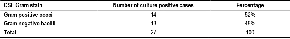
Table 1: CSF Gram’s stain smear study among the culture positive cases.
CSF Gram stain Number of culture positive cases Percentage
Gram positive cocci 14 52%
Gram negative bacilli 13 48%
Table 2: Organism isolated in culture positive cases
Etiological agent 0-1month 1-12months. 1yrs. – 5 yrs. 5 yrs. – 14 yrs. Total N(%)
Escherichia coli 03 02 00 00 5 (18.5%)
CONS 02 01 00 00 6 (22.2%)
Streptococcus agalactiae 01 00 00 00 1 (3.7%)
Streptococcus pneumoniae 00 01 01 00 2 (7.4%)
Klebsiella pneumoniae 02 01 00 00 3 (11.1%)
Staphylococcus aureus 00 01 01 02 4 (14.8%)
Pseudomonas aeroginosa 00 00 02 01 3 (11.1%)
Acinetobacter spp 00 01 01 00 2 (7.4%)
Enterococcus 01 00 00 00 1 (3.7%)
Total 09 07 06 05 27
Table 3: Antibiotic sensitivity pattern of gram positive bacteria (% sensitivity)
Antibiotic CONS (06) Streptococcus
agalactiae
Staphylococcus aureus
Streptococcus pneumoniae
Entetococcus
Amoxyclav 64% 0% 50% 50% 100%
Ciprofloxacin 33.3% 0% 25% 50% -
Chloramphenicol 50% 0% 50% 50% 100%
Clindamycin 64% 0% 75% 50% 0%
Erythromycin 0% 0% 50% 50% 0%
Gentamicin 50% 100% 75% 50% 0%
Linezolid 100% 100% 100% 100% 100%
Vancomycin 100% 100% 100% 100% 100%
Tetracycline 66.6% 0% - 100% 0%
Netlimicin 64% 0% 75% 50% 0%
Co-trimoxazole 64% 0% 75% 50% -
Daptomycin 100%
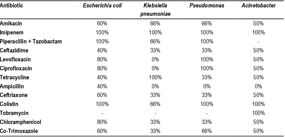
Table 4: Antibiotic sensitivity pattern of gram negative bacteria (% sensitivity)
Antibiotic Escherichia coli Klebsiella
pneumoniae
Pseudomonas Acinetobacter
Amikacin 60% 66% 66% 50%
Imipenem 100% 100% 100% 100%
Piperacillin + Tazobactam 100% 66% 100% -
Ceftazidime 40% 33% 33% 50%
Levofloxacin 80% 0% 100% 50%
Ciprofloxacin 80% 0% 100% 50%
Tetracycline 40% 100% 33% 50%
Ampicillin 40% 0% 0% 0%
Ceftriaxone 60% 33% 33% 50%
Colistin 100% 66% 100% 100%
Tobramycin - - - 100%
Chloramphenicol 80% 33% 33% 50%
Co-Trimoxazole 60% 33% 66% 50%
Highest number of suspected cases were found in the age group of 05 yrs. to 14 yrs. (33.9%), followed by 25% in age group of less than 01 months. Similarly the highest rate of culture positive cases were found in the age group of less
than 01 months (33.3%). Fever was the most common presentation (91%), followed by seizure (81.25%). The most common organism isolated was CONS (22.2%) followed
Fig 1: Distribution of cases according to age
Fig 2: Sex wise distribution of cases
Fig 3: Distribution of cases according to clinical features (sign and symptoms) 0
5 10 15 20 25 30 35
0 –1 Months 02 Months –01
Yrs. 02 Yr. –05 Yrs. 06 Yrs. –14 Yrs. 9
7 6
5 19
15
18
33
No
. o
f Case
s
Age group Culture Positive cases Culture negative cases
0 5 10 15 20 25 30 35 40 45
Culture positive cases (%) Culture negative cases (%) 16
45
11
40
No
. o
f Ca
se
s
Male Female
0 20 40 60 80 100 120
Fever Seizure Altered
sensorium Fefusal tofeed Vomiting Meningealsign fontanellBulging 102
91
66
44
34
25
13
Po
sit
ive
ca
se
s
Fig 4: Culture positive cases isolated in suspected pyogenic meningitis
Fig 5: Gross appearance of CSF
DISCUSSION
In the present study, 59% of cases were below one year of age. Similarly Shrestha et al7 in their study noted 55.6% of cases, Modi Gaurav B et al8 found 36.5% of cases and Kabra SK et al9 found 52% of cases below one year of age.
More than two third of cases of meningitis occur in first two years of life this is due to under developed immune system and high vascularity of brain.
In the present study out of 27 confirmed cases of acute bacterial meningitis male to female ratio was 1.47:1 in which male were 16 (59.25%) and female were 11 (40.75%) which correlate with study of Modi Gaurav B et al8 and Bijay Mirdha et al10 who also reported ratio of 1.5:1 in their studies. All the studies showed male preponderance.
The present study showed that fever was the most common clinical presentation (91%) followed by seizure (81.25%), altered sensorium (59.8%) and meningeal signs (22.3%) of cases. Similar observation were made by studies of Chinchankar N et al11, Attia Bari et al12, Fatima Zeeshan et al and Khan F et al.13
In the present study, among the 27 culture confirmed cases CSF appeared turbid in 12 (44.5%), clear in (40.7%), 7.4%have blood stained and 7.4% Xanthochromic. None of the samples showed cobweb formation.
In the present study out of 112 CSF samples collected 27 (24.1%) showed positive growth. Similar finding were observed by Rajesh Breja et al14, Dr. Sudharshan Raj C et al15, IMAD S. Mahmoud et al16 and Ramesh S. T et al17 who reported culture positivity in 23.58%, 19.1%, 34.5% and 25.1% of cases respectively.
In present study out of total culture positive cases gram positive cocci were 52% and gram negative bacilli were 48%. Similar observations were made by Fatima Khan et al, Rajesh Breja et al and Dr. Sudharshan Raj C et al.
In the present study coagulase negative staphylococcus (CONS) was the most common organism isolated in 22.2% cases of acute bacterial meningitis. Similar results were observed by R. Basri et al, Attia Bari et al and Amresh Kumar Singh et al18 who reported CONS to be 21.6%, 45% and 44.5% respectively. Utpola Devi et al19 16% CONS was detected.
0 10 20 30 40 50 60 70 80
Clear Turbid/Cloudy Blood stained Xanthochromic
11 12
2 2
75
2 6 2
No
. o
f Case
s
Confirmed cases Negative cases
24.1%
75.9%
E.coli was the etiological agent in 18.5% cases followed by 14.8% cases of Staphylococcus aureus of acute bacterial meningitis in the present study. Similar finding were observed by IMAD S. Mahmoud et al, Amresh Kumar Singh et al and Modi Gaurav B et al who reported Escherichia coli to be 16.2%, 19.2% and 14.15% respectively. R. Basri et al, Modi Gaurav B et al, Dr. Sudharshan Raj C et al and Amresh Kumar Singh et al who reported 13.6%, 19%, 11.4% and 19.83% was caused by staphylococcus aureus. In the present study Klebsiella spp. 11.1%, Pseudomonas spp. 11.1% and Acinetobacter 7.4% was isolated in acute bacterial meningitis cases. Similar observation was observed by studied of Utopala Devi et al, Dr. Sudharshan Raj C et al and Modi Gaurav B et al who observed that 12%, 18.1% and 22.92% of Klebsiella
spp. were isolated, 9%, 6.6% and 12.19% Pseudomonas spp. was isolated,18%, 1.4% and 11.2% acinetobacter spp. was isolated in acute bacterial meningitis cases.
In the present study all the gram positive cocci were highly sensitive to Linezolid and Vancomycin. They were highly resistant to Ciprofloxacin and Erythromycin. Mild resistance was seen with Choramphenicol and Clindamycin.This was similar to studies done by Amresh Kumar Singh et al and Dr. Sudharshan Raj C et al who also reported that gram positive cocci were highly sensitive to Vancomycin and Linezolid and highly resistant to Ciprofloxacin and Erythromycin.
In the present study all the gram negative bacilli were highly sensitive to Imipenem, and Colistin and highly resistant to Ampicillin, Ceftazidime and Co-Trimoxazole. Similar studies were done by Dr. Sudharshan Raj C et al, Amresh Kumar Singh et al and Modi Gaurav B et al, who observed thatgram negative bacilli were completely sensitive to Imipenem and colistin and highly resistant to Co-Trimoxazole, ceftazidime and Ampicillin.
CONCLUSION
This study may play an important role in the diagnosis and more accurate treatment to the patients suffering with acute bacterial meningitis. Regular prevalence and antibiotic susceptibility studies will help to enhance antimicrobial stewardship thus minimizing the emergence and spread of antimicrobial resistance and it would also be helpful for clinicians choosing an appropriate empirical antimicrobial. Hence, continued surveillance with more detailed studies is warranted to know the actual magnitude of problem and the spectrum of diseases and antimicrobial resistance caused by these pathogens.
REFERENCES
1. Ananthanarayan and Paniker’s Text book of Microbiology 9th
Edition, 2013, pg. 667.
2. Oostenbrink R, Moons KG, Theunissen CC, Derrksen-Lubsen G, Grobbee DE, Moll HA. Signs of meningial irritation at the emergency department: how often bacterial meningitis? Pediatrics Emerg Care 2001; 17:161-4.
3. Joseph Lascalzo, Dan L. Lango, Anthony S. Fauci, J. Larry
Jameson, Stephen L.Hauser Dennis L.Kasper, Harrison’s 19th Edition
principle of internal medicine, pg. 885, 2015.
4. Robert M. Kliegman, Bonita F. Stanton, Joseph W. St Geme III, Nina F. Schor, Richard E. Behrman Nelson Textbook of paediatrics 20th Edition, 2015, pg. 1371-72, 2939.
5. Koneman EW, Winn WC Jr, Allen SD et al (eds.). Charts. In:
Koneman’s Color Atlas and Textbook of Diagnostic Microbiology, 6th
ed. Lippincott Williams & Wilkins: Philadelphia: 2006: pp. 1437-1535.
6. Collee FG, Marmiom BP, Fraser AG et al (eds.). Mackie and Mc Cartney Practical Medical Microbiology. 14th Ed. Pub: Churchill Livingstone. Elsevier. New Delhi. India. 2008.
7. R. Basri et al, Burden of Bacterial meningitis: A retrospective review on laboratory parameters and factors associated with death in meningitis, Kelantan Malaysia.Nagoya Journal of Medical Science 2015 Feb;77(1-2): 59-68, PMCID 4361508.
8. Modi Gaurav B1 et al. Bacteriological Profile of Pyogenic meningitis in tertiary care hospitals Ahmedabad.National Journal of Medical research Volume 2 July-Sept 2012,ISSN 2249:2277 8810.
9. Levy C, Varon E, Picard C et al. Trends of pneumococcal meningitis in children after introduction of the 13-valent pneumococcal conjugate vaccine in France. Pediatr Infect Dis J 2014; 33:1216–21. 10. Bijay R. Mirdha, U. Gupta and R.A.Bhujwala. Latex agglutination test: An adjunct to the laboratory diagnosis of pyogenic bacterial meningitis. Indian J Pediatr 1991; 58:521-524.
11. Chinchankar N, Mane N, Bhave S, Bapat S, Bardekar A, Pandit A, et al. Diagnosis and outcome of acute bacterial meningitis in early childhood. Indian Pediatric 2002; 39: 914-92.
12. Attia Bari, Fatima Zeeshan et al. Childhood Acute Bacterial Meningitis: Clinical Spectrum, Bacteriological Profile and Outcome. Journal of the College of Physianss and Surgeons Pakistan 2016, 26(10):822-826.
13. Fatima Khan et al. Bacterial Meningitis in North India: Trends over a Period of eight years, Neurology Asia 2011;16(1): 47-56. JN Medical College Hospital, Aligarh Muslim University, Aligarh. 14. Rajesh Breja et al ,Trends in Bacterial etiology amongst cases of meningitis. J. Acad. Indus. Res. 1(12); May 2013 ISSN – 2278-5213. 15. Sudharshan Raj.C et al. Pattern and antiobiogram of bacterial meningitis in children at a tertiary care hospital. Journal of Scientific and Innovative Research 2013; 2 (6):1012-1016.
16. IMAD S. Mahmoud and Sanaa Kh. Jameel. Bacterial Meningitis in Children under 12 years of age. International Journal of Advanced Research, 4(12),2354-2359.Article DOI: 10.21474\IJAR01\2568 December 2016.
17. Ramesh S. T, Girish Babu R. J. Pathological and Microbiological Analysis of Cerebrospinal Fluid in Bacterial Meningitis. Tropical Journal of Pathology and Microbiology; 2(03) (2016).
18. Amresh Kumar Singh et al, Bacteriological Profile of Acute Bacterial Meningitis at a tertiary care hospital of North India. International Journal of Research in Medical Science 2016 Oct;4 (10):4387-4393.
19. Utpala Devi, Reeta Bora et al. Bacterial aetiology of neonatal meningitis: A study from north-east India. Indian Journal of Medical Research 2017 Jan;145 (1):138-143.
[
Source of Support: Nil.
Conflict of Interest: None Declared.
Copyright: © the author(s) and publisher. IJMRP is an official publication ofIbn Sina Academy of Medieval Medicine & Sciences, registered in 2001 under Indian Trusts Act, 1882. This is an open access article distributed under the terms of the Creative Commons Attribution Non-commercial License, which permits unrestricted non-commercial use, distribution, and reproduction in any medium, provided the original work is properly cited.