Original Article
and Memory Damage Induced by Global
Ischemia in Ovariectomised Rats
Farzaneh Vafaee1, Mahmoud Hosseini2, Hamid Reza sadegHnia3, Mosa Al-reza HadjzadeH1, Mohammad soukHtanloo4,
Motaharah RaHimi1
1 Neurocognitive Research Center, School of Medicine, Mashhad University of
Medical Sciences, Mashhad, Postal Code 9177948564, Iran
2 Neurogenic Inflammation Research Center, School of Medicine, Mashhad
University of Medical Sciences, Mashhad, Postal Code 9177948564, Iran
3 Pharmacological Research Center of Medicinal Plants and Department of
Pharmacology, School of Medicine, Mashhad University of Medical Sciences, Mashhad, Postal Code 9177948564, Iran
4 Department of Biochemistry, School of Medicine, Mashhad University of
Medical Sciences, Mashhad, Postal Code 9177948564, Iran
Submitted: 7 Sep 2013
Accepted: 5 Apr 2014
Abstract
Background: The effects of soy extract on memory as well as the oxidative damage to brain
tissue induced by ischemia was investigated in ovariectomised (OVX) rats.
Methods: The rats were divided into: 1) Sham; 2) OVX; 3) Sham‑Ischemia; 4) OVX‑Ischemia;
5) OVX‑Ischemia‑S 20; and 6) OVX‑Ischemia‑S 60. The common carotid artery was occluded (30 minutes), and it was then re‑perfused. The OVX‑Ischemia‑S 20 and OVX‑Ischemia‑S 60 groups received 20 or 60 mg/kg of soy extract for eight weeks before the ischemia.
Results: The Sham‑Ischemia and OVX‑Ischemia groups took a longer time to reach the
platform while, spent a shorter time in the target quadrant (Q1) than the Sham and OVX. The escape latencies in the OVX‑Ischemia‑S 20 and OVX‑Ischemia‑S 60 groups were lower while, time spent in the Q1 was higher than that of the OVX‑Ischemia. In the rotarod test, there were no significant differences between the groups. The hippocampal concentrations of malondialdehyde (MDA) in the Sham‑Ischemia and OVX‑Ischemia groups were higher than the Sham and OVX. Pre‑treatment by 20 and 60 mg/kg of the extract reduced the MDA.
Conclusion: It is suggested that soy prevents memory impairment and brain tissue oxidative
damage due to ischemia in OVX rats.
Keywords: soy, ischemia, ovariectomy, rat, memory, learning, malondialdehyde
Introduction
Stroke is the third leading cause of death and one of the most important factors of disability in the world (1,2). Transient global ischemia occurs due to cardiac arrest, cardiac surgery, extreme bleeding, a near drowning, and carbon monoxide toxicity, and a large number of people experience it each year (1–3).
Global ischemia induced experimentally by the occlusion of the carotid artery leads to a reduction in the oxygen supply to the brain, brain cell death, and functional and structural damage to different brain regions, including the hippocampus (4,5). In global ischemia,
have been shown to prevent memory impairment in various experimental models (14,15). The most vulnerable area to oxidative damage of the brain is the hippocampal CA1 region, which is largely associated with spatial memory in rats and humans (16–18).
Malondialdehyde (MDA), the final product of lipid peroxidation, is well known as a marker of oxidative stress and the antioxidant status (19). It has been well documented that lipid peroxidation occurs after brain ischemia and reperfusion (20– 22). In the present study, the four-vessel occlusion model of transient ischemia which produces lipid peroxidation in brain regions including hippocampus was used (23).
Much data have shown that estrogen and progesterone are effective in neuronal survival after brain injury (24). Especially the neuroprotective effects of estrogen have been well documented (25,26). The memory protective effects of estrogen in several animal models have been reported (27). The beneficial effects of estradiol against neuronal damage in animal models of cerebral ischemia have also been reported (28). Ovarian steroid hormones have been shown to inhibit apoptotic pathways while, activating the anti-apoptotic mechanisms, and interact with growth factors to reduce excitotoxicity and increase neuronal survival after brain injury (29,30). Results of studies indicate that the administration of both estradiol and estrogen receptor agonists before a brain ischemia reduces neuronal death in the hippocampus and increases the number of intact synapses in the brain of ovariectomised rats (31–33). Interestingly, the antioxidant actions of estrogens have long been recognised in a variety of in vitro and in vivo models (34). Estrogen has been found to protect against a wide range of toxic insults, including free radical generators (34,35) and excitotoxicity (36,37). The beneficial effects of estrogen on learning and memory have also been frequently attributed to the protective properties of estrogen against oxidative damage (38).
Phytoestrogens are plant-derived molecules similar to estrogen in structure. These compounds have a two-phenolic structure that connects directly to estrogen receptors. They also seem to have antioxidant effects (39–41) and provide neuronal protection against the toxic effects of glutamate, thapsigargin, and β-Amyloid (42– 46). They also protect the nerve cells against Parkinson's disease, hypoxia, and focal ischemia as well as in kainic acid-induced seizure model (47–52).
The aim of the present study was to evaluate the effects of soy extract on spatial learning and
memory and brain tissue oxidative damage induced by global ischemia in ovariectomised rats.
Materials and Methods
Plant extracts
Soy was procured from Gorgan City, Golestan Province, in the north of Iran, and was scientifically identified by the Department of Botany of Ferdowsi University of Mashhad, Iran, and the voucher specimen of the soybean was deposited. To prepare the hydroalcoholic extract, 50 g of the crumbled, dried plant was extracted with 300 mL ethanol-water (70/30, v/v), using a rotary vacuum evaporator in order to reduced to the dryness of the extracts (53,54).
Animals and experimental groups
Female Wistar rats weighing 200–250 g were kept in the animal house of Mashhad University of Medical Sciences, Mashhad, Iran, under standard conditions with a 12 h light/ dark cycle and at room temperature (24 ± 1 °C). They had free access to food and water. The animal groups (n = 6–8) were: (1) Sham: The animals in this group underwent anesthesia, but the ovaries were not removed. In this group, global ischemia was not carried out. (2) Ovariectomised (OVX): The animals in this group were ovariectomised, but global ischemia was not carried out. (3) Sham-Ischemia: The animals in this group underwent anesthesia, but the ovaries were not removed. In this group, global ischemia was carried out. (4) Ovariectomised-Ischemia (OVX-Ischemia): The ovaries of the rats in this group were removed under anesthesia. The animals were then anesthetized after 8 weeks and a global ischemia was carried out. (5) Ovariectomised-Ischemia-Soy 20 (OVX-Ischemia-S 20): The ovaries of the rats in this group were removed under anesthesia. The animals received 20 mg/ kg of soy extract daily for eight weeks. Then they were anesthetised and the global ischemia was carried out (5). Ovariectomised-Ischemia-Soy 60 (OVX-Ischemia-S 60): The rats in this group had their ovaries removed under anesthesia. The animals received 60 mg/kg of soy extract daily for eight weeks. Then they were anesthetised and global ischemia was carried out.
Ovariectomised surgery
ovariectomy was preceded by a midline dorsal skin incision, and each ovary and a part of the oviduct was removed. After surgery, the muscles, and the skin were sutured and the animals were kept warm. In the Sham groups, the surgical procedures were exactly like those of the OVX groups, but the ovaries were not removed.
Induction of transient global ischemia
Transient global cerebral ischemia was induced using the two-vessel occlusion technique (55). Eight weeks after their ovariectomy, the animals were anesthetised by an injection of chloral hydrate (350 mg/kg, ip), the skin of the neck was then split and the carotid arteries were carefully exposed. Both carotid arteries were blocked with two little clamps for 30 minutes, and then reperfusion was carried out 3 days after surgery, behavioral tests were conducted.
Rotarod test
The rotarod test was used to measure motor resistance and coordination in the test animals. This is a test of motor coordination and motor learning (56–58). The latency to fall from a rotating rod is scored automatically. Motor coordination can be tested by comparing the latency to fall on the very first trial between treatment groups (56–59). Motor learning can also be assessed by comparing the first trial with subsequent trials and is evident as an increased latency to fall over time (60–64).
In this study, speed was increased from 4 to 40 rmp. The experimental procedure for learning and adaptation was begun 3 days before the two-vessel ischemia surgery. The rotarod test was also performed 3, 7, and 14 days after the global ischemia. The rats were placed on a turntable for a period of 300 seconds (maximum). The length of time they could maintain their balance on the turntable against the movement's strength was recorded as resistance time. The experiments were repeated 3 times for each rat, and the average was calculated (65).
Morris water maze
Morris water maze is an experimental method which is commonly used to evaluate spatial learning and memory in animal models. In this test, the rodents try to find the platform hidden beneath the water using the cues which is located on the around space (66–68). The water maze was a circular pool 136 cm diameter, 60 cm high, and 30 cm deep. The pool was filled with water (24–25 ˚C), and a circular Plexiglas platform (10 cm diameter, 28 cm high) was
placed inside it, 2 cm below the water surface in the central part of the southwest quadrant (69). Various visual signs were placed around the water maze pool, and a camera was mounted above the center of the pool with which the rats’ motion was recorded and then transferred to a computer. The motions of the rats were analysed by Radyab software and the time latency to reach the platform was recorded.
The experiment was started three days after ischemia and performed four times per day (20 seconds interval), and each time the animal gave up in the water maze. Thus, the water maze was divided into four equal parts of north, south, east, and west. In each experiment the rats were released into the pool from one of these points. During the experiment the animal was allowed to swim freely, find the platform, and remain on the platform for 20 seconds. The animal was then taken out of the water. 20 seconds later, it was dropped into the water at another point. The time it took the animal to find the platform represented the amount of learning and memory and was recorded by camera. The animals were then dried and returned to the cage (70,71). One day after the last test, the platform was removed from the pool and each animal was allowed to swim in the pool for 60 seconds. The time spent in the target area was recorded by camera (72).
Biochemical assessment
At the end of the behavioral tests, the animals were put under deep anesthesia, and their brains were removed. Different parts of the brain such as the hippocampus, cortex, and cerebellum were isolated. The brain tissue with which a 10% homogeneous solution was to be created was homogenised with 1.5% Potassium chloride (KCL). For MDA levels, which represent lipid peroxidation, a 2 mL combination of thiobarbituric acid (TBA)/trichlorooacetic acid (TCA)/hydrochloric acid (HCL) was added to the 1 mL homogeneous solution and boiled at 100 °C for 45 min until a pink complex was formed. After cooling it was centrifuged at 1000 g for 10 min. The absorbance was measured at 535 nm. The MDA concentration was calculated as follows (73). C(m) = Absorbance/(1.56 × 105).
Statistical analysis
from the probe trial was compared using one-way ANOVA and post-hoc test. Data for MDA concentrations were evaluated by one-way ANOVA and post-hoc test. Differences were considered statistically significant when P < 0.05.
Results
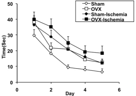
Escape latencies in the OVX group were significantly higher than in the Sham group (F 3,14 = 16.502, P < 0.01; Figure 1). The animals of the Sham-Ischemia group took a significantly longer time to reach the platform than those of the Sham group (F 3, 14 = 16.502, P < 0.01; Figure 1). In the OVX-Ischemia group, the escape latencies were also significantly higher than in the OVX group (F 3, 14 = 16.502, P < 0.001; Figure 1). The escape latencies in the OVX-Ischemia group were also significantly higher than those of the Sham-Ischemia group (F 3, 14 = 16.502,
Figure 1: The effects of ischemia on time
latency to reach the platform in Morris water maze (learning) of Sham and ovariectomised rats. Data are presented as mean SD (n= 6–8 in each group). The time latencies of the OVX group were significantly higher than those of the Sham group (P < 0.01). The animals of the Sham-Ischemia group took a significantly longer time to reach the platform compared to those of the Sham group (P < 0.01). In the OVX-Ischemia group, the escape latencies were also significantly higher than in the OVX group (P < 0.001). The escape latencies in the OVX-Ischemia group were also significantly higher than that of the Sham-Ischemia group (P < 0.05).
P < 0.05; Figure 1). In the probe trial, there were no significant differences in the time spent in the target quadrant (Q1) between the Sham and OVX groups (P > 0.05, Figure 2). However, the animals of the OVX group spent more time in the non- target quadrant (Q3) in comparison with those of the Sham group (F 3, 14 = 3.442, P < 0.05; Figure 2). The results also showed that the animals of the OVX-Ischemia group spent shorter times in the target quadrant (Q1) compared to those of the OVX group (F 3, 14 = 2.932, P < 0.05; Figure 2). In the rotarod test there were no significant differences between the Sham, OVX, Sham-Ischemia, and OVX-Ischemia groups when the animals were examined on days 3, 7, and 14 after ischemia (P > 0.05, Figure 3).
The results also showed that the escape latency to reach the platform in both the OVX- Ischemia-S 20 and OVX-Ischemia-S 60 groups were significantly lower than that of the OVX-Ischemia group (F 2, 14 = 20.792, P < 0.001; Figure 4). There was no significant difference between Ischemia-S 20 and OVX-Ischemia-S 60 groups (P > 0.05). In the probe trial, the animals of the OVX-Ischemia-S 60 group spent more time in the target quadrant (Q1) than those of the OVX-Ischemia group (F 2, 14 = 8.354, P < 0.05; Figure 5) but there was no significant difference between OVX-Ischemia-S 20 and OVX-Ischemia groups (P > 0.05). There was also no significant difference between Ischemia-S 20 and OVX-Ischemia-S 60 groups (P > 0.05). The animals of the OVX-Ischemia-S 60 group also spent shorter periods of time in the non-target quadrants (Q4) than those of the OVX-Ischemia group (F 2, 14 = 3.562, P < 0.05; Figure 5) but there was no significant difference between OVX-Ischemia-S 20 and OVX-Ischemia groups(P > 0.05). There was no significant difference between Ischemia-S 20 and OVX-Ischemia-S 60 groups (P > 0.05). In the rotarod test there were no significant differences between the Ischemia, OVX-Ischemia-S 20 and OVX-OVX-Ischemia-S 60 groups when the animals were examined on days 3, 7, and 14 after ischemia (P > 0.05, Figure 6).
Figure 2: The effects of ischemia on the time (second) spent in each quadrant during the probe trial in Morris water maze test (memory) of Sham and ovariectomised rats. Data are shown as mean SD of 6–8 animals per group. *P < 0.05 compared to Sham group,
+P < 0.05 compared to OVX group.
Figure 3: The effects of ischemia on rotarod test
performance (motor performance) of sham and ovariectomised rats. Data are shown as mean SD of 6–8 animals per group. There was no significant difference between groups.
Figure 4: The effects of soy extract on impaired
learning of ovariectomised rats induced by ischemia (time latency to reach the platform in Morris water maze). Data are presented as mean SD (n = 6–7 in each group). The results showed that the escape latency to reach the platform in both the OVX- Ischemia-S 20 and OVX-Ischemia-S 60 groups were significantly lower than that of the OVX-Ischemia group (both P < 0.001).
Figure 5: The effects of soy extract on impaired
Figure 6: The effects of soy extract on impaired rotarod performance (motor performance) of ovariectomised rats induced by ischemia. Data are shown as mean SD of 6–8 animals per group. There was no significant difference between groups.
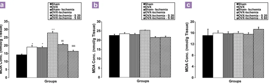
Figure 7: The malondialdehyde (MDA) concentrations in hippocampal (a), cortical (b), and cerebellar
(c) tissues were compared between groups using one way ANOVA test. Data are shown as mean SD of 6 – 8 animals per group *P < 0.05 compared to the OVX group, ++P < 0.01 compared to
the OVX group, $&P< 0.01 and $$$P < 0.01 compared to the OVX–Ischemia group.
30 = 28.24, P < 0.01 and P < 0.001, respectively; Figure 7a).
As figure 7b shows, there were no significant differences in cortical tissue MDA concentrations between the Sham-Ischemia and OVX-Ischemia groups compared to the Sham and OVX groups. There were also no significant differences between the OVX-Ischemia-S 20, OVX-Ischemia-S 60, and OVX-Ischemia groups (P > 0.05, Figure 7b). There were also no significant differences in MDA concentrations in cerebellum tissue between all groups (P > 0.05, Figure 7c).
Discussion
The results of the current study showed that deprivation of endogenous ovarian hormones impaired spatial learning and memory. The escape latencies to find the platform in ovariectomised rats were higher than those in the Sham-operated ones. The ovariectomised rats also spent shorter times in the target quadrant during the probe trial, as noted by the animals' ability to recall the situation of the platform. The results were in concordance with our previous studies which experimentally showed that deletion of ovarian hormones impaired learning and memory in rats (70,74–77). However, it was found that surgical menopause has no effect on cognitive function (78). Estrogen replacement therapy has been reported to prevent or delay cognitive decline in post-menopausal women and in estrogen-depleted animals (76,78). When the estrogen levels in older female rats were low, a significant reduction in synapses was seen in the hippocampus (79).
Regarding the neuroprotective effects of estrogens and their enhancing effects on memory which have been previously reported (29,30,83,84), it was assumed that the impairment in learning and memory due to cerebral ischemia may be different in the presence and absence of ovarian hormones. The results showed that the animals of the OVX-Ischemia group had higher latencies for finding the platform compared to other groups. They also spent shorter times in the target quadrant compared to all other groups. It does not seem that the Morris water maze impairments are due to the effects of motor movement; global ischemia did not affect the animals’ performance on the rotarod test.
Jover and colleagues also showed that estradiol in young males and female Jibril had a neuroprotective effect; however, they did not find this effect in OVX female rats (29,30). Moreover, other researchers have shown that estradiol in young rodents reduced the occurrence of brain damage and death of neurons in the hippocampus caused by global ischemia (29,30,83,84).
The molecular structure of soy phytoestrogens (SPs) is similar to that of estrogen therefore, they bind to estrogen receptors and mimic their functions (85). In concurrence with this idea, it has been previously shown that soy extract mimics the proconvulsant effects of estrogen in the pentylenetetrazole (PTZ)-induced seizure model (53,54). It has also been shown that these herbal compounds have beneficial effects on memory and the cognition of both menopausal women and OVX rats (45,86). They also have neuroprotective effects in vitro (87,88). Furthermore, it has been shown that these herbal compounds have behavioral benefices and neuroprotective effects against brain diseases such as Alzheimer’s, Parkinson’s, and cerebral stroke (42–44,89). Schreihofer et al. also showed that dietary soy (which contains high levels of phytoestrogens) have neuroprotective effects similar to estrogen and reduce the extent of brain injury in an OVX rat model of focal cerebral ischemia (89). Thus, we hypothesized that SPs would also affect learning and memory impairment due to cerebral ischemia. Interestingly, our results demonstrated that both the low (20 mg/kg) and high (60 mg/kg) doses of soy extract improved the Morris water maze task impairments due to cerebral ischemia in ovariectomised rats; however, there were no significant differences between the groups in the rotarod test results. In fact, our results confirmed that SPs can mimic the beneficial effects of ovarian hormones on memory, especially in the
global ischemia model. Azcoitia et al. also showed that an acute administration of soy extract or genistein prevented hippocampal neuronal loss against kainic acid-induced seizures (52). Although the exact mechanism(s) are not well-known, it might be suggested that soy extract (or the phytoestrogens of soy extract) also have these beneficial effects via estrogen receptors (ERs). As mentioned above, SPs bind to ERs and mimic the actions of endogenous estrogen. Cerebral vasodilatation may be another explanation for the effects of both endogenous estrogens and phytoestrogens, which were seen in the current results (90,91). Apoptosis is also an important cause of cell death after cerebral ischemia. Ischemia causes the mitochondria to activate intracellular pathways related to apoptosis (92). Besides the prevention of excessive glutamate release, manipulation of the balance between anti-apoptoic genes and proapototic ones has also been suggested (29,30,93). The antiapoptotic effect of estrogen is at least in part due to the decrease in caspase-3 after global ischemia (94).
Therefore, the antioxidant effects of soy might be suggested as a mechanism for the beneficial effects of soy in learning and memory impairment. The results of the present study confirmed the antioxidant effects as well the beneficial effects of soy and other phytoestrogens on the nervous system functions, including learning and memory (103,104).
Conclusion
In conclusion, it was shown that global ischemia impaired the learning and memory in both the presence and absence of ovarian hormones. The results also showed that soy extract attenuated learning and memory impairments due to brain ischemia in ovariectomised rats. The antioxidant effects of soy extract might be suggested as a possible mechanism.
Acknowledgment
The authors would like to thank the Vice Chancellor of Research Affairs of Mashhad University of Medical Sciences for financial assistance.
Conflicts of Interest
None.
Funds
Vice Chancellor of Research Affairs of Mashhad University of Medical Sciences for financial.
Authors’ Contributions
Conception and design and critical revision of the article for the important intellectual content: MH Analysis and interpretation of the data: MS Drafting of the article: FV
Statistical expertise: MAH
Administrative, technical or logistic support: HRS Collection and assembly of data: FV, MR
Correspondence
Dr Mahmoud Hosseini
PhD (Mashhad University of Medical Sciences) Neurogenic Inflammation Research Center School of Medicine
Mashhad University of Medical Sciences Mashhad, Postal Code 9177948564 Iran
Tel: +98-511-8828 565 Fax: +98-511-8828 564 Email: [email protected]
Refrences
1. Feigin VL, Lawes CM, Bennett DA, Barker-Collo SL, Parag V. Worldwide stroke incidence and early case fatality reported in 56 population-based studies: a systematic review. Lancet Neurol. 2009;8(4):355– 369.
2. Strong K, Mathers C, Bonita R. Preventing stroke: saving lives around the world. Lancet Neurol.
2007;6(2):182–187.
3. Etgen AM, Jover-Mengual T, Suzanne Zukin R. Neuroprotective actions of estradiol and novel estrogen analogs in ischemia: Translational implications. Front Neuroendocrinol. 2011;32(3):336–352.
4. Hua F, Ma J, Li Y, Ha T, Xia Y, Kelley J, et al. The development of a novel mouse model of transient global cerebral ischemia. Neurosci Lett. 2006;400 (1–2):69–74.
5. Schaller B, Graf R. Cerebral Ischemia and Reperfusion: The Pathophysiologic Concept as a Basis for Clinical Therapy. J Cereb Blood Flow Metab. 2004;24(4):351–371.
6. Cui K, Luo X, Xu K, Ven Murthy MR. Role of oxidative stress in neurodegeneration: recent developments in assay methods for oxidative stress and nutraceutical antioxidants. Prog Neuropsychopharmacol Biol Psychiatry. 2004;28(5):771–799.
7. Mršić-Pelčić J, Pelěić G, Peternel S, Pilipović K, Simonić A, Župan G. Effects of the hyperbaric oxygen treatment on the Na+, K+-ATPase and superoxide dismutase activities in the optic nerves of global cerebral ischemia-exposed rats.
Prog Neuropsychopharmacol Biol Psychiatry.
2004;28(4):667–676.
8. Halliwell B. Oxygen radicals as key mediators in neurological disease: fact or fiction? Annals of neurology. 1992;32(S1):S10–S15.
9. Coyle JT, Puttfarcken P. Oxidative stress, glutamate, and neurodegenerative disorders. Science. 1993;
262(5134):689–695.
10. Frlich L, Riederer P. Free radical mechanisms in dementia of Alzheimer type and the potential for antioxidative treatment. Arzneimittel for schung.
1995;45(3A):443–446.
11. Qureshi GA, Baig S, Sarwar M, Parvez SH. Neurotoxicity, oxidative stress and cerebrovascular disorders. Neurotoxicology. 2004;25(1–2):121–138. 12. Sharma DR, Sunkaria A, Bal A, Bhutia YD, Vijayaraghavan R, Flora SJ, et al. Neurobehavioral impairments, generation of oxidative stress and release of pro-apoptotic factors after chronic exposure to sulphur mustard in mouse brain. Toxicol Appl Pharmacol. 2009;240(2):208–218.
14. Bickford PC, Gould T, Briederick L, Chadman K, Pollock A, Young D, et al. Antioxidant-rich diets improve cerebellar physiology and motor learning in aged rats. Brain Res. 2000;866(1–2):211–217. 15. Baydas G, Ozer M, Yasar A, Tuzcu M, Koz ST.
Melatonin improves learning and memory performances impaired by hyperhomocysteinemia in rats. Brain Res. 2005;1046(1–2):187–194.
16. Dahlqvist P, Ronnback A, Bergstrom SA, Soderstrom I, Olsson T. Environmental enrichment reverses learning impairment in the Morris water maze after focal cerebral ischemia in rats. Eur J Neurosci. 2004;19(8):2288–2298.
17. Kiray M, Bagriyanik HA, Pekcetin C, Ergur BU, Uysal N. Protective effects of deprenyl in transient cerebral ischemia in rats. Chin J Physiol. 2008;51(5):275– 281.
18. Voneuler M, Bendel O, Bueters T, Sandin J, Voneuler G. Profound but transient deficits in learning and memory after global ischemia using a novel water maze test. Behavioural Brain Research.
2006;166(2):204–210.
19. Gawel S, Wardas M, Niedworok E, Wardas P. [Malondialdehyde (MDA) as a lipid peroxidation marker]. Wiad Lek. 2004;57(9–10):453–455. 20. Flamm ES, Demopoulos HB, Seligman ML, Ransohoff
J. Possible molecular mechanisms of barbiturate-mediated protection in regional cerebral ischemia.
Acta Neurol Scand Suppl. 1977;64:150–151.
21. Kogure K, Watson BD, Busto R, Abe K. Potentiation of lipid peroxides by ischemia in rat brain. Neurochem Res. 1982;7(4):437–454.
22. Yamamoto M, Shima T, Uozumi T, Sogabe T, Yamada K, Kawasaki T. A possible role of lipid peroxidation in cellular damages caused by cerebral ischemia and the protective effect of alpha-tocopherol administration.
Stroke. 1983;14(6):977–982.
23. Bromont C, Marie C, Bralet J. Increased lipid peroxidation in vulnerable brain regions after transient forebrain ischemia in rats. Stroke. 1989;20(7):918–924.
24. McCullough LD, Zeng Z, Blizzard KK, Debchoudhury I, Hurn PD. Ischemic nitric oxide and poly (ADP-ribose) polymerase-1 in cerebral ischemia: male toxicity, female protection. J Cereb Blood Flow Metab. 2005;25(4):502–512.
25. Strom JO, Theodorsson A, Theodorsson E. Mechanisms of Estrogens’ Dose–Dependent Neuroprotective and Neurodamaging Effects in Experimental Models of Cerebral Ischemia. Int J Mol Sci. 2011;12(3):1533–1562.
26. Alkayed NJ, Murphy SJ, Traystman RJ, Hurn PD, Miller VM. Neuroprotective Effects of Female Gonadal Steroids in Reproductively Senescent Female Rats. Editorial Comment. Stroke. 2000;31(1):161–168. 27. Daniel J. Estrogen replacement in ovariectomized
rats affects strategy selection in the Morris water maze. Neurobiol Learn Mem. 2004;82(2):142–149.
28. Plamondon H, Morin A, Charron C. Chronic 17β-estradiol pretreatment and ischemia-induced hippocampal degeneration and memory impairments: A 6-month survival study. Horm Behav. 2006;50(3):361–369.
29. Jover-Mengual T, Miyawaki T, Latuszek A, Alborch E, Zukin RS, Etgen AM. Acute estradiol protects CA1 neurons from ischemia-induced apoptotic cell death via the PI3K/Akt pathway. Brain Res. 2010;1321:1– 12.
30. Jover-Mengual T, Zukin RS, Etgen AM. MAPK Signaling Is Critical to Estradiol Protection of CA1 Neurons in Global Ischemia. Endocrinology. 2006;148(3):1131–1143.
31. Bagetta G, Chiappetta O, Amantea D, Iannone M, Rotiroti D, Costa A, et al. Estradiol reduces cytochrome c translocation and minimizes hippocampal damage caused by transient global ischemia in rat. Neurosci Lett. 2004;368(1):87–91.
32. Gulinello M, Lebesgue D, Jover-Mengual T, Zukin RS, Etgen AM. Acute and chronic estradiol treatments reduce memory deficits induced by transient global ischemia in female rats. Horm Behav.
2006;49(2):246–260.
33. Rau SW, Dubal DB, Bottner M, Gerhold LM, Wise PM. Estradiol attenuates programmed cell death after stroke-like injury. J Neurosci. 2003;23(36):11420– 11426.
34. Behl C, Widmann M, Trapp T, Holsboer F. 17-beta estradiol protects neurons from oxidative stress-induced cell death in vitro. Biochem Biophys Res Commun. 1995;216(2):473–82.
35. Green PS, Gordon K, Simpkins JW. Phenolic A ring requirement for the neuroprotective effects of steroids. J Steroid Biochem Mol Biol. 1997;63 (4–6):229–235.
36. Singer CA, Figueroa-Masot XA, Batchelor RH, Dorsa DM. The mitogen-activated protein kinase pathway mediates estrogen neuroprotection after glutamate toxicity in primary cortical neurons. J Neurosci.
1999;19(7):2455.
37. Singh M, Sétáló G Jr, Guan X, Warren M, Toran– Allerand CD. Estrogen-induced activation of mitogen-activated protein kinase in cerebral cortical explants: convergence of estrogen and neurotrophin signaling pathways. J Neurosci. 1999;19(4):1179.
38. Green PS, Gridley KE, Simpkins JW. Nuclear estrogen receptor-independent neuroprotection by estratrienes: a novel interaction with glutathione.
Neuroscience. 1998;84(1):7–10.
39. Casini ML, Marelli G, Papaleo E, Ferrari A, D'Ambrosio F, Unfer V. Psychological assessment of the effects of treatment with phytoestrogens on postmenopausal women: a randomized, double-blind, crossover, placebo-controlled study. Fertil Steril. 2006;85(4):972–978.
41. Rodriguez-Landa JF, Hernandez-Figueroa JD, Hernandez-Calderon Bdel C, Saavedra M. Anxiolytic-like effect of phytoestrogen genistein in rats with long-term absence of ovarian hormones in the black and white model. Prog Neuropsychopharmacol Biol Psychiatry. 2009;33(2):367–372.
42. Sarkaki A, Amani R, Badavi M, Moghaddam AZ, Aligholi H, Safahani M, et al. Pre-treatment effect of different doses of soy isoflavones on spatial learning and memory in an ovariectomized animal model of Alzheimer's disease. Pak J Biol Sci. 2008;11(8):1114– 1119.
43. Sarkaki A, Badavi M, Aligholi H, Moghaddam AZ. Preventive effects of soy meal (+/- isoflavone) on spatial cognitive deficiency and body weight in an ovariectomized animal model of Parkinson's disease.
Pak J Biol Sci. 2009;12(20):1338–1345.
44. Liu LX, Chen WF, Xie JX, Wong MS. Neuroprotective effects of genistein on dopaminergic neurons in the mice model of Parkinson's disease. Neurosci Res.
2008;60(2):156–161.
45. Pan Y, Anthony M, Watson S, Clarkson TB. Soy phytoestrogens improve radial arm maze performance in ovariectomized retired breeder rats and do not attenuate benefits of 17beta-estradiol treatment.
Menopause. 2000;7(4):230–235.
46. Pan M, Li Z, Yeung V, Xu RJ. Dietary supplementation of soy germ phytoestrogens or estradiol improves spatial memory performance and increases gene expression of BDNF, TrkB receptor and synaptic factors in ovariectomized rats. Nutr Metab (Lond). 2010;7:75. doi: 10.1186/1743-7075-7-75.
47. Grady D, Gebretsadik T, Kerlikowske K, Ernster V, Petitti D. Hormone replacement therapy and endometrial cancer risk: a meta-analysis. Obstet Gynecol. 1995;85(2):304–313.
48. Hammond CB. Women's concerns with hormone replacement therapy--compliance issues. Fertil Steril.
1994;62(6 Suppl 2):157S–160S.
49. Linford NJ, Dorsa DM. 17beta-Estradiol and the phytoestrogen genistein attenuate neuronal apoptosis induced by the endoplasmic reticulum calcium-ATPase inhibitor thapsigargin. Steroids. 2002;67(13–14):1029–1040.
50. Peterson G, Barnes S. Genistein inhibition of the growth of human breast cancer cells: independence from estrogen receptors and the multi-drug resistance gene. Biochem Biophys Res Commun. 1991;179(1):661–667.
51. Schreihofer DA, Redmond L. Soy phytoestrogens are neuroprotective against stroke-like injury in vitro.
Neuroscience. 2009;158(2):602–609.
52. Azcoitia I, Moreno A, Carrero P, Palacios S, Garcia-Segura LM. Neuroprotective effects of soy phytoestrogens in the rat brain. Gynecol Endocrinol.
2006;22(2):63–69.
53. Ebrahimzadeh Bideskan AR, Hosseini M, Mohammadpour T, Karami R, Khodamoradi M, Nemati Karimooy H, et al. Effects of soy extract on pentylenetetrazol-induced seizures in ovariectomized rats. Zhong Xi Yi Jie He Xue Bao. 2011;9(6):611–618. 54. Mohammadpour T, Hosseini M, Karami R, Sadeghnia HR, Ebrahimzadeh Bideskan AR, Enayatfard L. Estrogen-dependent effect of soy extract on pentylenetetrazole-induced seizures in rats. Zhong Xi Yi Jie He Xue Bao. 2012;10(12):1470–1476.
55. Bueters T, von Euler M, Bendel O, von Euler G. Degeneration of newly formed CA1 neurons following global ischemia in the rat. Exp Neurol.
2008;209(1):114–124.
56. Jones BJ, Roberts DJ. The quantiative measurement of motor inco-ordination in naive mice using an acelerating rotarod. J Pharm Pharmacol. 1968;20 (4):302–304.
57. Jones BJ, Roberts DJ. A rotarod suitable for quantitative measurements of motor incoordination in naive mice. Naunyn Schmiedebergs Arch Exp Pathol Pharmakol. 1968;259(2):211.
58. Pritchett K, Mulder GB. The rotarod. Contemp Top Lab Anim Sci. 2003;42(6):49.
59. Lalonde R, Hayzoun K, Selimi F, Mariani J, Strazielle C. Motor coordination in mice with hotfoot, Lurcher, and double mutations of the Grid2 gene encoding the delta–2 excitatory amino acid receptor. Physiol Behav. 2003;80(2–3):333–339.
60. Buitrago MM, Schulz JB, Dichgans J, Luft AR. Short and long-term motor skill learning in an accelerated rotarod training paradigm. Neurobiol Learn Mem. 2004;81(3):211–216.
61. Kuhn PL, Petroulakis E, Zazanis GA, McKinnon RD. Motor function analysis of myelin mutant mice using a rotarod. Int J Dev Neurosci. 1995;13(7):715–22. 62. Ogura T, Ogata M, Akita H, Jitsuki S, Akiba L, Noda
K, et al. Impaired acquisition of skilled behavior in rotarod task by moderate depletion of striatal dopamine in a pre-symptomatic stage model of Parkinson's disease. Neurosci Res. 2005;51(3):299– 308.
63. Rozas G, Guerra MJ, Labandeira-Garcia JL. An automated rotarod method for quantitative drug-free evaluation of overall motor deficits in rat models of parkinsonism. Brain Res Brain Res Protoc.
1997;2(1):75–84.
64. Watzman N, Barry H, 3rd, Kinnard WJ, Jr., Buckley JP. Influence of certain parameters on the performance of mice on the rotarod. Arch Int Pharmacodyn Ther.
1967;169(2):362–374.
66. Morris R. Developments of a water-maze procedure for studying spatial learning in the rat. J Neurosci Methods. 1984;11(1):47–60.
67. Nunez J. Morris Water Maze Experiment. J Vis Exp.
2008;19:897. doi: 10.3791/897.
68. Vorhees CV, Williams MT. Morris water maze: procedures for assessing spatial and related forms of learning and memory. Nat Protoc. 2006;1(2):848– 858.
69. Hosseini M, Dastghaib SS, Rafatpanah H, Hadjzadeh MA, Nahrevanian H, Farrokhi I. Nitric oxide contributes to learning and memory deficits observed in hypothyroid rats during neonatal and juvenile growth. Clinics (Sao Paulo). 2010;65(11):1175–1181. 70. Hosseini M, Feizpour A, Rezaeipour M, Amani A,
Saffarzadeh F, Farrokhi E. Chronic Treatment by L-NAME differently Affects Morris Water Maze Tasks in Ovariectomized and Naïve Female Rats. Basic Clin Neurosci. 2011;2(4):47–52.
71. Monteiro SC, Matté C, Bavaresco CS, Netto CA, Wyse ATS. Vitamins E and C pretreatment prevents ovariectomy-induced memory deficits in water maze.
Neurobiol Learn Mem. 2005;84(3):192–199. 72. Li L-X, Cheng Y-F, Lin H-B, Wang C, Xu J-P, Zhang
H-T. Prevention of cerebral ischemia-induced memory deficits by inhibition of phosphodiesterase-4 in rats. Metab Brain Dis. 2011;26(1):37–47.
73. Hosseini M, Pourganji M, Khodabandehloo F, Soukhtanloo M, Zabihi H. Protective Effect of L-Arginine against Oxidative Damage as a Possible Mechanism of its Bene. cial Properties on Spatial Learning in Ovariectomized Rats. Basic Clin Neurosci. 2012;3(5):36–44.
74. Azizi-Malekabadi H, Hosseini M, Saffarzadeh F, Karami R, Khodabandehloo F. Chronic treatment with the nitric oxide synthase inhibitor, L-NAME, attenuates estradiol-mediated improvement of learning and memory in ovariectomized rats. Clinics (Sao Paulo). 2011;66(4):673–679.
75. Azizi-Malekabadi H, Hosseini M, Soukhtanloo M, Sadeghian R, Fereidoni M, Khodabandehloo F. Different effects of scopolamine on learning, memory, and nitric oxide metabolite levels in hippocampal tissues of ovariectomized and Sham-operated rats.
Arq Neuropsiquiatr. 2012;70(6):447–452.
76. Hosseini M, Headari R, Oryan S, Hadjzadeh MA, Saffarzadeh F, Khazaei M. The effect of chronic administration of L-arginine on the learning and memory of estradiol-treated ovariectomized rats tested in the morris water maze. Clinics.
2010;65(8):803–807.
77. Saffarzadeh F, Eslamizade MJ, Nemati Karimooy HA, Hadjzadeh MA, Khazaei M, Hosseini M. The effect of L-arginine on Morris water maze tasks of ovariectomized rats. Acta Physiol Hung.
2010;97(2):216–223. doi: 10.1556/APhysiol.97.2010. 2.8.
78. Vearncombe KJ, Pachana NA. Is cognitive functioning detrimentally affected after early, induced menopause? Menopause. 2009;16(1):188–198. 79. Adams MM, Shah RA, Janssen WGM, Morrison JH.
Different modes of hippocampal plasticity in response to estrogen in young and aged female rats. Proc Natl Acad Sci U S A. 2001;98(14):8071–8076.
80. Harukuni I, Bhardwaj A. Mechanisms of Brain Injury after Global Cerebral Ischemia. Neurol Clin.
2006;24(1):1–21.
81. Huang Y, McNamara JO. Ischemic stroke: "acidotoxicity" is a perpetrator. Cell. 2004;118(6): 665–666.
82. Min D, Mao X, Wu K, Cao Y, Guo F, Zhu S, et al. Donepezil attenuates hippocampal neuronal damage and cognitive deficits after global cerebral ischemia in gerbils. Neurosci Lett. 2012;510(1):29–33. doi: 10.1016/j.neulet.2011.12.064.
83. De Butte-Smith M, Nguyen AP, Zukin RS, Etgen AM, Colbourne F. Failure of estradiol to ameliorate global ischemia-induced CA1 sector injury in middle-aged female gerbils. Brain Res. 2007;1153:214–220. 84. Miller NR, Jover T, Cohen HW, Zukin RS, Etgen
AM. Estrogen can act via estrogen receptor alpha and beta to protect hippocampal neurons against global ischemia-induced cell death. Endocrinology. 2005;146(7):3070–3079.
85. Molteni A, Brizio-Molteni L, Persky V. In vitro hormonal effects of soybean isoflavones. J Nutr.
1995;125(3 Suppl):751S–756S.
86. Kritz-Silverstein D, Von Muhlen D, Barrett-Connor E, Bressel MA. Isoflavones and cognitive function in older women: the SOy and Postmenopausal Health In Aging (SOPHIA) Study. Menopause.
2003;10(3):196–202.
87. Zhao L, Chen Q, Brinton RD. Neuroprotective and neurotrophic efficacy of phytoestrogens in cultured hippocampal neurons. Exp Biol Med (Maywood). 2002;227(7):509–519.
88. Gélinas S, Martinoli MG. Neuroprotective effect of estradiol and phytoestrogens on MPP+ induced cytotoxicity in neuronal PC12 cells. J Neurosci Res.
2002;70(1):90–96.
92. Niizuma K, Yoshioka H, Chen H, Kim GS, Jung JE, Katsu M, et al. Mitochondrial and apoptotic neuronal death signaling pathways in cerebral ischemia.
Biochim Biophys Acta. 2010;1802(1):92–99. 93. Qian Y, Guan T, Huang M, Cao L, Li Y, Cheng H, et
al. Neuroprotection by the soy isoflavone, genistein, via inhibition of mitochondria-dependent apoptosis pathways and reactive oxygen induced-NF-kappaB activation in a cerebral ischemia mouse model.
Neurochem Int. 2012;60(8):759–767. doi: 10.1016/j. neuint.2012.03.
94. Jover T, Tanaka H, Calderone A, Oguro K, Bennett MV, Etgen AM, et al. Estrogen protects against global ischemia-induced neuronal death and prevents activation of apoptotic signaling cascades in the hippocampal CA1. J Neurosci. 2002;22(6):2115– 2124.
95. Pan Y, Anthony M, Clarkson TB. Evidence for up-regulation of brain-derived neurotrophic factor mRNA by soy phytoestrogens in the frontal cortex of retired breeder female rats. Neurosci Lett.
1999;261(1–2):17–20.
96. Occhiuto F, Zangla G, Samperi S, Palumbo DR, Pino A, De Pasquale R, et al. The phytoestrogenic isoflavones from Trifolium pratense L. (Red clover) protects human cortical neurons from glutamate toxicity. Phytomedicine. 2008;15(9):676–682. 97. Lee YW, Lee WH. Protective effects of genistein
on proinflammatory pathways in human brain microvascular endothelial cells. J Nutr Biochem.
2008;19(12):819–825.
98. Czlonkowska A, Ciesielska A, Gromadzka G, Kurkowska-Jastrzebska I. Estrogen and cytokines production - the possible cause of gender differences in neurological diseases. Curr Pharm Des.
2005;11(8):1017–1030.
99. Culmsee C, Vedder H, Ravati A, Junker V, Otto D, Ahlemeyer B, et al. Neuroprotection by estrogens in a mouse model of focal cerebral ischemia and in cultured neurons: evidence for a receptor-independent antioxidative mechanism. J Cereb Blood Flow Metab. 1999;19(11):1263–1269.
100. Muley MM, Thakare VN, Patil RR, Bafna PA, Naik SR. Amelioration of cognitive, motor and endogenous defense functions with silymarin, piracetam and protocatechuic acid in the cerebral global ischemic rat model. Life Sci. 2013;93(1):51–57.
101. Saito T, Nito C, Ueda M, Inaba T, Kamiya F, Muraga K, et al. Continuous oral administration of atorvastatin ameliorates brain damage after transient focal ischemia in rats. Life Sci. 2014;94(2):106–114. 102. Viswanatha GL, Shylaja H, Mohan CG. Alleviation of
transient global ischemia/reperfusion–induced brain injury in rats with 1,2,3,4,6-penta-O-galloyl-beta-d-glucopyranose isolated from Mangifera indica. Eur J Pharmacol. 2013;720(1–3):286–23. doi: 10.1016/j. ejphar.2013.10.016.
103. Bansal N, Parle M. Soybean supplementation helps reverse age- and scopolamine-induced memory deficits in mice. J Med Food. 2010;13(6):1293–1300. 104. Ferrari CK. Functional foods, herbs and nutraceuticals: towards biochemical mechanisms of healthy aging.