149
Clarithromycin Resistance And Genetic Pattern
Of Helicobacter Pylori In A Group Of Patients
With Peptic Ulcer Disease In Alexandria, Egypt
Mohamed A Alhammad, Hadir EL-Kady, Yaman Hamed
Abstract: Clarithromycin resistance is one of the main predictors of eradication treatment failure in Helicobacter pylori (H. pylori) infections. Clarithromycin-based regimens were commonly used as a first-line therapy for H. pylori-positive patients. Lately, cure rates of H. pylori infection are decreasing to as low as 60% and are inversely correlated with antibiotic resistance rates that have crossed the 15-20% threshold. Monitoring of antibiotic susceptibility of H. pylori can be achieved through molecular methods; which stand out as an attractive alternative to conventional culture-based methods. The 23S rRNA Real-time PCR has several advantages in detection of H. pylori resistance to antibiotics; such as short working time, a high specificity up to 100% and low risk of contamination. This study aimed to detect clarithromycin resistance and genetic pattern of H. pylori in a group of 50 patients suffering from symptoms suggestive of gastrointestinal diseases. Gastric biopsy specimens were taken by endoscopy at the Gastroenterology Department of Alexandria Main University Hospital. Genotyping of H. pylori strains using multiplex PCR to detect CagA and VacA genes and detection of point mutations conferring clarithromycin resistance using a 23 S rRNA real time PCR was carried out. The majority (98%) of H. pylori strains detected in patients were CagA positive while only 28/50 (56%) were VacA positive. Most of the strains (67.86%) expressed the s2 (non toxigenic) allele and the most common genotype was VacA s2m1; expressed by 39.3% of strains. All H. pylori strains of the control group were sensitive to clarithromycin while resistance was detected in 26% of strains recovered from cases. The majority (77%) of point mutations responsible for resistance to clarithromycin were due to A-G transition at position 2143 while only 23% of which were due to A-C transition at position 2142.
Key words: H. pylori, PUD, genotyping, CagA, VacA, 23S Rrna, clarithromycin resistance, multiplex PCR, real-time PCR
————————————————————
1
INTRODUCTION:
Helicobacter pylori (H. pylori) is a Gram-negative bacterium that causes chronic stomach infections and is known as the main cause of gastric ulcers and subsequently may lead to development of gastric cancer if untreated [1]. The eradication therapy of H. pylori does not only heal gastritis of peptic ulcer disease (PUD) but it also prevents the spread and recurrence of infection and reduces the risk of development of gastric cancer; thus saving further costs required for treatment [2]. Virulence genes of H. pylori as cytotoxin-associated A (CagA ) gene and vacuolating toxin A (VacA ) gene are implicated in epithelium damage of gastric mucosa leading to gastric atrophy that can later progress on to gastric carcinoma. Therefore, it is important to identify patients who harbor these pathogenic strains to properly manage and limit this progression [3]. H. pylori clinical isolates are classified according to the presence or absence of Cag pathogenicity island (PAI); into Type I; associated with severe disease pathology, expresses functional VacA and contains the Cag PAI and Type II ; which lacks Cag PAI and has a non toxic form of VacA and is thus regarded as less virulent [4].
VacA gene comprises variable regions (s, m and i) and it encodes a vacuolating cytotoxin; which leads to epithelial cell damage. The s region (encoding the signal peptide) exists as either s1 or s2 allele and the m (middle) region presents as m1 or m2 allele. The mosaic combination of s and m alleles determines the level of cytotoxin production; which is associated to the degree of pathogenicity of H. pylori [5]. The first-line empirical treatment for an H. pylori infection; proposed at the First Maastricht conference, employs a triple drug regimen using one of the following antibiotics (tetracycline, amoxicillin or clarithromycin), along with metronidazole and a proton pump inhibitor (PPI) or bismuth salt. A quadruple regimen; with combined PPI and bismuth salt is employed when triple therapy regimens have failed [6]. In Egypt, the standard therapy for H. pylori infections combines PPI and metronidazole with one antibiotic; that is either clarithromycin or amoxicillin [7]. The causes of treatment failure in H. pylori infections can be grouped into microorganism-related factors, host-related factors and treatment-related factors. The most common factors include: ineffective penetration of antibiotics into the gastric mucosa, antibiotic inactivation by low stomach pH, lack of patient compliance and the emergence of acquired resistance to antibiotics by H. pylori [8]. Worldwide, H. pylori resistance towards different antibiotics is increasing and it is the key factor affecting efficacy of the current therapeutic regimens. Antibiotic resistance should be considered seriously; since its prevalence varies not only among diverse countries but also between two different periods in the same area [9]. H. pylori resistance to antibiotics has been attributed to the widespread use of certain antibiotics for infections other than H. pylori by the general population (i.e. metronidazole for parasitic and dental infections, tetracycline for respiratory and bowel diseases, amoxicillin for streptococcal pharyngitis and urinary tract infections and clarithromycin for respiratory infections) [10]. Clarithromycin is a macrolide antibiotic that inhibits protein synthesis of bacteria by binding to the 50 s subunit of bacterial _________________________
Mohamed A Alhammad: Medical Technology Department, Faculty of Public Health, Benghazi University. Telephone: +218944916865 Email:
alhammad_70@hotmail.com
Hadir El-Kady: Medical Laboratory Technology Department, Faculty of Allied Medical Sciences, Pharos University, Alexandria, Egypt. Telephone: +201146000465 Email: Hadir_elkady@yahoo.com Yaman Hamed: Fundamental and Applied Sciences
ribosomes. Acid stability and good absorption in the gastric mucosa renders it a good choice for H. pylori eradication when administered in high doses [11]. Some H. pylori strains have developed resistance to clarithromycin by mutation of the genetic sequence in the peptidyl transferase loop of the 23S rRNA. H. pylori resistance to clarithromycin is mainly due to an adenine-to-guanine (A-G) transition at positions 2142 and 2143 and to an adenine-to-cytosine (A-C) transversion at position 2142 [12]. Global claritrhomycin-resistance rates are contrasted among various countries from 1% up to above 65% [13,14]. Accordingly, it was recommended that in high-clarithromycin resistance regions (>15% resistance prevalence or dual clarithromycin and metronidazole resistance >15%); bismuth-containing quadruple therapy is the best first-line treatment [15]. The cure rate of PUD is between 0 and 50% when the H. pylori strain involved is resistant to clarithromycin, whereas it is around 90% when the strain is susceptible [12]. The need for rapid resistance screening procedures that replace the conventional cultural methods is mandatory as H. pylori is a fastidious slow growing bacterium; thus culture- based susceptibility testing is a time consuming and challenging task [16]. Numerous molecular-based techniques have been recommended as possible alternatives to conventional H. pylori detection because of their high sensitivity, rapid results and accuracy; although at a higher price [17].These methods include fluorescence in-situ hybridization, analysis of polymerase chain reaction (PCR) products by DNA enzyme immunoassay, restriction fragment length polymorphism (RFLP), oligonucleotide ligation assay (OLA) and reverse hybridization line probe assay [18]. More recently, real-time PCR methods combined with melting curve analysis by biprobes and hyprobes were performed on gastric biopsies in order to determine H. pylori susceptibility to clarithromycin [19]. This study aimed to detect the prevalence of H. pylori infection in DNA samples extracted directly from stomach biopsy specimens taken by endoscopy at the Gastroenterology Department of Alexandria Main University Hospital. The study also aimed to detect the point mutations that confer resistance to clarithromycin; using a 23S rRNA real-time PCR assay and to detect the genotype of H. pylori strains.
2
MATERIALS
AND
METHODS:
2.1Study design, sample size and study setting The present study was conducted on 50 Egyptian patients having upper gastrointestinal (GIT) symptoms as (epigastric pain, heartburn, dyspepsia, vomiting, hematemesis, melena and loss of weight) besides being infected with H. pylori. Also 20 asymptomatic controls; who had H. pylori infection and who accepted to participate in the present work, were enrolled in the study. They were all submitted to upper GIT endoscopy at the Gastroenterology Department of Alexandria Main University Hospital in the duration from October 2017 to March 2018.
2.2Selection criteria:
2.2.1Inclusion criteria
Positive for H. pylori infection; documented by a previous stool antigen test [20].
2.2.2Exclusion criteria a. Age > 70 years old.
b. Suffering from cardiac, hepatic or renal failure. c. History of bleeding and/or coagulation disorders.
All patients were asked to stop any anti-PUD drugs or antibiotics for at least two weeks before endoscopy. An informed consent was obtained from all enrolled subjects and the study was approved by the ethics committee of Alexandria University. Full history was taken from all cases covering their complaint and clinical condition and previous history of treatment of gastric ulcer or H. pylori infection.
2.3Samples’ collection
Upper GIT endoscopic examination was done to all subjects and gastric biopsy specimens were taken from the antrum of the stomach within 2 cm of the pyloric channel for PCR assay (one tissue biopsy) and for histopathological examination (three fragments).
2.4 Processing of samples
2.4.1Histopathological examination
All biopsy specimens for histological examination were fixed in 10% formalin, embedded in paraffin wax on the oriented edge and cut into 5 μm thick sequential sections. All tissue sections were stained with hematoxylin and eosin (H&E) for histological examination and also Giemsa staining was carried out to ascertain presence of H. pylori. Endoscopic observation and histopathologic confirmation were used to determine pathologies in the gastric mucosa.
2.4.2DNA extraction
DNA was extracted from gastric biopsy samples using QIAmp DNA Mini Kit according to a tissue DNA extraction protocol (Qiagen, Hilden, Germany). One hundred eighty microliter (µl) of ATL buffer and 20 µl of proteinase K were added to the sample and then incubated at 56°C for overnight with occasional vortexing until the pellet was completely lysed. After lysis of the sample, 200 µl of buffer AL were added to the sample and the mixture was incubated for 10 minutes at 70°C. The mixture was then combined with 200 µl of absolute ethanol and mixed by pulse-vortexing for 15 seconds. After that, the mixture was applied to a spin column which holds a silica gel membrane and was spun for 1 minute at 6,000 × g. The spin column was washed with 500 µl of buffer AW1 and then AW2 by centrifugation at 12,000 × g for 1 and 3 minutes, respectively. The DNA bound on the membrane was eluted by centrifugation with 50 µl of buffer AE after 5 minutes incubation at room temperature. The resulting DNA extracts were stored at -20°C until PCR assessment [19].
2.4.3PCR amplification for detection of H. pylori DNA PCR was performed with primers for urease gene (Ure C) [136 bp] 5’- AAGCTTTTAGGGGTGTTAGGGGTTT – 3' and 5' – CGCAATGCTTCAATTCTAAATCTTG – 3' indicative of H. pylori infection. Amplification was performed in a final volume of 50 µl of PCR mixture containing 0.8 µm of each primer, 10 mM of each deoxynucleotide triphosphate (dATP, dGTP, dTTP and dCTP), 10 mM tris HCl, 50 mM KCl. 0.1% triton X–100, 1.5 mM MgCl2, 1 unit of Taq DNA
151 Thermo Scientific, Fermentas) and10 µl of template DNA.
DNA amplification was carried out as follows: denaturation at 94°C for 5 minutes in the first cycle, followed by annealing for 30 seconds at 60°C, extension for 2 minutes at 72°C and denaturation for 30 seconds at 94°C for a total of 40 PCR cycles. The extension for the last cycle was increased to 5 minutes to ensure complete extension of the amplified fragment. Amplifications were performed with a thermal cycler (Genius Techne, England). The PCR products were resolved by 1.5 % agarose gel electrophoresis and were visualized after ethidium bromide (0.5 µg / ml) staining; using an UV transilluminator and photographed by Polaroid camera [19].
2.4.3.1 Multiplex PCR to detect H. pylori genotypes PCR was performed to detect CagA and VacA( s1/s2, m1/m2) alleles using primers for CagA, the signal (s1 & s2) and mid regions (m1 & m2) alleles of the VacA gene as shown in table I. Amplification was performed in a final volume of 50 µl of PCR mixture containing 0.5 µM of each primer, 10 mM of each deoxy nucleotide triphosphate (dATP, dGTP, dTTP and dCTP), 10 mM tris HCl, 50 mM KCl, 0.1% triton X– 100, 1.5 mM MgCl2, 1 unit of DNA
polymerase (Maxima Hot Start Green PCR Master Mix, Thermo Scientific, Fermentas) and 10 µl of template DNA. DNA amplification was carried out under the following general conditions: 30 cycles of 94° C for 1 minute, 55°C for 1 minute and 72°C for 1 minute. The amplified genes were detected by electrophoresis in a 1.5% agarose gel with ethidium bromide and bands were visualized using an UV transilluminator and photographed by Polaroid camera [21].
Table I:Nucleotide sequence of primers used in this study:
[21]
Size (bp) PCR product Nucleotide sequence
Primer Region
s1 259 ATG GAA ATA CAA CAA
AÇA CAC Va1-F
VacA s1 & VacA
s2 CTG CTT GAA TGC GCC s2 289
AAA C Va1-R
290 GGT CAA AAT GCG GTC
ATG G Va3-F
VacA
m1a CCA TTG GTA CCT GTA
GAA AC Va3-R
352 GGA GCC CCA GGA AAC
ATT G Va4-F
VacA m2
CAT AAC TAG CGC CTT GCA C Va4-R
349 GAT AAC AGG CAA GCT
TTT GAGG
CagA-F CagA
CTG CAA AAG ATT GTT TGG CAGA
CagA-R
Figure 1: Agarose gel showing specific bands of H. pylori ure C gene PCR products in lane 2,3 & 6. Lane M: 100 bp
DNA marker.
Figure 2: Agarose gel showing CagA (lane 2), Vac s2 (lane 2) & Vac m1 (lane 3) genes PCR products specific bands.
Lane M: 100 bp DNA marker.
2.4.3.1 Real-time PCR for clarithromycin susceptibility testing
A 23S rRNA real-time PCR assay has been used both for specific confirmed detection of H. pylori infection and for determination of point mutations in the 23S rRNA gene; responsible for clarithromycin resistance. Amplifications were performed with a light cycler (Applied Biosystem, One Step). For real-time PCR assay, 20µl reaction mixture was prepared as follows: Maxima SYBR Green/ROX q PCR Master Mix (Thermo Scientific, Fermentas), 4 mM MgCl2, 0.5
acquisition. A final cooling step was performed at 40°C for 10 seconds. Samples were considered to be H. pylori positive on determination of a specific melting curve. In the presence of mismatched bases between the probe and the target, melting curve analysis revealed a lower melting temperature than in case of a perfectly matched sequence. DNA extracts of H. pylori strains produced melting curves with melting temperatures of 63°C for the wild type (sensitive), 58°C for the A2142C mutant, and 54°C for the A2142G and A2143G mutants[19].
2.5Statistical analysis
This study was conducted on a PUD case sample of 50 patients besides an asymptomatic control sample of 20 patients. The data were categorized in respect to the patients, gender and age groups. The statistical analysis was done using the statistical package for social sciences program SPSS v.24[22].The difference in the average age of both case and control groups was tested using the two tailed Student t-test with a critical level . Chi-square test of association was done to determine the relationship between resistance to clarithromycin and the presence of the CagA and VacA genes. The case sample was tested against the control sample for all parameters of the study using the Chi-square test of independency. Each association test was considered statistically significant if the related p-value was less than 0.05.
3
RESULTS:
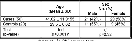
A total of 50 patients suffering from upper GIT symptoms and 20 asymptomatic control cases were enrolled in the current work. All the participants were positive for H. pylori Ag in stool and were submitted to upper GIT endoscopy. The mean age of the studied cases was 41.02 years with a standard deviation of 11.91. The studied controls mean age was 29.5 years with a standard deviation of 6.62. The mean age was significantly higher in the cases than in controls (p<0.001). The higher percentage of cases (58%) was females, while the higher percentage of controls (55%) was males. No significant difference between both groups regarding sex was noted (Table II).
Table II: Comparison of cases and controls according to their age and sex
Age (Mean SD)
Sex No. (%)
Male Female
Cases (50) 41.02 11.9155 21 (42%) 29 (58%) Controls (20) 29.5 6.62 11 (55%) 9 (45%) Test
(p-value)
t-test (p<0.001)*
2 p=0.32 t: t-test : Chi-square test
p: probability value *: Statistically significant at p ≤ 0.05
Histopathological examination of gastric biopsy specimens revealed normal gastric mucosa in controls. On the other hand, manifestations of benign PUD including gastric and duodenal ulcers were detected in all cases. No malignant changes were detected. PCR confirmed the presence of H. pylori infection in 100% of gastric biopsy samples taken by endoscopy from both cases and controls. Using multiplex
PCR, genotyping of H. pylori revealed that the majority of strains detected in 98% of cases were CagA positive and 56% of them were VacA positive. On the other hand, the majority of strains detected in 80% of controls were CagA negative and 95% were VacA negative. The difference between both groups was highly significant (p<0.001) (Table III).
Table III: Comparison of cases and controls regards the virulence factors of H. pylori strains
CagA VacA
+ve -ve +ve -ve
Cases (50) 49 (98%) 1(2%) 28 (56%) 22 (44%) Controls (20) 4 (20%) 16 (80%) 1 (5%) 19 (95%) Test
(p-value)
2 (p<0.001)*
2 (p<0.001)*
Only one of the 20 H. pylori strains that were detected in the control group was VacA positive and it was a VacA s2/m2 (non toxigenic) strain. Genotyping showed that the majority (67.86%) of H. pylori strains detected among cases expressed the s2 (non toxigenic) allele and that the VacA s2m1 genotype was the most common genotype expressed by 39.3% of strains (Table IV).
Table IV: Distribution of H. pylori strains detected in cases of PUD according to VacA genotyping
VacA gene Total
N %
Negative 22 44
S1m1 5 10
S1m2 4 8
S2m1 11 22
S2m2 8 16
Total 50 100
Using the PCR-guided genotyping, the prevalence of CagA and VacA gene expression was used to define the types of H. pylori strains. Accordingly, the strains were categorized into 3 groups: Type I (expressing both CagA and VacA genes), Type II (no expression of CagA or VacA genes) and Type III (expressing CagA gene only). The predominant H. pylori type in our cases was Type I (56%) while in controls H. pylori Type II was the most common type (80%). A significant difference between both groups was recorded (p<0.001) (Table V).
Table V: Classification of cases and controls according to genotypes of H. pylori strains
Type I Type II Type III Total Cases (50) 28 (56%) 1 (2%) 21 (42%) 50 (100%) Controls (20) 1 (5%) 16 (80%) 3 (15%) 20 (100%) Test
(p-value)
2 (p<0.001)*
Type I: CagA+ve /VacA+ve Type II: CagA-ve /VacA-ve Type III: only CagA +ve
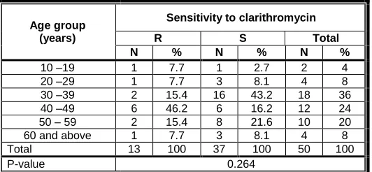
153 to clarithromycin while resistance was detected in 13/50
(26%) of H. pylori strains of the cases. The majority (77%) of the detected point mutations responsible for resistance to clarithromycin were due to A-G transition at position 2143 while only 23% were due to A-C transition at position 2142. 46.2% of the strains resistant to clarithromycin were detected among cases of the age group of 40-49 years and 53.8% of them were detected among males. Table VI and table VII show no significant difference among the cases harbouring clarithromycin resistant H. pylori strains regarding their age or sex.
Table VI: Distribution of clarithromycin resistant strains of H. pylori among PUD cases according to age of cases
Age group (years)
Sensitivity to clarithromycin
R S Total
N % N % N %
10 –19 1 7.7 1 2.7 2 4
20 –29 1 7.7 3 8.1 4 8
30 –39 2 15.4 16 43.2 18 36
40 –49 6 46.2 6 16.2 12 24
50 – 59 2 15.4 8 21.6 10 20
60 and above 1 7.7 3 8.1 4 8
Total 13 100 37 100 50 100
P-value 0.264
Table VII: Distribution of clarithromycin resistant strains of H. pylori among PUD cases according to sex
Sex
Sensitivity to larithromycin
R S Total
N % N % N %
Female 6 46.2 15 40.5 21 42
Male 7 53.8 22 59.2 29 58
Total 13 100 37 100 50 100
P-value 0.724
Also no significant association between the expression of either CagA or VacA genes and the resistance to clarithromycin was recorded. Nevertheless, the majority (92.3%) of resistant H. pylori strains that were detected in the cases were CagA positive and 53.8% were VacA positive; as shown in Table VIII and Table IX.
Table VIII: Association between clarithromycin resistance and CagA gene in H. pylori strains detected in PUD cases
CagA gene
Sensitivity to clarithromycin
R S Total
N % N % N %
Negative 1 7.7 0 0 1 2
Positive 12 92.3 37 100 49 98
Total 13 100 37 100 50 100
P-value 0.088
Table IX: Association between clarithromycin resistance and VacA gene in H. pylori strains detected in PUD patients
VacA gene
Sensitivity to clarithromycin
R S Total
N % N % N %
Negative 6 46.2 16 43.2 22 44
Positive 7 53.8 21 56.8 28 56
Total 13 100 37 100 50 100
P-value 0.856
4
DISCUSSION:
m1/m2) alleles using multiplex PCR. Virulence factors as CagA and VacA genes were identified in 98% and 56%, respectively of H. pylori strains recovered from cases of PUD compared to 80% and 95%, respectively of H. pylori strains detected in the control group. The predominant H. pylori genotype in our cases was type I (expressing both CagA and VacA genes); detected in 56% of cases. In controls; type II H. pylori (no expression of CagA or VacA genes) was the most common type detected in 80% of whom. This finding highlights that there is a significant positive association between H. pylori virulence factors (CagA and VacA) and the disease status of the examined individuals. The range of detection of the CagA gene among H. pylori strains varies between 17% up to 100% in different geographical regions [31,32]. The prevalence of CagA positive strains in our study (98%) ; which is considerably high, is in line with those obtained in similar Mexican and Japanese studies (86%) and (90%), respectively [33,34]. On the other hand, our results are remarkably higher than those of other studies in different countries including Pakistan (56%) [35], Gambia (58.3%) [21], Morocco (59.6%) [5], Bangladesh (70%) [36], India (77.27%) [37] and even in our country (Egypt) but in a different location; Mansoura governorate (62%) [30]. It is worth note that some studies, including the current one, reported a significantly higher prevalence of CagA virulent strains in older ages; which may suggest that this population may be at higher risk for developing more serious pathology of the gastric mucosa [21,37]. Regards VacA gene, it was detected at a nearly similar rate (58%) to the current findings (56%) among cases investigated by Zaki et al., in Egypt, (2016) [30]. Unlikely Saeidi et al., (2017) in Iran detected VacA gene in 100% of H. pylori strains [38]. H. pylori management stands out as a major concern for both microbiologists and gastroenterologists. The use of appropriate antibiotics is crucial for the success of treatment and recovery from H. pylori-related diseases. There is strong evidence showing that eradication of H. pylori infection reduces the risk of peptic and duodenal ulcers and likely gastric cancer; if treated early in the course of the disease [39]. The decision regarding which H. pylori treatment regimen to use is more difficult than in most other infections in that culture and susceptibility testing is often not available and physicians are required to make empiric choices (triple or quadruple drug regimens) [40]. However, recent reports show that the efficacy of these combinations has decreased; with successful cure in only 70% of cases [41]. According to the recent Kyoto Global Consensus Meeting, only regimens expected to result in at least a 90% eradication rate in a particular region should be adopted as empiric therapy [42]. Therefore the goal in designing a treatment regimen for H. pylori should focus on a strategy which results in a cure rate approaching 100% [43]. Eradication failure is alarming at the present time and is attributed to infection with an antimicrobial resistant H. pylori strain or emergence of a new resistant strain from a susceptible ancestor. As reported globally, the successful eradication attempts are inversely correlated with the antimicrobial resistance rates [29]. In 2017, the World Health Organization (WHO) published a list of bacteria for which new antibiotics are urgently needed. A total of 12 families were included; categorized according to their priority into: critical, high, and medium.
155 reported H. pylori resistance to clarithromycin at rates lower
than 20%. On the contrary, resistance rates higher than 20% were reported in Austria [19], Spain [59], Germany [60], Japan [61], Chile [62], Israel [63], Iran [64], Morocco [65], Jordan [39]and KSA [14]. Detection of low resistance rates to clarithromycin among H. pylori strains can be attributed to the mixed infection with susceptible and resistant genotypes; where up to a certain percentage the susceptible genotype suppresses the amplification of the mutant genotype [19]. H. pylori strains have been categorized according to geographical associations into several groups (East Asian type, south/central Asian type, Iberian/African type and European type). Thus, geographic differences associated with the presence of phylogeographic features of H. pylori may explain the existing variable antibiotic resistance rates [66]. The Toronto Consensus for the treatment of H. pylori infection (2016) recommended that optimal treatment of H. pylori infection requires careful attention to local antibiotic resistance and eradication patterns [67]. Another factor that can contribute to different resistance rates is the previous antibiotic treatment for H. pylori infection (higher resistance rates develop in treated than in naı¨ve patients) [68].The variance in the rates of resistance to clarithromycin among H. pylori strains could also be explained by time difference, use of different susceptibility tests, sample size, use of fresh or frozen biopsies and incubation conditions [69]. In the present work, 46.2% of clarithromycin resistant strains of H. pylori were detected in the group of patients ≥ 40 years of age. This could be attributed to recombination of the H. pylori genome over the course of decades and/or re-exposure to novel strains. Unlikely, gender didn’t have a significant association with the prevalence of resistance to clarithromycin among the current cases, whereas Diab et al., (2016) [39] reported male predominance and De Francesco et al., (2011) reported female predominance of clarithromycin resistant H. pylori strains [70] In the current study mutation site of 23S rRNA associated with clarithromycin resistance was determined as A2143G in the majority (77%) of H. pylori strains while the point mutation A2142C was detected in only 23% of which. Vala et al., (2016) [69] and Diab et al., (2018) [29] detected A2143G point mutation in 100% of H. pylori strains recovered from their studied patients in Iran and Egypt, respectively. A2143G mutation is strongly associated with failure of eradication therapy. Nevertheless, although most studies reported A2143G as the most common point mutation in clarithromycin resistant strains of H. pylori, still A2142G point mutation was reported by Naserpour et al., (2013) at an incidence higher than all other point mutations [71]. It is worth mentioning that despite absence of any significant association between the expression of virulence genes and the resistance to clarithromycin among H. pylori strains, yet the majority of resistant H. pylori strains detected in our cases were CagA positive (92.3%) and VacA positive (53.8%). This variability of clarithromycin resistance reported in different regions emphasizes the need to examine resistance rates in each geographic area. Pretreatment susceptibility testing for clarithromycin has become imperative specially if the prevalence of primary resistance in a community reaches 15%-20% [62].
5
CONCLUSION
Genotyping of H. pylori in patients may be a useful strategy for identifying those at high risk of PUD and gastric cancer. Clarithromycin triple therapy should be confined to patients with no previous history of macrolide exposure and who reside in areas where clarithromycin resistance is known to be low. Quantitative 23S rRNA real-time PCR assay is highly recommended for the accurate detection as well as quantification of H. pylori in gastric biopsy samples and for clarithromycin antibiotic susceptibility testing. Extended large scale studies are required to screen for antibiotic resistance pattern of H. pylori in the Egyptian population. This will have considerable cost/benefit implications because it will save the National Health System and patient resources; in terms of drugs, diagnostic tests and medical examination expenses.
Acknowledgment:
The authors would like to express special appreciation to all specialists at the Gastroenterology Department of Alexandria Main University Hospital for their great efforts in the selection of cases and for their continuous support and cooperation.
REFERENCES:
[1] Y.Thaker, A. Moon and A. Afzali. "Helicobacter pylori:A Review of Epidemiology,Treatment,and Management, " J. Clin. Gastroenterol. Treat., vol. 2, no. 2, pp.019, 2016.
[2] S. Shiota and Y. Yamaoka. "Strategy for the treatment of Helicobacter pylori infection," Curr. Pharm. Des., vol. 20, no. 28, pp.4489 – 4500, 2014.
[3] V. Shetty, M. Ballal, R, Lingadakai and A. Mukhopadhyay. "Determination of Helicobacter pylori Virulence Genes in Clinical Isolates of Symptomatic Patients from South Coastal Region of Karnataka – A Preliminary Work". Austin J. Gastroenterol., vol. 2, no. 1, pp.1031, 2015.
[4] A.S.M. Ali, I.M.S. Al-Kadmy, M.R. Ali, S.S. Al-Jubori. "Emergency of Helicobacter pylori resistance isolates obtained from Iraqi patients suffering Acute and chronic gastritis, " World J. Pharm. Sci. vol. 4, no. 7, pp. 18 – 23, 2016.
[5] S. Alaoui Boukhris, A. Amarti, K. El Rhazi, M. El Khadir, D.A. Benajah, S.A. Ibrahimi, C. Nejjari, M. Mahmoud, A. Souleimani and B. Bennani. "Helicobacter pylori Genotypes Associated with Gastric HistoPathological Damages in a Moroccan Population," PLoS ONE, vol. 8, no. 12, pp. e82646, 2013.
[7] N. Toshihiro, S. Hidekazu and H. Toshifumi. "Quinolones-Based third-line therapy for Helicobacter pylori eradication". J. Clin. Biochem. Nutr., vol. 44, no. 3,pp.119 – 124, Mar 2009.
[8] R. Ghotaslou, H.E. Leylabadlo and Y.M. Asl. " Prevalence of antibiotic resistance in Helicobacter pylori: A recent literature review". World J. Methodol.,vol.5, no.3, pp.164 – 174, Sep. 2015.
[9] D. Boltin, H, Ben-Zvi, T.T. Perets, Z. Kamenetsky, Z. Samra, R. Dickman and Y. Niv. "Trends in secondary antibiotic resistance of Helicobacter pylori from 2007 to 2014: has the tide turned," J. Clin. Microbiol., vol. 53, pp. 522 – 527, 2015.
[10] V. Papastergiou, S.D. Georgopoulos and S. Karatapanis. "Treatment of Helicobacter pylori infection: Past, present and future," World J. Gastrointest. Pathophysiol. vol. 5, pp. 392 – 399, 2014.
[11] Y-T Chu, Y-H. Wang, J-J. Wu, H-Y. Lei. "Invasion and multiplication of Helicobacter pylori in gastric epithelial cells and implications for antibiotic resistance," Infect. Immun. Vol. 78, no. 10, pp. 4157 – 4165, 2010.
[12] M. Kargar, S. Ghorbani-Dalini, A. Doosti and N. Souod. "Real-time PCR for Helicobacter pylori quantification and detection of clarithromycin resistance in gastric tissue from patients with gastrointestinal disorders," Res. Microbiol. Vol. 163, pp. 109 – 113, 2012.
[13] V. De Francesco, F. Giorgio, C. Hassan, G. Manes, L. Vannella and C. Panella. "Worldwide H. pylori antibiotic resistance: a systematic review," J. Gastrointestin. Liver Dis., vol. 19, pp. 409 – 414, Dec.2010.
[14] M. Bakri. "Prevalence of Helicobacter pylori infection and the incidence of urea and clarithromycin resistance gene 23S rRNA genotypes status in Saudi Arabia," Saudi. J. Biol. Sci., vol. 20, no, 1, pp. 75 – 78, 2013.
[15] R. Rana, S.L.Wang, J. Li, Y.X. Wang, Q.W. Rao and C.Q. Yang. "Helicobacter pylori infection: A recent approach to diagnosis and management". J. Biomed.,vol. 2, pp. 45 – 56, 2017.
[16] J.J. Redondo, P.M. Keller, R. Zbinden and K. Wagner. (2018). "A novel RTPCR for the detection of Helicobacter pylori and identification of clarithromycin resistance mediated by mutations in the 23S rRNA gene," Diagn. Microbiol. Infect. Dis., vol. 90, no. 1, pp. 1 – 6, 2018.
[17] Y. Zhang, F. Zhao, M. Kong, S. Wang, L. Nan, B. Hu, M.A. Olszewski, Y. Miao, D. Ji, W. Jiang, Y. Fang, J. Zhang, F. Chen, P. Xiang, Y. Wu and H. Zhao. "Validation of a high-throughput multiplex genetic detection system for Helicobacter pylori identification, quantification, virulence, and resistance analysis," Front. Microbiol., vol. 7, pp. 1401, 2016.
[18] C. Albaa, A. Blancoa and T. Alarco. "Antibiotic
resistance in Helicobacter pylori," Curr. Opin. Infect. Dis. vol. 30, pp. 489 – 497, 2017.
[19] C. Schabereiter-Gurtner, A.M. Hirschl, B. Dragosics, P. Hufnagl, S. Puz, Z. Kova´ch, M. Rotter and A. Makristathis. "Novel Real-Time PCR Assay for Detection of Helicobacter pylori Infection and Simultaneous Clarithromycin Susceptibility Testing of Stool and Biopsy Specimens," J. Clin. Microbiol.,vol. 42, no. 10, pp. 4512 – 4518, Oct. 2004.
[20] ABON Biopharm Co., 2012. ACON H. Infectious disease antigen detection. China: ABON BIOPHARM (HANGZHOU) CO., LTD. available from: http://www.abon.com.cn/Product/Index/
15080715055791.
[21] O. Secka, M. Antonio, M. Tapgun, D.E. Berg, C. Bottomley, V. Thomas, R. Walton, T. Corrah, R.A. Adegbola and J.E. Thomas. "PCR-based genotyping of Helicobacter pylori of Gambian children and adults directly from biopsy specimens and bacterial cultures," Gut. Pathog. Vol. 3, no. 1, pp. 5, 2011.
[22] IBM Corp. Released 2016. IBM SPSS Statistics for Windows, Version 24.0. Armonk, NY: IBM Corp .
[23] D.C. Angol, P. Ocama, T. Ayazika Kirabo, A. Okeng, I. Najjingo and F. Bwanga. "Helicobacter pylori from Peptic Ulcer Patients in Uganda Is Highly Resistant to Clarithromycin and Fluoroquinolones: Results of the GenoType HelicoDR Test Directly Applied on Stool," BioMed. Res. Int. Vol. 2017, pp. 5430723, 2017.
[24] S.G. Kim, H.K. Jung, H.L. Lee, J.Y. Jang, H. Lee, C.G. Kim, W.G. Shin, E.S. Shin and Y.C. Lee. "Guidelines for the diagnosis and treatment of Helicobacter pylori infection in Korea,". J. gastroenterol. hepatol. Vol. 29, pp. 1371 – 1386, 2014.
[25] P. Kjaeldgaard, A.P. Nielsen and M. Chen. Real Time PCR assay for detection of Helicobacter pylori infection and clarithromycin susceptibility in biopsy specimens in southern Denmark," JSM. Gastroenterol. Hepatol. vol. 4, no. 1, pp. 1052, 2016.
[26] R. Monno, F. Giorgio, P. Carmine, L. Soleo, V. Cinquepalmi and E. Ierardi. "Helicobacter pylori clarithromycin resistance detected by Etest and TaqMan real-time polymerase chain reaction: a comparative study," APMIS, vol. 120, pp. 712 – 717, 2012.
[27] Y.K. Wang, F.C. Kuo, C.J. Liu, M.C. Wu, H.Y. Shih, S.S. Wang, J.Y. Wu, C.H. Kuo, Y.K. Huang and D.C. Wu. "Diagnosis of Helicobacter pylori infection: Current options and developments," World J. Gastroenterol. Vol. 21, no. 40, pp. 11221 – 11235, 2015.
157 York: Springer Science Business Media, pp.13 -30,
2013.
[29] M. Diab, A. El-Shenawy, M. El-Ghannam, D. Salema, M. Abdelnasser, M. Shaheen, M. Abdel-Hady, E. El-Sherbini and M. Saber. "Detection of antimicrobial resistance genes of Helicobacter pylori strains to clarithromycin, metronidazole, amoxicillin and tetracycline among Egyptian patients," Egypt. j. med. hum. genet, vol.19, pp. 417–423, 2018.
[30] M.S. Zaki, A. Elewa, M.A. Ali and A. Shehta. "Study of Virulence Genes Cag A and Vac A in Helicobacter pylori Isolated from Mansoura University Hospital Patients by Multiplex PCR", Int. J. Curr. Microbiol. App. Sci., vol. 5, no. 2, pp. 154 – 160, 2016.
[31] F.A. Amer, R.H. El-Sokkary, M. Elahmady, T. Gheith, E.H. Abdelbary, Y. Elnagar and W.M. Abdalla. "Helicobacter pylori genotypes among patients in a university hospital in Egypt: identifying the determinants of disease severity, " JMID, vol. 3, pp. 109 – 115, 2013.
[32] N.R. Hussein. "Helicobacter pylori and gastric cancer in the Middle East: A new enigma?," World J. Gastroenterol. 2010;16:3226–3234.
[33] R. González-Vázquez, S. Herrera-González, M.G. Cordova-Espinoza, G. Zúñiga, S. Giono-Cerezo, J.M. Hernández-Hernández, G. León-Ávila. " Helicobacter pylori: detection of iceA1 and iceA2 genes in the same strain in Mexican isolates, " Arch. Med. Res. Vol. 43, no. 5, pp. 339 – 346, 2012.
[34] S. Maeda, K. Ogura, H. Yoshida, F. Kanai, T. Ikenoue, N. Kato, Y, Shiratori, M. Omata. " Major virulence factors, VacA and CagA, are commonly positive in Helicobacter pylori isolates in Japan," Gut, vol. 42, no. 3, pp. 338 – 343, 1998.
[35] J.Yakoob, S.Abid, Z.Abbasetal." Distribution of Helicobacter pylori virulence markers in patients with gastroduodenal diseases in Pakistan," BMC. Gastroenterol., vol. 9, no 87, pp. 1–7, 2009.
[36] T. Essawi, W. Hammoudeh, I. Sabri, W. Sweidan and M.A. Farraj. "Determination of Helicobacter pylori virulence genes in gastric biopsies by PCR," ISRN. Gastroenterol. Vol. 2013, pp. 606258, 2013.
[37] H.B. Pandya, H.H. Agravat and J.S. Patel. Prevalence of Specific Helicobacter Pylori CagA, VacA, iceA, UreC Genotypes and its Clinical Relevance in the Patients with Acid-Peptic Diseases. J. Clin. Diagn. Res. vol. 11, no. 8, pp. DC23-DC26, 2017.
[38] Y. Saeidi, A. Pournajaf, M. Gholami, M. Hasannejad-Bibalan, S. Yaghoubi, M. Khodabandeh, B. Emadi, E. Ferdosi-Shahandashti and R. Rajabnia. "Determination of Helicobacter pylori virulence-associated genes in duodenal ulcer and gastric biopsies," Med. J. Islam. Repub. Iran. vol. 31, pp. 95, 2017.
[39] A.F. Diab, F.H. Diab and S.S. Nassar. " Prevalence of Helicobacter pylori resistance to clarithromycin determined by 23S ribosomal RNA analysis in Jordan, " IAJAA, vol. 6, no. 2,pp. 4, 2016.
[40] Y. Li, E. Rimbara, S. Thirumurthi, A. Trespalacios, R. Reddy, S. Sabounchi, T.A. Attumi, D.Y. Graham. "Detection of clarithromycin resistance in Helicobacter pylori following noncryogenic storage of rapid urease tests for 30 days, " J. Dig. Dis., vol. 13, no. 1, pp. 54 – 59, Jan. 2012.
[41] T. Nishizawa, T. Maekawa, N. Watanabe, N. Harada, Y. Hosoda, M. Yoshinaga, T. Yoshio, H. Ohta, S. Inoue, T. Toyokawa, H. Yamashita, H. Saito, T. Kuwai, S. Katayama, E. Masuda, H. Miyabayashi, T. Kimura, Y. Nishizawa, M. Takahashi and H. Suzuki. " Clarithromycin Versus Metronidazole as First-line Helicobacter pylori Eradication: a multicenter, prospective, randomized controlled study in Japan," J. Clin. Gastroenterol. Vol. 49, no. 6, pp. 468 – 471, 2015.
[42] K. Sugano, J. Tack, E.J. Kuipers, D.Y. Graham, E.M. El-Omar, S. Miura, K. Haruma, M. Asaka, N. Uemura and P. Malfertheiner." Kyoto global consensus report on Helicobacter pylori gastritis," Gut, vol. 64, pp. 1353 – 1367, 2015.
[43] N. Stollman. " Helicobacter pylori Infection in the Era of Antibiotic Resistance, " Gastroenterol. Hepatol. vol. 12, no. 2, pp. 122 – 125, 2016.
[44] World Health Organization (WHO), "Global priority list of antibiotic-resistant bacteria to guide research, discovery and development of new antibiotics", Geneva: WHO, 2017.
[45] T. P. Van Boeckel, S. Gandra, A. Ashok, Q. Caudron, B. T. Grenfell, S. A. Levin, and R. Laxminarayan, "Global antibiotic consumption 2000 to 2010: an analysis of national pharmaceutical sales data", Lancet. Infect. Dis., vol. 14, pp. 742-750, Aug 2014.
[46] I. Thung, H. Aramin, V. Vavinskaya, S. Gupta, J.Y. Park, S.E. Crowe and M.A. Valasek, "Review article: the global emergence of Helicobacter pylori antibiotic resistance," Aliment. Pharmacol. Ther., vol. 43, pp. 514-533, Feb 2016.
[47] T. Champathai, S. Gonlachanvit and N. Chaichanawongsaroj, "Detection of A2143Gmutation
in23S rRNA geneassociated with
clarithromycinresistant H. pylori by Loop mediated isothermal amplification", J. Chem. Pharmac. Res., vol. 6, pp.148-155, 2014.
[49] D. Chen, S.A. Cunningham, N.C. Cole, P.C. Kohner, J.N. Mandrekar, and R. Patel, "Phenotypic and Molecular Antimicrobial Susceptibility of Helicobacter pylori," Antimicrob. Agents. Chemother., vol. 61, Apr 2017.
[50] S.A. Chisholm and R.J. Owen, "Application of polymerase chain reaction-based assays for rapid identification and antibiotic resistance screening of Helicobacter pylori in gastric biopsies," Diagn. Microbiol. Infect. Dis., vol. 61, pp. 67-71, May 2008.
[51] A.K. Lins, R.A. Lima, and M. Magalhaes, "Clarithromycin-resistant Helicobacter pylori in Recife, Brazil, directly identified from gastric biopsies by polymerase chain reaction," Arq. Gastroenterol, vol. 47, pp. 379-382, Oct-Dec 2010.
[52] D. Ghaith, M. Elzahry, G. Mostafa, S. Mostafa, R. Elsherif, and I. Ramzy, "Mutations affecting domain V of the 23S rRNA gene in Helicobacter pylori from Cairo, Egypt," J. Chemother., vol. 28, pp. 367-370, Oct 2016.
[53] A.I. Gunnarsdottir, H. Gudjonsson, H. Hardardottir, K.D. Jonsdottir, and E.S. Bjornsson, "Antibiotic susceptibility of Helicobacter pylori in Iceland," Infect. Dis. (Lond)., vol. 49, pp. 647-654, Sep 2017.
[54] V. De Francesco, A. Zullo, E. Ierardi, F. Giorgio, F. Perna, C. Hassan, S. Morini, C. Panella, and D. Vaira, "Phenotypic and genotypic Helicobacter pylori clarithromycin resistance and therapeutic outcome: benefits and limits," J. Antimicrob. Chemother., vol. 65, pp. 327-332, Feb 2010.
[55] M. Miftahussurur, A.F. Syam, I.A. Nusi, D. Makmun, L.A. Waskito, L.H. Zein, F. Akil, W.B. Uwan, and D. Simanjuntak, "Surveillance of Helicobacter pylori Antibiotic Susceptibility in Indonesia: Different Resistance Types among Regions and with Novel Genetic Mutations," PLoS One, vol. 11, pp. e0166199, 2016.
[56] N.R. Hussein, I. Tunjel, H.S. Majed, S.T. Yousif, S.I. Aswad, and M.S. Assafi, "Duodenal ulcer promoting gene 1 (dupA1) is associated with A2147G clarithromycin-resistance mutation but not interleukin-8 secretion from gastric mucosa in Iraqi patients," New Microbes New Infect., vol. 6, pp. 5-10, Jul 2015.
[57] K. Ben Mansour, C. Burucoa, M. Zribi, A. Masmoudi, S. Karoui, L. Kallel, S. Chouaib, S. Matri, and M. Fekih, "Primary resistance to clarithromycin, metronidazole and amoxicillin of Helicobacter pylori isolated from Tunisian patients with peptic ulcers and gastritis: a prospective multicentre study," Ann. Clin. Microbiol. Antimicrob., vol. 9, pp. 22, Aug 13 2010.
[58] M.S. Alfaresi and A.A. Elkoush, "Characterization of clarithromycin resistance in isolates of Helicobacter pylori from the UAE," Indian J. Gastroenterol., vol. 29, pp. 116-120, Jun 2010.
[59] S. Agudo, G. Perez-Perez, T. Alarcon, and M. Lopez-Brea, "High prevalence of clarithromycin-resistant Helicobacter pylori strains and risk factors associated with resistance in Madrid, Spain," J. Clin. Microbiol., vol. 48, pp. 3703-3707, Oct 2010.
[60] T. Regnath, O. Raecke, A. Enninger, and R. Ignatius, "Increasing metronidazole and rifampicin resistance of Helicobacter pylori isolates obtained from children and adolescents between 2002 and 2015 in southwest Germany," Helicobacter, vol. 22, Feb 2017.
[61] K. Murakami, T. Furuta, T. Ando, T. Nakajima, Y. Inui, T. Oshima, T. Tomita, K. Mabe, and M. Sasaki, "Multi-center randomized controlled study to establish the standard third-line regimen for Helicobacter pylori eradication in Japan," J. Gastroenterol., vol. 48, pp. 1128-1135, Oct 2013.
[62] P. Gonzalez-Hormazabal, M. Musleh, S. Escandar, H. Valladares, E. Lanzarini, V.G. Castro, L. Jara, and Z. Berger, "Prevalence of clarithromycin resistance in Helicobacter pylori in Santiago, Chile, estimated by real-time PCR directly from gastric mucosa," BMC. Gastroenterol., vol. 18, pp. 91, Jun 20 2018.
[63] N. Pastukha, A. Peretza, D. Brodskya, N. Isakovicha, M. Azrada and A. Onb. "Antimicrobial Susceptibility Of Helicobacter Pylori Strains Isolated From children in Israel," JGAR, vol. 12, pp. 175–178, 2018.
[64] M. Goudarzi, M. Heidary, M. Azad, M. Fazeli and H. Goudarzi. " Evaluation of antimicrobial susceptibility and integron carriage in Helicobacter pylori isolates from patients," Gastroenterol. Hepatol. Bed. Bench., vol. 9, pp. S47–S52, 2016.
[65] N. Bouihat, C. Burucoa, A. Benkirane, H. Seddik, S. Sentissi, A. Al Bouzidi, M. Elouennas, A. Benouda. "Helicobacter pylori primary antibiotic resistance in 2015 in Morocco: a phenotypic and genotypic prospective and multicenter study," Microb. Drug. Resist., vol. 23, no. 6, pp. 727 – 732, 2016.
[66] W. Wu, Y. Yang and G. Sun. " Recent Insights into Antibiotic Resistance in Helicobacter pylori Eradication. Review article," Gastroenterol. Res. Pract., vol. 2012, pp. 723183, 2012.
[67] C.A. Fallone, N. Chiba, S.V. van Zanten, L. Fischbach, J.P. Gilbert and R.H. Hunt. "The Toronto Consensus for the Treatment of Helicobacter pylori Infection in Adults," Gastroenterology, vol. 151, no. 1, pp. 51 – 69, Jul. 2016.
159 [69] M. Hakemi Vala, S. Eyvazi, H. Goudarzi, H.R. Sarie
and M. Gholami. Evaluation of Clarithromycin Resistance Among Iranian Helicobacter pylori Isolates by E-Test and Real-Time Polymerase Chain Reaction Methods. Jundishapur. J. Microbiol., vol. 9, no.5, pp. e29839, 2016.
[70] V. De Francesco, F. Giorgio, E. Ierardi, M. Zotti, M. Neri, A. Milano, V. Varasano, F. Luzza, E. Suraci, R. Marmo, A. Marone, R. Manta, V.G. Mirante, M. de Matthaeis, A. Pedroni, G. Manes, S. Pallotta, P. Usai, M. Liggi, G. Gatto, V. Peri, R. Sacco, G. Bresci, F. Monica, C. Hassan and A. Zullo. " Primary clarithromycin resistance in Helicobacter pylori: the Multicentric Italian Clarithromycin Resistance Observational (MICRO) study, " J. Gastrointestin. Liver Dis., vol. 20, no. 3, pp. 235 – 239, 2011.