http://www.ijcmph.com pISSN 2394-6032 | eISSN 2394-6040
Review Article
Nipah virus in India: past, present and future
Prarthana M. S.*
INTRODUCTION
Nipah virus (NiV) is an emerging, highly pathogenic, zoonotic virus from the Paramyxoviridae family (order-
Mononegavirales) and genus- Henipavirus. It is
designated as category C pathogen by US National Institute of Allergy and Infectious diseases and classified as Biosafety level 4 pathogen as currently no prophylactic or therapeutic treatment is available against this deadly infection.1,2 The virus has high mortality rate ranging from 40% to 75%, to up to 100% in some outbreaks. This novel Paramyxovirus was first isolated from Kampung Sungai Nipah (Nipah river village), Malaysia in 1998 and was named as Nipah virus.3 The family Paramyxoviruses
in the past was denoted as a group of viruses with narrow host range and typically caused outbreaks with low mortality rates. But with the emergence of highly
pathogenic Hendra virus and closely related NiV as a cause of fatal encephalitis has marked the changing trends of Paramyxoviridae.4
The first outbreak of NiV in Malaysia and Singapore from 1998-99 was associated with severe febrile encephalitis infecting 265 patients, with a case fatality rate of 38%.5,6 Three years later in 2001, a new strain independently emerged in India as well as Bangladesh. Since then cases have been reported every yearly in Bangladesh.7 Following the 2001 outbreak the second outbreak of the virus was reported in India in 2007. Both the outbreaks had occurred in the Eastern states of West Bengal, which shared its borders with Bangladesh. 45 people succumbed to the virus in Siliguri town in West Bengal in 2001. 66 cases of encephalitis were identified and the case fatality ratio was around 74%. 45 of the
ABSTRACT
Nipah virus (NiV) is one of the emerging highly pathogenic virus. Like Ebola and Zika viruses, NiV too is threatening the integrity of the mankind. The family Paramyxoviruses has been traditionally associated with a group of viruses with narrow host range and typically causes outbreaks with low mortality rates. But with the emergence of highly pathogenic Hendra virus and closely related NiV, they have evolved as a cause of fatal encephalitis across broad range of vertebrate species including humans. The natural reservoir of NiV is Pteropus bat, which is apparently distributed all over the South East Asia. The bat population from North East to North West states in India have NiV antibodies which mean there is active NiV infection among Indian bats. As NiV is associated with high morbidity and mortality they pose a risk from natural outbreaks, laboratory accidents or deliberate misuse. The development of effective prevention and treatment strategies is very crucial. Preparedness, surveillance, constant vigil needs to be carried out continuously in the country. The present outbreak in India after nearly eleven years with a high case fatality rate indicate that there is a total lack of health care systems preparedness and surveillance strategy. The anthropogenic and environmental changes occurring due to rapid urbanization and massive deforestation has made India now even more vulnerable for such recurrent outbreaks. This review highlights the changing trend of the NiV outbreaks in the past and the current outbreak in India.
Keywords: Nipah virus, Paramyxoviruses, Pteropus bat, Kozhikode Department ofMedical Laboratory, Qassim University, Buraydah, KSA
Received: 02 August 2018
Accepted: 18 August 2018
*Correspondence:
Dr. Prarthana M. S.,
E-mail: [email protected]
Copyright: © the author(s), publisher and licensee Medip Academy. This is an open-access article distributed under the terms of the Creative Commons Attribution Non-Commercial License, which permits unrestricted non-commercial use, distribution, and reproduction in any medium, provided the original work is properly cited.
(75%) of the 60 patients had a history of hospital exposure, indicating human to human transmission. However, there was no indication of how or where the zoonotic transmission occurred that started the outbreak.8,9 The second outbreak in Nadia district of West Bengal in 2007 was an interfamilial outbreak, involving 5 deaths and person-to-person transmission was attributed to the spread of infection. The index case was a farmer addicted to country liquor derived from palm juice. Hundreds of bats were observed hanging from the trees around his residence.10
CURRENT OUTBREAK OF NIPAH VIRUS IN INDIA - 2018
The present outbreak seems to be a re–emergence of the NiV in India. NiV infection was not reported since 2007 until the current outbreak which started in the month of May 2018. The epicenter of the third outbreak in India is Southern state of Kerala, Kozhikode district. 16 deaths are reported out of the 18 confirmed cases and 17 more suspected cases on treatment as on 2nd June 2018, which includes a nurse named Lini who was attending the infected patients had succumbed to death.11 The case fatality rate reaching up to 45.7% (Figure 1). In Karnataka, the neighboring state of Kerala, 2 suspected cases of NiV were reported. Both of them gave the history of visiting to the affected area.12 The first few cases were reported in a family from Kozhikode district which included 2 brothers in their late 20s and father 51 years and their aunt who was 50 years old. 13 The source was found to be a Well which was housed with several bats from where the family was drawing water. Many dead bats and bred rabbits were found near the well recently.14
Figure 1: Mortality and morbidity in humans due to nipah virus-India.
Human infection with NiV in India: number of reported cases and deaths by year.
Phylogenetic analysis of NiV transmission
The evolutionary analysis reveals that the natural reservoir of the NiV are bats from Pteropus genus. These are the fruit eating species of bat, popularly called as
flying foxes. The distribution of Pteropus bats as projected by CDC ranges over: Bhutan, Brunei, China, India, Indonesia, Laos, Madagascar, Myanmar, Nepal, Philippines, PNG, Singapore, Taiwan, Thailand and Vietnam (Figure 2). Other species which are found to be susceptible to NiV infection includes pigs, cats and dogs. Experimentally bats, pigs, cats, Syrian hamsters, ferrets, guinea pigs, squirrels, monkeys and African green monkeys can be infected.6,9,15-17
Figure 2: Nipah virus outbreak and pteropus distribution map.11,17
Nipah virus outbreak Blue line indicates Pteropus home range.
The white shaded region/countries as shown in the Figure 2 represent the countries with reported outbreaks or at risk based on serological evidence or molecular detection in Pteropus bats.
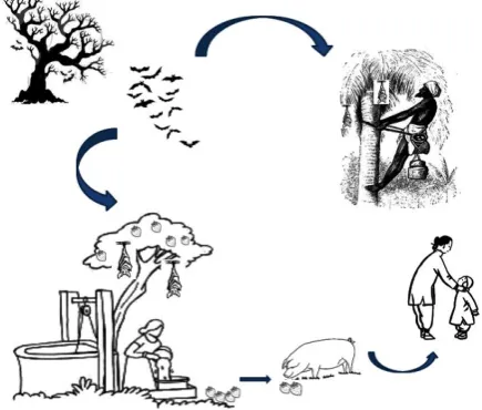
Figure 3: Transmission cycle of NiV.18
Transmission to humans mostly occurs by close contact with infected animals, consumption of contaminated food such as fruits eaten by the Pteropus bats or consumption of contaminated date palm saps (Figure 3).18 So far the transmission has not been efficient enough to maintain human-to-human transmission. But, worrying human to human transmission is common in Bangladesh as outbreaks of NiV occur annually. The present outbreak in
0 10 20 30 40 50 60 70
2001 2007 2018
Year of outbreak
Kerala, though the primary source seems to be bats in a well from where the family consumed the water. Human to human transmission can be attributed to the spread of the infection.14
[Rapid urbanization associated with massive deforestation has forced the movement of bats in to cities and towns with resultant spread of virus to the humans. Contaminated date palm saps and direct contact with the bats and human to human transmission have been the modes of transmission of NiV in India so far. But the chances of livestock getting infected and transmitting the disease to humans (as in case of outbreak in Malaysia) should also be kept in mind and proper measures taken to prevent future outbreaks].
PROPERTIES OF NIPAH VIRUS
NiV is an envelope virus with a filamentous nucleocapsid. Its genome is non-segmented, negative sense, single stranded RNA virus varying in size from 120 to 500 nm. The genome is large around 18,250 nucleotides as compared to other members of Paramyxoviruses due to an extended open read frame of the P gene and longer non- coding region for all genes except L gene.19 The genome consists of six major structural proteins, nucleocapsid (N), phosphoprotein (P), matrix protein (M), fusion protein (F), glycoprotein (G) and a large protein or RNA polymerase (L). The P gene produces non-structural accessory proteins known as C, V and W. NiV does not have the hemagglutinin and neuraminidase proteins as other Paramyxoviruses.20,21 The viral matrix protein (M) of the plasma membrane mediates the contact between the ribonucleoprotein (RNP) complex and the surface glycoprotein, virus particle formation and budding. Replication occurs in cytoplasm.22 NiV is encapsulated by nucleocapsid (N) and transcribed and replicated by the polymerase protein (L). The phosphoprotein (P) plays an essential role as a polymerase cofactor. It also acts as an immunosuppressor blocking interferon signaling by binding to host STAT-1.21
NiV- human interaction
The virus usually has a broad host range, with infection occurring predominantly in two cell types: neurons and endothelial cells.23 NiV enters the body via respiratory tract, then overcomes epithelial barrier and spreads systemically. The pathogenesis of NiV infection is mainly due to endothelial damage, multinucleate syncytia, vasculitis – induced thrombosis, ischemia and micro infarction in CNS, allowing the virus to overcome the blood-brain-barrier(BBB).24 The G and F proteins of NiV mediates viral entry in to the cell and also induce neutralizing antibodies.22 The entry of Paramyxovirus in to host cell requires the merging of viral lipid envelope and the host cell plasma membrane.25 In viruses of
Henipavirinae genus, this process is triggered by specific
binding of G protein to the host cell receptor ephrin B2/3, which induces G protein conformational changes that triggers F protein refolding drawing the host and viral membrane together to promote bilayer fusion.26 The viral matrix (M) protein mediates contact between the ribonucleoprotein complex and the surface glycoprotein brings about virus particle formation and budding.27
Clinical features
After an incubation period of 5 to 14 days, patients present with fever and headache followed by drowsiness, disorientation and mental confusion. It can progress to coma within 24-48 hours. Some patients may have respiratory illness during the early part of infection.17 The symptoms observed in patients during Siliguri outbreak were fever, headache, myalgia, vomiting, altered sensorium, respiratory symptoms (tachycardia to acute respiratory distress) and involuntary movements or convulsions.28 Long term squeal includes persistent convulsions and personality changes. Latent infection with subsequent reactivation of Nipah virus and death has also been reported months to years after exposure.
Laboratory diagnosis
The OIE reference laboratory for Henipaviruses in Asia-Pacific region is located at Australian Animal health Laboratory, Gee-long. In India BSL4 lab at National Institute of Virology (ICMR), Pune has got all the preparedness for the diagnosis of NiV in the Country. High Security Animal Disease Laboratory, Bhopal with BSL 3+ facility caters the need for exotic animal disease diagnosis.16
TREATMENT AND PREVENTION
Treatment is limited to supportive care. There is no approved or licensed therapeutic for treating NiV infection or disease in people.33 Invitro studies have shown that Ribavirin is effective against Nipah virus replication, but its clinical usefulness is remains uncertain.17 Passive immunization using a human monoclonal antibody targeting the Nipah G glycoprotein has been evaluated in the post-exposure therapy in the ferret model and is found to be beneficial.17 Standard infection control practices, proper barrier nursing are important in preventing nosocomial infections. Avoiding exposure to sick pigs and bats in endemic areas, not drinking raw date palm sap. Active surveillance, early detection of disease in community and livestock. Raising awareness of transmission, symptoms is important to reinforce standard infection control practices.
Current status of vaccine
All the candidate vaccines for NiV are in the pre-clinical stage, most of them being tested in the hamster, ferret and aorta gonad mesonephros (AGM) pre-clinical models. A subunit vaccine utilizing Hendra sG protein and adjuvant such as Alhydrogel and CpG oligodeoxynucleotide has shown to be protective against NiV in multiple animal models. The vaccine has been shown to produce cross protective antibodies against NiV and Hendra virus and offers great potential for Henipavirus protection in humans as well.34 Other various vaccines vector candidates are been tested in animal models using NiV outer- membrane G and/or F protein as target antigens to elicit neutralizing antibodies. Vaccine vectors included in different studies are vascular stomatitis virus (VSV), Rabies virus (RABV), Canary pox vaccine virus (CNPV ALVAC), Adeno associated virus (AAD), Measles virus (MV), Venezuelan equine encephalitis virus (VEE) and Newcastle disease virus (NDV).35-37
Possible causes of re-emergence of nipah virus in india
South–East Asia host wide variety of bats and is habitat for 30% of the known global bat fauna.38 Anthropogenic and environmental changes may impact the dynamic of virus transmission and public health.38 India is facing rapid growth, economic development, speedy urbanization associated with massive deforestation, overcrowding of cities, movement of migrant workers. Political instability is associated with decrease in nations output, Economic crisis, currency devaluation, and inflation leading to decrease in funding for health care
infrastructure.39 Existing weak surveillance, lack of awareness, lack of facilities for early diagnosis. Facilities if available are restricted to one place alone. Expanding population and diversity and poor infrastructure are possible causes of re-emergence of the NiV in India.
CONCLUSION
Nipah virus is zoonotic Paramyxovirus with potential of causing lethal disease across broad range of vertebrate species including humans. They are present in wide variety of bat reservoirs; they can be isolated and propagated. And as it is associated with high morbidity and mortality they pose a risk from natural outbreaks, laboratory accidents or deliberate misuse. The potential of NiV as a biological warfare agent cannot be ruled out, though the pandemic potential is poorly defined. The development of effective prevention and treatment strategies is very crucial.
Future challenges
Major challenge for the future is to develop and ensure a supply of robust, specific and affordable reagents to establish a rapid diagnostic capability in laboratories in regions where the virus is expected to occur in wildlife reservoir host. There is a need for international collaboration to develop systems of Quality control for laboratories conducting tests. Bat population from North East to North West states like Haryana has NiV antibodies which mean there is active NiV infection among Indian bats. 16 Preparedness, surveillance, constant vigil needs to be carried out continuously in the country. Inspection of all livestock at the point of origin, and when they arrive facilities for their slaughter to be maintained.
In endemic areas all facilities for the care of the patients infected with dangerous organisms should be made available. Neglect in use of safety precautions at anytime by the Healthcare workers should be taken care of. There is a need to improve communication between medical and veterinary health departments. Active interinstitutional and international co-ordination among human - animal virologist to facilitate early detection of outbreaks and to institute preventive measures as soon as possible is essential. Also there is a need for exchange of knowledge between human - animal virologist to understand how and when the shedding of virus by bats occurs. And last but not the least, there is an increasing need for educating the common people about personal and food hygiene.
Funding: No funding sources Conflict of interest: None declared Ethical approval: Not required
REFERENCES
1. Luby SP. The pandemic potential of Nipah virus. Antiviral Res. 2013;100(1):38-43.
glycoprotein using a phage display system. J Virological Methods. 2017;243:1-9.
3. Chua KB, Goh KJ, Wong KT, Kamarulzaman A, Tan PS, Ksiazek TG, Zaki SR, Paul G, Lam SK, Tan CT. Fatal encephalitis due to Nipah virus among pig-farmers in Malaysia. Lancet. 1999;354(9186):1257-9.
4. Virtue ER, Marsh GA, Wang LF. Paramyxoviruses infecting humans: the old, the new and the unknown. Future Microbiol. 2009;4(5):537-54. 5. Paton NI, Leo YS, Zaki SR, Auchus AP, Lee KE,
Ling AE, et al. Outbreak of Nipah-virus infection among abattoir workers in Singapore. Lancet. 1999;354(9186):1253-6.
6. Vera-Velasco NM, García-Murria MJ, del Pino MM, Mingarro I, Martinez-Gil L. Proteomic composition of Nipah virus-like particles. J Proteomics. 2018;172:190-200.
7. Mire CE, Satterfield BA, Geisbert JB, Agans KN, Borisevich V, Yan L, et al. Pathogenic differences between Nipah virus Bangladesh and Malaysia strains in primates: implications for antibody therapy. Scientific Rep. 2016;6:30916.
8. Chadha MS, Comer JA, Lowe L, Rota PA, Rollin PE, Bellini WJ, et al. Nipah virus-associated encephalitis outbreak, Siliguri, India. Emerging Infect Dis. 2006;12(2):235.
9. de Wit E, Munster VJ. Nipah Virus Emergence, Transmission, and Pathogenesis. In: Global Virology I-Identifying and Investigating Viral Diseases. Springer, New York, NY: 2015: 125-146. 10. Arankalle VA, Bandyopadhyay BT, Ramdasi AY, Jadi R, Patil DR, Rahman M, et al. Genomic characterization of nipah virus, west bengal, India. Emerging Infectious Dis. 2011;17(5):907.
11. Hariharan R. Fresh Nipah outbreak scare, schools, colleges to stay shut in Kozhikode till June 12: 10 points. Available at: https://www.ndtv.com/kerala- news/kerala-warns-of-second-wave-of-nipah-virus- killed-16-10-points-1861308?pfrom=home-topstories. Accessed on 05 October 2017.
12. Reuters. Nipah virus ourbreak: Two suspected cases of Nipah virus reported from Karnataka. The Times of India. Available at: https://timesofindia.indiatimes.com/india/two- suspected-cases-of-nipah-virus-reported-from-karnataka/articleshow/64285807.cms. Accessed on 05 May 2018.
13. Suri M. 10 Confirmed deaths from Nipah virus outbreak in India, including nurse treating patients.
Available at:
https://edition.cnn.com/2018/05/22/health/nipah-virus-death-toll-rises-intl/index.html. Accessed on 22 May 2018.
14. Xinhua. Bat infected well liked to 3rd outbreak of Nipah virus in India since 2001: officials. Asia and Pacific. Available at:
http://www.xinhuanet.com/english/2018-05/23/c_137200667.htm. Accessed on 23 May 2018.
15. Angeletti S, Presti AL, Cella E, Ciccozzi M. Molecular epidemiology and phylogeny of nipah virus infection: a mini review. Asian Pacific J Tropical Med. 2016;9(7):630-4.
16. Kulkarni DD, Tosh C, Venkatesh G, Kumar DS. Nipah virus infection: current scenario. Indian J Virol. 2013;24(3):398-408.
17. Center for Disease control and Prevention. Nipah Virus (NiV). Available at: https://www.cdc.gov/vhf/nipah/. Accessed on 30 May 2018.
18. Clayton BA. Nipah virus: transmission of a zoonotic paramyxovirus. Current Opinion Virol. 2017;22:97-104.
19. Lam SK. Nipah virus—a potential agent of bioterrorism? Antiviral Res. 2003;57(1-2):113-9. 20. Sugai A, Sato H, Yoneda M, Kai C. Gene end-like
sequences within the 3′ non-coding region of the Nipah virus genome attenuate viral gene transcription. Virology. 2017;508:36-44.
21. Bruhn JF, Barnett KC, Bibby J, Thomas JM, Keegan RM, Rigden DJ, et al. Crystal structure of the Nipah virus phosphoprotein tetramerization domain. J Virol. 2014;88(1):758-62.
22. Subramanian SK, Tey BT, Hamid M, Tan WS. Production of the matrix protein of Nipah virus in Escherichia coli: Virus-like particles and possible application for diagnosis. J Virological Methods. 2009;162(1-2):179-83.
23. Talekar A, Pessi A, Porotto M. Infection of primary neurons mediated by nipah virus envelope proteins: role of host target cells in antiviral action. J Virology. 2011;85(16):8422-6.
24. Erbar S, Maisner A. Nipah virus infection and glycoprotein targeting in endothelial cells. Virology J. 2010;7(1):305.
25. Bose S, Jardetzky TS, Lamb RA. Timing is everything: Fine-tuned molecular machines orchestrate paramyxovirus entry. Virology. 2015;479:518-31.
26. Wong JJ, Young TA, Zhang J, Liu S, Leser GP, Komives EA, et al. Monomeric ephrinB2 binding induces allosteric changes in Nipah virus G that precede its full activation. Nature Communications. 2017;8(1):781.
27. Dietzel E, Kolesnikova L, Sawatsky B, Heiner A, Weis M, Kobinger GP, et al. Nipah virus matrix protein influences fusogenicity and is essential for particle infectivity and stability. J Virol. 2016;90(5):2514-22.
28. Chadha MS, Comer JA, Lowe L, Rota PA, Rollin PE, Bellini WJ, et al. Nipah virus-associated encephalitis outbreak, Siliguri, India. Emerging infectious diseases. 2006;12(2):235.
29. Daniels P, Ksiazek T, Eaton BT. Laboratory diagnosis of Nipahand Hendra virus infections. Microbes and infection. 2001;3(4):289-95.
Nipah virus using real-time RT-PCR (TaqMan). J Virol Methods. 2004;120(2):229-37.
31. Wacharapluesadee S, Hemachudha T. Duplex nested RT-PCR for detection of Nipah virus RNA from urine specimens of bats. J Virological Methods. 2007;141(1):97-101.
32. Kaku Y, Noguchi A, Marsh GA, Barr JA, Okutani A, Hotta K, et al. Second generation of pseudotype-based serum neutralization assay for Nipah virus antibodies: sensitive and high-throughput analysis utilizing secreted alkaline phosphatase. J Virological Methods. 2012;179(1):226-32.
33. Broder CC, Xu K, Nikolov DB, Zhu Z, Dimitrov DS, Middleton D, et al. A treatment for and vaccine against the deadly Hendra and Nipah viruses. Antiviral Res. 2013;100(1):8-13.
34. Satterfield BA, Dawes BE, Milligan GN. Status of vaccine research and development of vaccines for Nipah virus. Vaccine. 2016;34(26):2971-5.
35. Kurup D, Wirblich C, Feldmann H, Marzi A, Schnell MJ. Rhabdovirus-based vaccine platforms against henipaviruses. J Virol. 2015;89(1):144-54.
36. Poulet H, Minke J, Pardo MC, Juillard V, Nordgren B, Audonnet JC. Development and registration of recombinant veterinary vaccines: the example of the canarypox vector platform. Vaccine. 2007;25(30):5606-12.
37. Ploquin A, Szécsi J, Mathieu C, Guillaume V, Barateau V, Ong KC, et al. Protection against henipavirus infection by use of recombinant adeno-associated virus–vector vaccines. J Infectious Dis. 2012;207(3):469-78.
38. Afelt A, Lacroix A, Zawadzka-Pawlewska U, Pokojski W, Buchy P, Frutos R. Distribution of bat-borne viruses and environment patterns. Infection, Genetics Evolution. 2018;58:181-91.
39. Pang T. Vaccination in the third world. Contemporary issues. Comptes Rendus de l'Académie des Sciences-Series III-Sciences de la Vie. 1999;322(11):995-7.